

To the Editor:
High-resolution peripheral quantitative computed tomography (HR-pQCT; Scanco Medical AG, Brüttisellen, Switzerland) is a novel peripheral CT instrument capable of accurately and reproducibly imaging bone microstructure at great resolution (isotropic voxel dimension of 82 μm). It provides precise measures of 3-D microstructural morphometric details and volumetric density of the cortical and trabecular components of bone (Figure 1), with minimal radiation exposure (< 3 μSv per scan)1. Therefore, HR-pQCT has the potential to identify and quantify early microstructural bone quality changes before permanent bone damage has occurred. To date, HR-pQCT has been used to assess bone quality in a variety of metabolic bone conditions. Image acquisition and analysis protocols are well defined for the assessment of systemic bone density and microarchitecture of the distal radius and tibia. This has largely been semiautomated to promote reproducibility and observer independence across investigational sites.
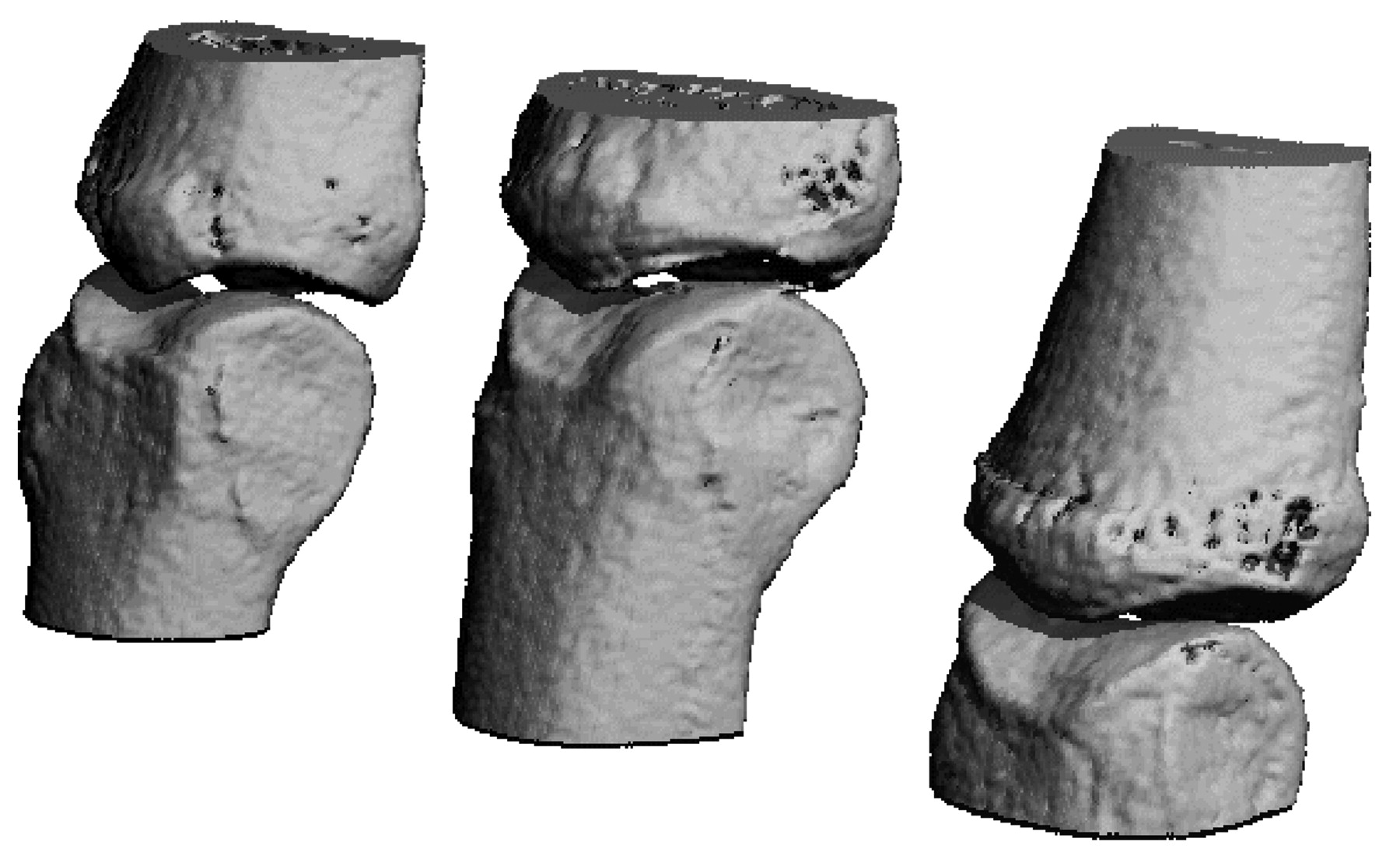
3-D reconstruction of the second, third, and fourth metacarpophalangeal joints using high-resolution peripheral quantitative computed tomography.
A new proposed application of HR-pQCT is in the study of inflammatory arthritis, to determine alterations to systemic bone density and periarticular bone such as erosions, joint space narrowing, and localized osteoporosis. The initial publication on using HR-pQCT to study rheumatoid arthritis (RA) metacarpophalangeal (MCP) joint changes was by Stach, et al2. This was followed by further studies to characterize erosions in RA and psoriatic arthritis3, erosion identification comparing high-resolution ultrasound and HR-pQCT4, and erosion healing with RA treatment5. Others have assessed erosion identification and periarticular bone density in RA6. One study has assessed systemic bone density in Chinese women using corticosteroids for lupus7. Researchers from sites in Calgary and Vancouver, Canada; Zurich, Switzerland; and San Francisco, California, USA, have also initiated studies and presented abstracts at international meetings, with publications in peer review and in press. An international collaboration meeting was held in Calgary on November 10-11, 2011, to share work completed to date, discuss image acquisition and analysis protocols, and develop new collaborative projects. Participants agreed on a standard image acquisition protocol for MCP joint analysis in inflammatory arthritis (Table 1).
Image acquisition protocol for metacarpophalangeal joint analysis in inflammatory arthritis using high-resolution peripheral quantitative computed tomography.
From this meeting, the SPECTRA, or Study GrouP for XTrEme-CT in RA, was established. Working groups were established to focus on priority areas for research and agreement, such as consensus on defining erosions, measuring joint space width, comparing HR-pQCT imaging with standard imaging techniques (e.g., plain radiographs, magnetic resonance imaging, and ultrasound), analysis methodology, and cortical bone evaluation analysis protocols.
The SPECTRA group will initially focus on HR-pQCT imaging in early and established RA, but future avenues of research should include HR-pQCT evaluation of early microstructural periarticular MCP joint bone changes in other systemic inflammatory arthritis conditions. The SPECTRA group plans to convene regularly around the European League Against Rheumatism and American College of Rheumatology annual meetings. Any interested investigators are welcome to join the collaboration.
Acknowledgment
The co-chairs thank the following participants who attended the first SPECTRA meeting: Susan Barr, University of Calgary, Calgary, Canada; Stephanie Boutroy, Université de Lyon, Lyon, France; Andrew Burghardt, University of California San Francisco, San Francisco, USA; Roland Chapurlat, Université de Lyon; Stephanie Finzel, University of Erlangen-Nuremberg, Nuremberg, Germany; Anne Fouque-Aubert, Université de Lyon; Tanja Harrison, University of Calgary; Xiaojuan Li, University of California San Francisco; Hubert Marotte, Sainte-Etienne, France; Liam Martin, University of Calgary; Kathryn Stok, ETH Zurich, Zurich, Switzerland; Martin Zulliger, Scanco Medical, Bassesdorf, Switzerland.


