Article Text

Statistics from Altmetric.com
IL-18 is a pleiotropic immunoregulatory cytokine that has been described and implicated in the pathogenesis of a variety of inflammatory diseases.1–4
Studies in murine models of arthritis and clinical studies suggest that dendritic cells, macrophages and synoviocites within the synovial membrane can produce IL-18.1 5–7 IL-18 expression has in turn been implicated in the reciprocal regulation of other pro-inflammatory cytokines, such as tumour necrosis factor alpha.8
Recent data clearly demonstrated that IL-18 serum levels were significantly elevated in adult-onset Still’s disease (AOSD) and correlated with disease activity and serum ferritin levels.2–4 AOSD is characterised by substantial and dysregulated cytokine production, with higher levels of IL-18 messenger RNA expression detected in skin and synovial membrane biopsies of active AOSD compared with controls.9
However, the main source of IL-18 expression is as yet poorly understood.
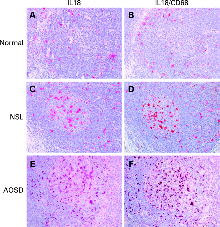
We had the opportunity to investigate the expression of IL-18 in two lymph nodes of AOSD and thereby define this site as a critical tissue of origin for IL-18 hyperproduction. The expression was compared with non-specific lymphadenitis (NSL; no systemic inflammatory disorder present) and two lymph nodes from “normal” controls obtained during vascular surgery. Local IL-18 expression was analysed by immunohistochemistry using an IL-18 monoclonal antibody (clone 2D3B6; MD Biosciences, Switzerland) as previously described.10 Greater expression of IL-18 was observed in AOSD lymph nodes compared with NSL and normal lymph nodes (fig 1). IL-18 was particularly overexpressed in hyperplastic, dysmorphic germinal centres detected in AOSD-derived tissue (fig 1E). Commensurate with our previous observations, IL-18 was also detected in germinal centres of mature follicles in NSL.10 Of interest, whereas IL-18 was scarcely detected in the mantle zone in normal lymph nodes (fig 1A), high levels of expression were observed in the mantle zone of AOSD follicles (fig 1E). In addition, numerous IL-18-producing cells were observed in afferent lymphatics in biopsies obtained from AOSD and NSL patients compared with normal controls. Finally, we performed double immunohistochemistry for IL-18 and CD68, and found that IL-18 co-localised almost exclusively with CD68-positive cells (fig 1F), indicative of a monocyte–macrophage lineage origin in lymph nodes.
We next confirmed increased serum IL-18 levels in nine cases of AOSD compared with patients with rheumatoid arthritis, primary Sjögren’s syndrome and healthy subjects, as previously described (fig 2).10 Moreover, IL-18 serum levels in AOSD patients correlated with serum ferritin levels (data not shown).

Although the importance of systemic IL-18 upregulation in AOSD is well documented, no data were previously available regarding the nodal expression of IL-18. We provide evidence that IL-18 is highly expressed at the protein level in the germinal centres and in the mantle zone of AOSD lymph nodes compared with non-specific lymphadenitis and “normal” lymph nodes as controls.
The increased systemic production of IL-18, observed in AOSD patients, could reflect the high local expression of this cytokine in the lymph node as an important site for such activation. This observation now needs to be confirmed in larger groups of patients to validate the hypothesis that the lymph node represents a major source of IL-18 in AOSD.
REFERENCES
Footnotes
Competing interests: None.
Funding: PC is receiving fellowship support from the University of Glasgow. MB is receiving support from the Arthritis Research Campaign.