Article Text

Statistics from Altmetric.com
A 19 year old man attended hospital after his recent discharge from the navy owing to his hand arthropathy. His symptoms began aged 14 years with swelling but no pain or disability of his distal interphalangeal (DIP) and proximal interphalangeal (PIP) finger joints. At the age of 17 he joined the navy and 1 year later began to experience considerable pain in his hands while carrying out his duties. He was reassigned and after 3 months of light duties was symptom free, although his joints remained swollen and deformed (fig 1). Flexion at the PIP and DIP joints was limited (fig 2), and the hands were functionally normal with an average grip and pinch strength. He continued to work as a gardener with no joint symptoms. Both his father and paternal grandfather had had the same condition.

The PIP and DIP joints of the fingers of both hands are swollen.
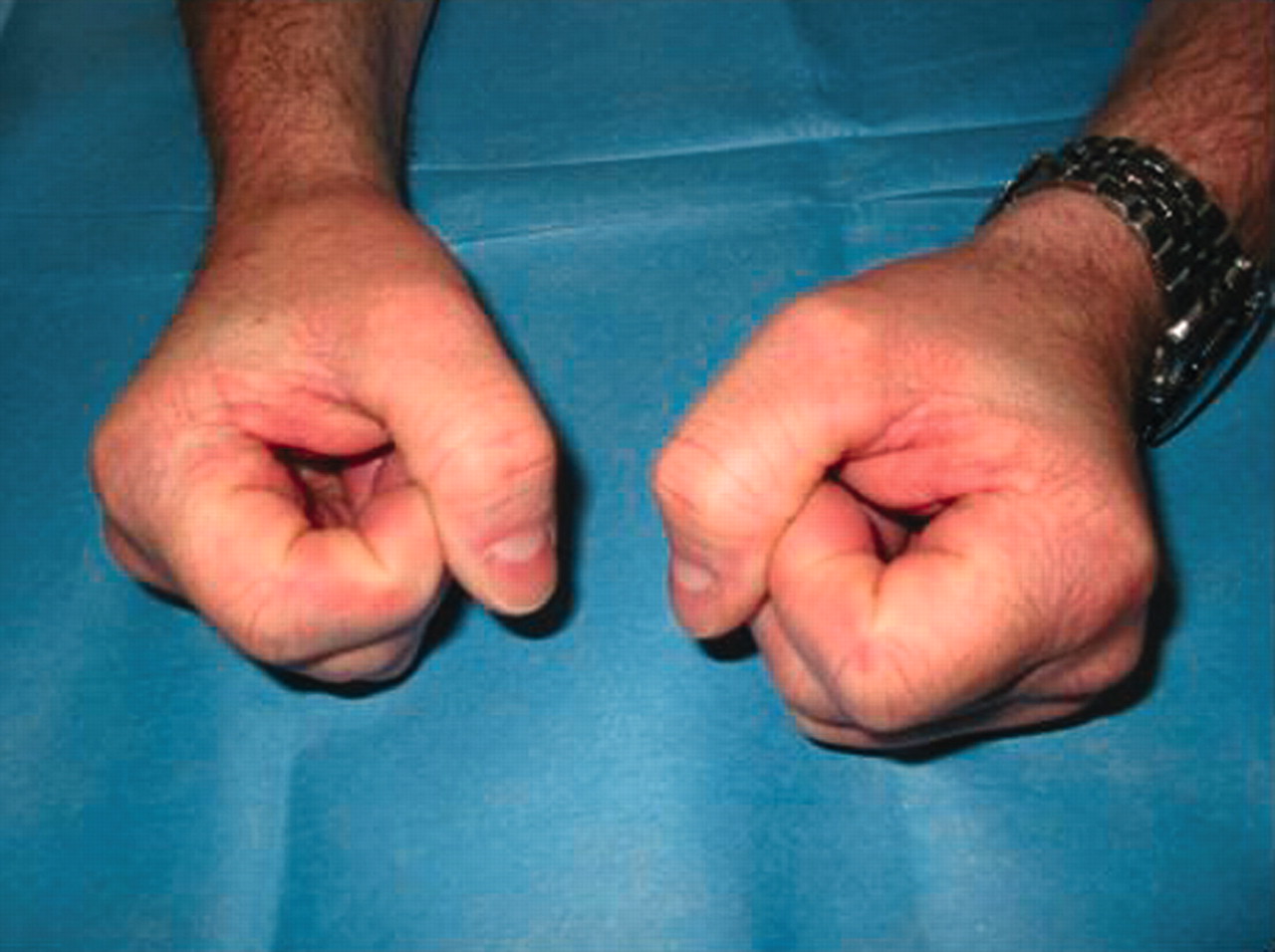
Flexion at the PIP and DIP joints is limited.
Blood tests showed normal haemoglobin, white cell count, and erythrocyte sedimentation rate (ESR). A hand x ray examination showed the classical radiological features of Thiemann’s disease with flattening, broadening, fragmentation, and irregular opacification of the phalangeal epiphyses (fig 3).

Flattening, broadening, fragmentation, and irregular opacification of the phalangeal epiphyses.
Thiemann’s disease was first described in 1909 in a 16 year old boy who had noted progressive enlargement of his PIP joints.1 It manifests as painless swelling of the PIP joints of the fingers occurring before 25 years of age. Blood tests, including acute phase markers, are normal. The radiological findings are initially those of flattening, broadening, fragmentation, and irregular opacification of the proximal phalangeal epiphyses, followed by late osteoarthritic changes.2 Differential diagnosis includes osteochondrosis (infarct of epiphysis secondary to a blood dyscrasia), rheumatoid arthritis, osteoarthritis, an infective lesion, trauma, and metabolic or endocrine disorders.3
The condition has been reported as an autosomal dominant hereditary disease with strong penetrance, resulting in an equal sex distribution,4 and also as sporadic cases showing a threefold male dominance.2 Histological investigation has shown aseptic necrosis of the bone with normal vessels and no inflammation.5 Thiemann’s disease often has a benign course; a follow up of seven patients presenting in adolescence found only two had continuing long term symptoms, and radiographs after growth plate closure showed normal phalangeal dimensions without arthrosis in four patients.6 Trauma may worsen the prognosis by deformation of a susceptible epiphysis.6
Treatment comprises reduction in those activities producing symptoms,7 with good response as illustrated by the sailor.