Article Text

Abstract
Objective: To carry out a prospective two year follow up study comparing conventional radiography, three-phase bone scintigraphy, ultrasonography (US), and three dimensional (3D) magnetic resonance imaging (MRI) with precontrast and dynamic postcontrast examination in detecting early arthritis. The aim of the follow up study was to monitor the course of erosions during treatment with disease modifying antirheumatic drugs by different modalities and to determine whether the radiographically occult changes like erosive bone lesions of the finger joints detected by MRI and US in the initial study would show up on conventional radiographs two years later. Additionally, to study the course of soft tissue lesions depicted in the initial study in comparison with the clinical findings.
Methods: The metacarpophalangeal, proximal interphalangeal, and distal interphalangeal joints (14 joints) of the clinically more severely affected hand (soft tissue swelling and joint tenderness) as determined in the initial study of 49 patients with various forms of arthritis were examined twice. The patients had initially been divided into two groups. The follow up group I included 28 subjects (392 joints) without radiographic signs of destructive arthritis (Larsen grades 0–1) of the investigated hand and wrist, and group II (control group) included 21 patients (294 joints) with radiographs showing erosions (Larsen grade 2) of the investigated hand or wrist, or both, at the initial examination.
Results: (1) Radiography at the two year follow up detected only two erosions (two patients) in group I and 10 (nine patients) additional erosions in group II. Initial MRI had already detected both erosions in group I and seven (seven patients) of the 10 erosions in group II. Initial US had depicted one erosion in group I and four of the 10 erosions in group II. (2) In contrast with conventional radiography, 3D MRI and US demonstrated an increase in erosions in comparison with the initial investigation. (3) The abnormal findings detected by scintigraphy were decreased at the two year follow up. (4) Both groups showed a marked clinical improvement of synovitis and tenosynovitis, as also shown by MRI and US. (5) There was a striking discrepancy between the decrease in the soft tissue lesions as demonstrated by clinical findings, MRI, and US, and the significant increase in erosive bone lesions, which were primarily evident at MRI and US.
Conclusions: Despite clinical improvement and a regression of inflammatory soft tissue lesions, erosive bone lesions were increased at the two year follow up, which were more pronounced with 3D MRI and less pronounced with US. The results of our study suggest that owing to the inadequate depiction of erosions and soft tissue lesions, conventional radiography alone has limitations in the intermediate term follow up of treatment. US has a high sensitivity for depicting inflammatory soft tissue lesions, but dynamic 3D MRI is more sensitive in differentiating minute erosions.
- magnetic resonance imaging
- ultrasonography
- x rays
- scintigraphy
- arthritis, finger joints
- ACR, American College of Rheumatology
- CRP, C reactive protein
- 3D, three dimensional
- DIP, distal interphalangeal
- DMARDs, disease modifying antirheumatic drugs
- ESR, erythrocyte sedimentation rate
- IV, intravenous
- MCP, metacarpophalangeal
- MIP, maximum intensity projection
- MRI, magnetic resonance imaging
- PIP, proximal interphalangeal
- RA, rheumatoid arthritis
- RF, rheumatoid factor
- US, ultrasonography
Statistics from Altmetric.com
- ACR, American College of Rheumatology
- CRP, C reactive protein
- 3D, three dimensional
- DIP, distal interphalangeal
- DMARDs, disease modifying antirheumatic drugs
- ESR, erythrocyte sedimentation rate
- IV, intravenous
- MCP, metacarpophalangeal
- MIP, maximum intensity projection
- MRI, magnetic resonance imaging
- PIP, proximal interphalangeal
- RA, rheumatoid arthritis
- RF, rheumatoid factor
- US, ultrasonography
The therapeutic options available for rheumatoid arthritis (RA) have recently been supplemented by targeted treatments that stop clinical progression and the process of joint destruction.1–6 These newer and more expensive treatments crucially rely on the early and reliable diagnosis of RA. Conventional radiography is currently regarded as the standard of reference for detecting and quantifying destructive joint processes in arthritis, although its sensitivity limits its usefulness in depicting early arthritic joint lesions.7–12 Ultrasonography (US) and magnetic resonance imaging (MRI) are alternative diagnostic modalities for demonstrating early arthritis.12–14
This article reports the first prospective long term study comparing different novel and conventional imaging modalities for diagnosing early arthritis of the finger joints. The second part of this study presents the follow up results of our initial study and uses the same study design.12 The follow up study aimed at investigating the course of erosions in arthritic patients treated with disease modifying antirheumatic drugs (DMARDs) and determining whether the radiographically occult changes like bone lesions of the finger joints detected by MRI and US in the initial study would show up on conventional radiographs two years later. In addition, we wanted to study the course of soft tissue lesions depicted in the initial study in comparison with the clinical findings. Another aim was to identify the optimal imaging modality for detecting both early erosions and acute inflammatory joint changes (synovitis, tenosynovitis) compared with conventional radiography and clinical findings. Seronegative spondyloarthropathies with involvement of peripheral joints are an important differential diagnosis in RA. They include psoriatic arthritis with a polyarticular course and HLA-B27 associated arthritis with involvement of the finger joints. We therefore included patients with these disorders both in our initial study and in the follow up examination.
PATIENTS AND METHODS
Patients
Forty nine of a total of 60 patients investigated initially underwent follow up assessment two years later (1998). The patients met established diagnostic criteria of rheumatoid arthritis (RA),15 spondyloarthropathy, especially psoriatic arthritis,16 and arthritis associated with connective tissue disease.17 The results of the initial comparison of the imaging procedures have been reported before.12
Table 1 summarises the clinical characteristics of the patients participating in the follow up study.
Number of patients, sex, age, duration of disease, CRP, and ESR in both groups in relation to different diagnoses. Results shown as number or median (range)
The follow up study used the two groups into which the patients had been divided for the initial study: group I, included 28 patients (20 women, eight men; median age 41 years, range 20–66; disease duration of 42.5 months, range 24–221) of the 32 patients initially assigned to this group. Assignment to this group was based on the absence of radiographic signs of erosive arthritis (Larsen stage 0 or 1)18,19 of the hand and wrist investigated in the initial study. Group II, our control group, included 21 patients (13 women, eight men; median age 58 years, range 35–86; disease duration 114 months, range 32–330) of the 28 patients included initially. The initial radiographs of these patients had shown erosions (erosions affecting <25% of the joint area, corresponding with the radiographic Larsen stage 2) of the hand and/or wrist examined.
Group I included 16 patients with RA who met the revised American College of Rheumatology (ACR) criteria.15 Five patients with RA already had radiographic signs of erosions in other joints. Seven patients in group I fulfilled the criteria for spondyloarthropathy. Six patients had psoriatic arthritis,16 and one patient had ankylosing spondylitis. Two patients had polyarticular arthritis associated with connective tissue disease (one patient with systemic lupus erythematosus and one patient with undifferentiated connective tissue disease, both with polyarthritis) who were initially included in the study with the tentative diagnosis of RA. Three patients had undifferentiated oligoarthritis (one patient with psoriasis and arthralgia of distal interphalangeal (DIP) joints and two patients with HLA-B27 associated oligoarthritis without a history of gastrointestinal or urogenital infections).
Group II included 15 patients with RA and six patients with spondyloarthropathy, all having psoriatic arthritis.
Experienced investigators who were unaware of the clinical findings and diagnoses analysed all methods independently of each other. In all patients, the clinical examination was performed by AS, radiography by DL, scintigraphy by DS, US by MBa, and MRI by MBo. These observers were the same at baseline and after two years with the exception of the clinical investigator. Each investigator was completely unaware of the other investigators' findings. However, it is normal procedure that the doctor performing US also records the patient's clinical findings. For the purpose of the study, US was performed by MBa without knowledge of the clinical score assigned by AS. Fourteen metacarpophalangeal (MCP), proximal interphalangeal (PIP), and DIP joints of the dominant hand were analysed for each patient using conventional radiography, three-phase bone scintigraphy, US, and contrast-enhanced MRI. The clinically more severely affected hand as determined in the initial study was examined by MRI both initially and at follow up. This procedure was chosen to save time and costs and because it is not possible to examine both hands simultaneously at high resolution in a single contrast-enhanced MRI examination. All patients had been receiving stable treatment for a minimum of four weeks before the initial imaging was done. Drugs at entry into the follow up study included non-steroidal anti-inflammatory drugs (group I, 0% of patients; group II, 14% of patients), corticosteroids (group I, 46%; group II, 67%), methotrexate (group I, 46%; group II, 76%), sulfasalazine (group I, 4%; group II, 5%), aurothiomalate (group I, 0%; group II, 5%), and azathioprine (group I, 7%; group II, 9.5%). Seven patients of group I and no patients of group II received no drugs. As a rule, all imaging procedures were performed on the same day, with the possible exception of MRI which was performed 2–4 weeks before or after the other diagnostic procedures with the patients still receiving the same drugs. Of the 49 patients, seven patients underwent MRI more than one week before or after the other imaging modalities. This delay was due to organisational reasons. The remaining 42 patients were examined by all imaging modalities within one week..
The following laboratory parameters were included in the evaluation: erythrocyte sedimentation rate (ESR, Westergren method), C reactive protein (CRP, nephelometry), leucocytes, γ-globulin, and rheumatoid factor (RF, RF-IgM-ELISA). HLA alleles had already been determined by HLA-DRB1 and HLA-DQB1 sequence-specific oligonucleotide typing20 during the initial study.
The analysis of diagnostic procedures (clinical examination, standard radiography, three-phase bone scintigraphy, US, and MRI) was performed for each joint in the same way as described in the initial study.12 Only the dominant hand, which was also examined by MRI, was included in the analysis.
Clinical analysis
A binary scoring system (0–1) was used to assess each joint as normal (0) or abnormal (1) for joint tenderness and soft tissue swelling.
Standard radiographs
Standard radiographs of the hands and feet were obtained in two planes (dorsopalmar and zither player position) and evaluated according to the Larsen score.12,18,19 The presence of erosions, juxta-articular osteoporosis, and juxta-articular soft tissue swelling was recorded for each joint and graded as normal (0) or abnormal (1).
Three-phase bone scintigraphy
Three-phase bone scintigraphy21 was performed after intravenous (IV) injection of 600 MBq of technetium-99m methylene diphosphonate. Static images of the hands (five minute acquisition time) were obtained two minutes (phase II - “blood pool” images) and three hours (phase III - “bone” images) after injection. In addition, whole body scintigraphy was performed in phase III. Accumulation of the radiopharmaceutical drug was scored separately for each joint in phases II and III and graded as normal (0) or abnormal uptake (1).
Ultrasonography
Ultrasonography was performed with a 7.5 MHz linear array transducer in combination with an acoustic standoff (silicone) for better focusing (Ultramark 4, ATL, Bothel, USA). We deliberately used the same US system and transducer in both studies in order to have identical conditions for comparison, although high frequency 10–15 MHz transducers are available today for examination of the finger joints. Each finger joint was investigated in the sagittal plane from a dorsal and a palmar orientation with the hand in a neutral position. The bone surfaces of the finger joints were examined from the dorsal aspect during extension and flexion (>70°) and from the palmar aspect during extension. Synovitis was identified as a hypoechoic or anechoic area in the joint space of the MCP, PIP, or DIP joint. The synovial sheath of the flexor tendon, which was identified as a slightly hypoechoic area, was clearly detectable at the edge of the tendon's profile on the transverse scans. The presence of a well defined area of increased echogenicity within the tendon sheath was considered to indicate synovial thickening. Tendon sheath thickness was measured on longitudinal scans mainly at the MCP joint. Erosion was defined as a disruption of the bone surface by an indentation which gave the bone surface an irregular appearance. Abnormal findings were documented in two perpendicular planes. The MCP, PIP, and DIP joints were examined for capsular distention, synovial proliferation (indicating synovitis), erosions, and signs of tenosynovitis. The findings were also graded as normal (0) or abnormal (1).
Magnetic resonance imaging
MRI was performed in the same manner as in the initial examination12 with a 0.2 T imager (Magnetom Open, Siemens, Erlangen, Germany) with the patients in sitting position and using a small flex coil. The dominant hand was examined. Three dimensional (3D) T1 weighted gradient echo sequences were obtained before and after IV injection of the non-ionic paramagnetic contrast agent gadodiamide (Gd-DTPA-BMA (Omniscan), Nycomed, Oslo, Norway) at a dose of 0.3 mmol/kg of body weight. The high dose administration of gadodiamide was performed as part of a phase III contrast agent study initially investigating both the efficacy and safety of the contrast agent. The MRI contrast agent has since been approved for clinical use in Germany.
Patients were examined with an unenhanced T1 weighted 3D FLASH sequence (repetition time 34 ms; echo time 12 ms; flip angle 20°, matrix 256×256; field of view 144×230 mm; slice thickness 1 mm; two acquisitions) and a T1 weighted multislice dynamic 3D FLASH sequence with eight repetitions and 1.6 mm slice thickness. The latter was used for one precontrast acquisition and seven acquisitions after contrast administration. The contrast agent was injected as a bolus in the interval between the first and the second acquisition. Each acquisition took 55 seconds with a delay of two seconds between them.
MRI assessment
The magnetic resonance images were assessed from hard copies and on the monitor (Magic View, Siemens, Erlangen, Germany). A qualitative analysis of erosive joint lesions of the MCP, PIP, and DIP joints was performed using coronal sagittal reconstructions (multiplanar reconstruction). An erosive joint lesion was defined as a joint related cortical defect with or without a decrease in the signal intensity of the adjacent subchondral bone marrow on precontrast T1 weighted images. For qualitative analysis of inflammatory lesions such as synovitis and tenosynovitis, a maximum intensity projection (MIP) was generated from the fourth and seventh postcontrast acquisition. On MIP images, all regions showing contrast enhancement were added up and displayed in three dimensions. The inherently hyperintense bone segments were likewise displayed in the MIP and provided anatomical and morphological orientation.
Enhancement was assessed qualitatively by visual evaluation and quantitatively using a region of interest technique calculated by subtracting the precontrast image from the postcontrast image. All tendon sheaths or peritendinous tissue showing an enhancement of >50% were classified as tenosynovitis or tendinitis at MRI. The presence of tenosynovitis was assessed on coronal images and by analysing sagittal reconstructions of the fingers. The latter were nearly parallel to the course of the tendons. Similarly, a 50% signal enhancement was used as a threshold for identifying the presence of synovitis in assessing the joints. The number of erosions as well as the presence of synovitis and tenosynovitis were determined for each joint and graded as either normal (0) or abnormal (1).
Statistical analysis
The data were analysed by non-parametric methods. The McNemar test was used to analyse differences between groups of patients and imaging modalities.
RESULTS
Patient group
Laboratory findings (ESR, CRP, leucocytes, and γ-globulin) did not differ significantly between the groups (table 1).
Figures 1–4 show the percentages of pathological findings in groups I and II by the different imaging methods.

Detection of soft tissue lesions (%) by the different modalities in group I (n=392 finger joints = 100%).

Detection of bone lesions (%) by the different modalities in group I (n=392 finger joints = 100%).

Detection of soft tissue lesions (%) by the different modalities in group II (n=294 finger joints = 100%).

Detection of bone lesions (%) by the different modalities in group II (n=294 finger joints = 100%).
Clinical data
The soft tissue swelling and joint tenderness were decreased in both groups at the two year follow up (figs 1–4).
Conventional radiography
The number of erosions was increased in both groups at the two year follow up, whereas soft tissue swelling was decreased (figs 1–4).
No patient in group I had erosions of the finger joints on initial radiographs because this would have automatically assigned this person to group II (see “Patients and methods”) at the beginning of the study. Two years later only two erosions of the finger joints (PIP joint) were found in two patients with RA (fig 2). Both erosions had already been detected by initial MRI and were evident at follow up as well (figs 5A-D).

Images obtained by radiography (A-D), MRI (E-I), scintigraphy (J-M), ultrasound (N-P) of the clinically most severely affected left hand in a woman with RA aged 20 at the time of the initial examination (time 0) and 22 at the time of follow up. (A, B) Survey radiograph of the left hand at time 0 in dorsovolar projection (A) and with the hand in the volardorsal, semisupine (45°) position with abducted fingers, the so-called “zither player position” (B): demonstration of a narrowed joint cleft in PIP joint III and of a small cystoid brightening on the radial side without disruption of the border lamella at the head of the proximal phalanx of digit III (open arrow). There is partial loss of the subchondral border lamella at the ulnar base of the proximal phalanx of digit IV (closed arrow). These changes do not yet represent direct signs of arthritis (Larsen stage 1). (C, D) Follow up radiography of the left hand performed two years after the initial examination in the dorsovolar projection (C) and in the zither player position (D): the joint clefts of all PIP joints now clearly show narrowing. The cystoid brightening at the head of the proximal phalanx of digit III seen at time 0 is now definitely identified as an erosion, but only on the film obtained in the zither player position (thick arrow) and not on the dorsovolar projection. The partial loss of the subchondral border lamella at the ulnar base of the proximal phalanx of digit IV now shows recalcification. Newly developed cystoid lesions not disrupting the border lamellae (thin arrows) are seen on the radial side of the head of the proximal phalanx of digit II, on the ulnar side of the head of the proximal phalanx of digit V, at the metacarpal head of digit I, and in corresponding locations at the scaphotrapezoid joint. The new erosion in PIP joint III is a direct sign of arthritis affecting less than 25% of the joint area, corresponding to Larsen stage 2.

(contd) (E, F, G) MRI of the left hand performed at time 0 using an unenhanced T1 weighted 3D gradient echo sequence. The figure only shows one coronal section (E) obtained before contrast administration and a corresponding postcontrast section (F, G). (E) A large erosion on the radial side of the head of the proximal phalanx of digit III (open arrow) can be seen, which is demonstrated by MRI in association with extensive synovitis (asterisk) at PIP III already two years before there is clear cut radiographic evidence of its presence (fig 5D). After contrast medium administration (F), this erosion is partly obscured by the pronounced enhancement of the synovitis (*) at PIP III. Additional, smaller erosions—likewise not demonstrated radiographically—are seen in the metacarpal head of digit IV (arrow with loop) (more clearly seen in other images), on the ulnar side of the head of the proximal phalanx of PIP IV (thin, white arrow), and on the ulnar side of the metacarpal head of digit V (white arrow ) (G). Florid, contrast-enhanced synovitis (*, s) is depicted at PIP joints I–V, and at MCP joints I, II, IV, V. Florid, contrast-enhancing tenosynovitis (t) is seen affecting the flexor tendons of fingers 1–5. The radiographically demonstrated (fig 5A) partial loss of the subchondral border lamella at the ulnar base of the proximal phalanx of digit IV corresponds to contrast-enhancing osteitis affecting the entire base of the phalanx at MRI (black arrow). (H, I) After two years of DMARD treatment the patient underwent follow up MRI of the left hand using the same parameters as at time 0 using a comparable section orientation. The known lesion on the radial side of PIP joint III shows regression but still shows pronounced enhancement reflecting florid activity. The erosion on the ulnar side of PIP joint IV (open arrow (I) shows progression in the presence of more extensive synovitis (*, I). The tenosynovitis on the flexor side of digit III has a higher signal intensity on the postcontrast image than at time 0.

(contd) (J, K) Initial scintigraphy, phases II (J) and III (K), shows hot spots in PIP joints II, III, and IV and in the wrist (lunate bone or adjacent parts of ulnar and radial bones). (L, M) At follow up two years later, scintigraphy, phase II (L), shows hot spots in the wrist, MCP joints I and III, IP joint, PIP joints II and III. Scintigraphy, phase III (M), shows hot spots in the wrist, MCP joints I, III, and V, interphalangeal joint, and DIP joints II, III, and IV.

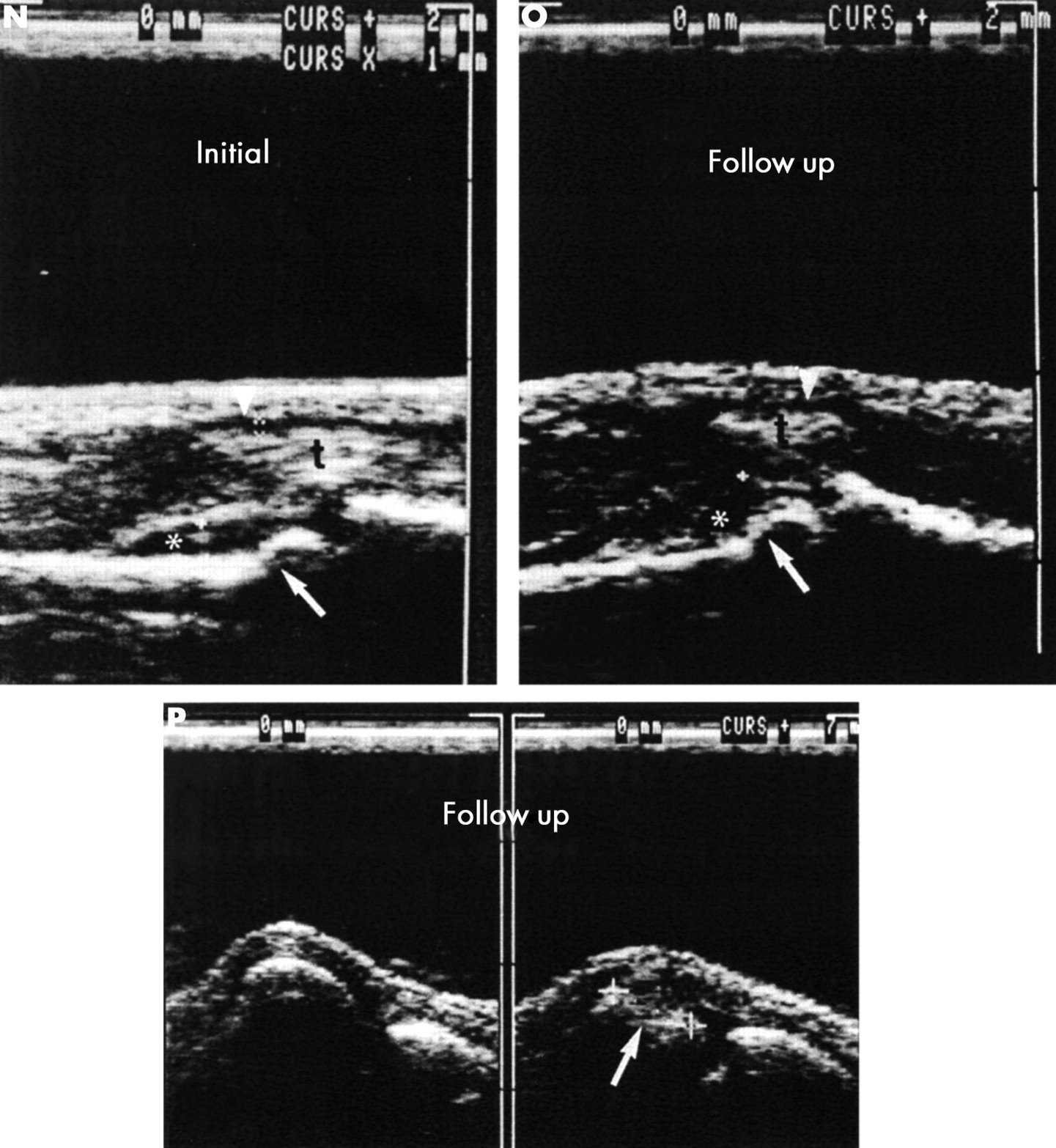
(contd) (N, O, P) (N) The initial ultrasound examination showed synovitis of the proximal interphalangeal (PIP) joints II, III. Ultrasound shows a hypointense line indicating synovitis (*) in PIP joints II (left) and III (right). The flexor tendon sheaths (arrowheads) of the second and third fingers are distended as an indication of tenosynovitis (t, flexor tendon). The step-like interruption of the bone margin (arrow) of the head of the proximal phalanx of digits II and III is indicative of erosion. (O) The figure shows a hypointense line indicating synovitis (*) in PIP joints II (left) and III (right). The flexor tendon sheaths (arrowheads) of the second and third fingers show minimal distention as an indication of tenosynovitis (t, flexor tendon). Irregularities of the joint contour were found at the heads of the proximal phalanx II and III (arrows). Ultrasound shows a large erosion at the head (arrow) of MCP joint V (P, right side) in the dorsal aspect and in the flexed MCP joint, which was not seen at conventional radiography (figs 5A-D) but in both MRI examinations (initially and at the two year follow up, fig 5G). The left side shows a normal joint contour of MCP joint IV.
Erosions in group II were found in 49 finger joints (17%; 17 patients) initially and in 53 finger joints (18%; 19 patients) two years later (web extra fig W1). Six erosions (four patients): three in MCP joints IV and V, two in PIP joints III and V, one in DIP joint II) were not detected by conventional radiography at follow up, whereas MRI identified four additional erosions (four patients) initially and five erosions (four patients) at follow up. Radiography detected only 10 (nine patients with RA) additional erosions at the two year follow up. Initial MRI had already detected seven of these 10 erosions (seven patients). Four patients had erosions only in the wrist, but not in the finger joints on initial radiographs. Two of these four patients had three erosions of the finger joints at follow up radiography. MRI detected bone lesions of the finger joints in all four of these patients both initially and two years later. Two of these three erosions (two patients) seen on conventional radiographs had already been identified by MRI initially as well as at the two year follow up.
Of the 49 erosions demonstrated initially, 14 were also depicted by US and 35 by MRI. Erosions detected by conventional radiography only, but not by MRI in the initial examination were present in 14 finger joints (five patients: four with RA and one with spondyloarthropathy). Two of these erosions were found in the MCP joints I of two patients, five erosions in the PIP joints III–V of three patients, and seven erosions in the DIP joints II–V of three patients. At follow up, 12 of the 14 erosions (five patients) were identified by conventional radiography and eight of them (five patients) by MRI. Of the 53 erosions demonstrated at follow up, 23 were observed by US and 44 by MRI (figs 5E-I). Seven erosions escaped detection by MRI (five patients: one with psoriatic arthritis, four with RA); two of these erosions were found in the PIP joints IV of two patients, five in DIP joints III–V of three patients.
Juxta-articular osteoporosis was increased in group I (17% v 35%) and decreased in group II (32% v 24%) at the two year follow up.
Web fig W1 shows the agreement for the presence of erosions (group II) among plain radiography, MRI, and US in the initial examination and at follow up.
Three-phase bone scintigraphy
Abnormal tracer accumulation was decreased in phases II and III, and the decrease was significant for phase II in group II and phase III in both groups at the two year follow up (figs 1–4, 5J–M).
Ultrasonography
Whereas synovitis was reduced significantly in both groups, the detection of erosions was increased at the two year follow up, but the difference was significant for group II only (figs 1–4). Seven patients of group I had no signs of erosions either initially or after two years. In group I one erosion detected by conventional radiography at follow up only, had already been seen initially (figs 5N-P). Two patients of group II showed no signs of erosion at either examination. In group II four erosions (four patients) which conventional radiography depicted at follow up only had already been seen initially. Of the 33 erosions detected by initial US in group II, 29 were also detected by MRI. Four erosions seen by US were not detected by MRI (two erosions in the MCP joints II and IV of one patient, two erosions in the PIP joint II of two patients) (web fig W1). Of the 72 erosions seen at follow up US, 64 were also seen by MRI. MRI did not detect eight erosions (two erosions of the MCP joints I and IV in two patients, three erosions of the PIP joints II–IV in two patients, three erosions of the DIP joints II, IV, and V in three patients). The tenosynovitis of the flexor tendon sheath was decreased significantly in both groups and the tendinitis of the extensor tendon was increased significantly in both groups compared with the initial examination. However, comparison of the absolute numbers showed no significant differences between US and MRI.
MRI
Whereas synovitis was reduced significantly in both groups, the detection of erosions was increased significantly in both groups at the two year follow up (figs 1–4). MRI detected erosions not seen on conventional radiographs in the finger joints of 24 patients (15 patients with RA, five patients with spondyloarthropathy, two patients with connective tissue disease, two patients with undifferentiated oligoarthritis) in the initial examination and in 27 patients at follow up. Erosions showing enhancement as a sign of a florid inflammatory process were increased in group I (not significant) and decreased in group II (not significant) at the two year follow up. Erosions without enhancement were increased in both groups (significant) two years later. Most erosions were found in the MCP joints, especially in the head of the os metacarpale. Web fig W2 shows the distribution of erosions in the different finger joints of all patients with RA detected by different imaging techniques.
Tenosynovitis of the flexor tendon sheath and tendinitis of the extensor tendon were decreased significantly in both groups at the two year follow up.
Statistical analysis
Group I
Fewer soft tissue lesions were identified at follow up. The difference was significant for conventional radiography (p=0.001), US (p<0.001), and MRI (p<0.001) (McNemar test). The results for phase II scintigraphy were not significant (p=0.065). Joint tenderness was also significantly decreased in the follow up study (p<0.001) (fig 1). The number of erosions/bone lesions detected was increased, especially on MRI (p<0.001), and decreased in phase III scintigraphy (p<0.001) (McNemar test) (fig 2).
Group II
The number of soft tissue lesions detected in the follow up study was decreased. The differences were significant for phase II scintigraphy (p<0.001), US (p<0.001), and MRI (p<0.001) (fig 3). Joint tenderness was also significantly reduced in the follow up study (p<0.001) (McNemar test) (fig 4). The number of erosions/bone lesions was increased, especially for MRI (p<0.001) and US (p<0.001), and decreased in phase III scintigraphy (p<0.001) (McNemar test) (fig 4).
DISCUSSION
The early detection of inflammatory joint changes is crucial for initiating treatment and influencing the further course of the disease.22 Recently published studies have shown that newly introduced drugs (biological agents) can stop both the clinical disease activity and the radiographically proven process of bone destruction in disorders such as RA.1,2,4–6 Signs of soft tissue inflammation like synovitis and tenosynovitis of the small and large joints are an early indication of articular inflammation, especially in RA and psoriatic arthritis. Such inflammatory signs escape detection by conventional radiography. Conventional radiography has so far been considered the standard of reference for detecting and quantifying destructive joint processes in arthritis. However, inflammatory changes of the synovial membrane are not reliably depicted on radiographs. Juxta-articular soft tissue swelling and juxta-articular demineralisation and cystoid brightening near the joint are used as early signs of arthritis but they are unspecific and not reliable in diagnosing early arthritis. The results of our initial study12 showed that MRI and US depict inflammatory soft tissue and bone changes in patients who have suspected arthritis but normal radiographic findings.
Our results at the two year follow up show that fewer soft tissue lesions were detected by all four imaging modalities and the clinical examination. The percentage of erosions at follow up, on the other hand, was higher with conventional radiography, US, and MRI in both groups, with the increase being significant in both groups for MRI and in group II for US. Although signs of inflammatory activity were decreased, disease activity was not suppressed completely as suggested by the increase in erosions. Therefore, it is not surprising that progressive joint destruction is found in many joints. This is the first longitudinal study of finger arthritis showing a striking increase in erosions detected by 3D MRI and US at follow up in contrast with the results of conventional radiography. Other authors including our group have shown that MRI detects erosions of MCP, PIP, and DIP joints earlier than conventional radiography.12,23–25 Several longitudinal studies have shown that MRI is more sensitive than radiography for follow up of bone damage.25,26
McGonagle et al suggest that radiographic erosions should be strictly distinguished from MRI bone lesions because the destruction of the bone cortex is the essential feature of radiographic erosion.27,28 The interpretation of the state of the bone cortex with MRI is complicated both by the limitations in the spatial resolution of this modality and by the partial volume effects occurring in the small joints of the hands. Under these conditions, it is difficult to clearly demonstrate a disruption of the bone cortex. However, because our results show that erosions already detected by initial MRI become visible on conventional radiographs at the two year follow up, we feel justified in designating such “bone lesions” as “early erosions”.
At follow up, MRI identified 30% more erosions of the finger joints in group I (the early arthritis group) than in the initial examination as opposed to only 2% by US. This is primarily due to the better identification of defects of the bone and joint contours resulting from the high soft tissue contrast of MRI and the use of a 3D MRI technique with a slice thickness of 1 mm. Ultrasonography clearly depicts contour defects located at the surface but is less reliable than MRI in demonstrating deeper erosions that have only a narrow connection to the joint surface. In group II, follow up MRI and US detected 17% and 13% more bone lesions, respectively. This is an expected result because erosive contour lesions increase in size at chronic disease stages and thus become detectable for both modalities. A possible explanation for the detection of more early erosions by MRI than by conventional radiography is that the erosions are too small to be visualised by conventional radiography, which only depicts erosions that are hit by the beam strictly tangentially. To depict such small lesions by MRI, a 3D technique with a slice thickness of 1 mm was used. Larger erosions demonstrated by MRI appear as cystoid brightening on conventional radiographs.
We found a pronounced increase in erosions at follow up, especially in group I, in contrast with the results presented by other authors.26 This is primarily due to the use of a 3D data set and the small slice thickness (1 mm). This 3D technique allows for the easy reconstruction of the joints, tendon sheaths, and tendons in any plane. It was thus possible to identify very small erosions that would be missed in other studies using 2D MRI in transverse orientation with a slice thickness of 3 mm. Our study uses this improved technique for detecting erosions of the hand. After administration of contrast medium, it was possible also to identify inflammatory activity in a small percentage of the erosions. The improvement resulting from monitor analysis and use of the 3D technique allows for the detection of minute erosive changes with a high sensitivity. This technique is thus far better than the conventionally used 2D technique.
McQueen et al also described progression of erosions of the wrist in early RA demonstrated by MRI despite clinical improvement in a longitudinal study.27,29 Other authors including our group have reported similar results for the early and sensitive detection of erosions by MRI compared with conventional radiography for the wrists,24–26,30–35 shoulders,36–40 and knees of patients with RA41–44 as well as for the sacroiliac joint of patients with spondyloarthropathies.45–46,47 Braun et al investigating the sacroiliac joints also found an increase in the proportion of bone lesions at repeat MRI in the course of the disease.48 The fact that MRI and US detect erosive bone lesions, especially of RA, before they are detected by conventional radiography12–14,26,29 gives rise to the question whether the detection of erosive changes by MRI and US should be included in the ACR criteria for RA.
All patients received DMARDs after completion of the initial diagnostic investigation. The efficacy of DMARD treatment is underlined by a decreased inflammatory activity as shown not only by the laboratory parameters but also by the clinical findings and a regression of soft tissue lesions, which are an indicator of active synovitis. This treatment appears to stop progression of the disease as suggested by the fact that only a few additional lesions were identified by the radiographic procedures at follow up, whereas the increased demineralisation noted in group I as well as the MR findings indicate insidious or latent progression of disease. DMARDs probably suppress the inflammatory soft tissue process and the development of larger erosions while the number of bone lesions seen at MRI and US in the follow up study show an increase. Ostergaard showed a highly significant correlation between short term (one year) MRI progression and long term (five year) erosive progression detected by conventional radiography.49
Demineralisation, however, is regarded as an unspecific feature by radiologists, whereas the higher rate of erosions concomitantly demonstrated by MRI is a clear cut indicator of progression. In patients with known arthritis, arthritic demineralisation is considered to indicate progression of the disease,50,51 which is also reflected by our MR and US findings. Armour et al could show in a mouse model that cytokine-induced nitric oxide synthase-mediated osteoblast apoptosis and depressed bone formation have important roles in the pathogenesis of inflammation-mediated osteoporosis.52 Remineralisation, on the other hand, is regarded as a sign of chronic disease or regeneration.
Inflammatory soft tissue lesions as an early sign of arthritis are excellently depicted by MRI and US. In the initial examination and in the follow up study, both groups showed a good correlation between joint pain or joint swelling and signs of synovitis depicted by US and MRI. The number of soft tissue lesions detected by MRI was slightly but not significantly higher than the number identified by US. This difference is primarily due to the use of a paramagnetic contrast medium, which is highly sensitive in detecting inflammatory soft tissue lesions. Moreover, one has to take into account the fact that MRI and US were performed at different times. However, drug treatment had to be kept constant during the study period until all examinations were completed. The close correlation we found between US and MRI in assessing synovitis suggests that no significant changes occurred between the two examinations. The detection of tendinitis of the extensor tendons by US was slightly increased at follow up compared with the initial examination, whereas the detection by MRI was decreased. Comparison of the absolute numbers shows that the difference between the two modalities is not significant. The higher rate of tendinitis of the extensor tendons detected by US at follow up is probably due to the higher experience of the examiner at follow up.
The results of our study further show that MRI may be positive despite negative findings at scintigraphy, especially for the depiction of erosions. This is apparently because radionuclide uptake relies on active bone metabolism.
Ultrasonography is an inexpensive and readily available diagnostic tool that has established itself as the “extended diagnostic finger”53 of rheumatologists in the clinic in some countries. Technical advances have led to the development of small high frequency US probes (10–15 MHz) for examining the finger joints. Ultrasonography reflects very well the early inflammatory soft tissue lesions such as synovitis and tenosynovitis. Ultrasonography is more sensitive than the clinical examination in detecting synovial thickening and more sensitive than conventional radiography in detecting erosions.12–14,54 The use of contrast-enhanced colour Doppler ultrasonography is helpful in differentiating between active and inactive synovitis. Klauser et al found that contrast-enhanced colour Doppler ultrasonography shows significant variations in intra-articular vascularisation of the finger joints in patients with RA depending on disease activity.55
The relative costs of these methods are as follows: the price of three-phase bone scintigraphy including the radionuclide and of MRI including the paramagnetic contrast medium is about 200 euros according to the current reimbursement plan in Germany. In contrast, ultrasonography costs about 27 euros and conventional radiography 33 euros. However, US can only be performed by a trained doctors, whereas the other three imaging modalities are performed by technologists under the supervision of a doctor. The time required for performing these examinations is 2×5 minutes for three-phase bone scintigraphy of the hands (both phases) and about 30 minutes for body scintigraphy, about 13 minutes for contrast-enhanced MRI, about 5 minutes for conventional radiography, and about 15 minutes for US. The time required for interpretation and reporting of the results by a doctor is about 10–15 minutes for each of the four procedures.
In summary, our results indicate that conventional radiography is not sensitive enough for assessing early arthritis. Three-phase bone scintigraphy is a good screening modality. Bone scintigraphy is very helpful in patients requiring whole body assessment. Because of its low specificity, bone scintigraphy is not suitable for the detection of early erosions. US is an excellent modality for depicting signs of soft tissue inflammation. It allows for the sensitive differentiation of effusion, synovial proliferation, and tenosynovitis. US demonstrates erosions before they are seen by conventional radiography, though not to the same extent as 3D MRI. Contrast-enhanced 3D MRI is a sensitive method for detecting both soft tissue lesions and early erosions in arthritis. Its role is in the diagnostic assessment of early arthritis in patients with negative radiographic findings in whom more aggressive treatment should be discussed. Because MRI has been proved to have a higher sensitivity than conventional radiography, it may furthermore have an important role in short term and intermediate term treatment studies with biological agents or other drugs. To elucidate the exact prognostic significance of the changes depicted by MRI, longitudinal studies have to be performed to determine if all MRI erosions detected at presentation of the disease have the same implications as radiographic erosions.
Acknowledgments
We thank B Herwig (Department of Radiology, Charité Medical School, Humboldt University of Berlin) for editorial help and G Siebert (Department of Medical Physics and Biophysics, Charité University Hospital, Humboldt University of Berlin) for statistical advice.
The study was supported by Nycomed (Oslo, Norwegia) and by university research funds of Charité Medical School, Humboldt University of Berlin.
REFERENCES
Supplementary materials
Prospective two year follow up study comparing novel and conventional imaging in patients with arthritic finger joints
M Backhaus, GR Burmester, Sandrock D et al.
Web-Only Figures
Figure W1 Agreement in the number of bone lesions detected by MRI, x ray examination, and ultrasound in group II; initial examination/two year follow up (n=294 finger joints)

Figure W2 Distribution of bone lesions in all patients detected by different imaging techniques (from left to right x ray scintigraphic phase III, ultrasound, MRI initially and two year follow up). Note the different scales on the y axes
