Article Text

Abstract
Objectives MRI is recommended in the diagnostic process of rheumatoid arthritis (RA) to detect joint damage early. MRI-detected erosions are also present in symptom-free controls, especially at older age. It is unclear if RA-specific MRI-detected erosions can be distinguished from ‘physiological’ erosions in symptom-free individuals. This study compared MRI-detected erosions of patients with RA with healthy controls and with other arthritides.
Methods 589 newly presenting patients with early arthritis (238 RA, 351 other arthritides) and 193 symptom-free controls underwent contrast-enhanced 1.5T MRI of unilateral metacarpophalangeal and metatarsophalangeal (MTP) joints. Total erosion score (according to the Rheumatoid Arthritis MRI Scoring System), number, severity, location of erosions and simultaneous presence of MRI-detected inflammation (synovitis and/or bone marrow oedema) were compared; participants were categorised in three age groups (<40, 40–59, ≥60).
Results Patients with RA had statistically significant higher total erosion scores than controls but scores of individual persons largely overlapped. Grade ≥2 erosions and MTP5 erosions were specific for RA (specificity 98%–100% and 90%–98% for different age groups). MTP1 erosions were only specific if aged <40 (specificity 98%) and erosions with inflammation if aged <60 (specificity 91%–100%). ≥1 of the mentioned erosion characteristics were present in 29% of patients with RA. Comparing patients with RA with other arthritides revealed that grade ≥2 erosions and MTP5 erosions remained specific for RA (specificity ≥89%) as well as MTP1 erosions if aged <40 (specificity 93%), in contrast to erosions combined with inflammation (specificity 49%–85%).
Conclusions Total erosion scores of individual persons were largely overlapping. Erosion characteristics specific for RA were identified, but were infrequently present. Caution is needed not to overestimate the value of MRI erosions in the diagnostic process.
- rheumatoid arthritis
- magnetic resonance imaging
- arthritis
Statistics from Altmetric.com
Introduction
Rheumatoid arthritis (RA) is characterised by joint inflammation that may lead to bone erosions. Traditionally, erosions are evaluated using conventional radiographs. Recently it has been recommended by the European League Against Rheumatism (EULAR) imaging task force that MRI is valuable to detect erosions early.1 Indeed, MRI has shown to be more sensitive for structural damage in early RA than conventional radiographs.2–9
Radiographic erosions specific for RA are defined in the 2010 American College of Rheumatology/EULAR criteria10 as erosions seen in at least three separate joints at the proximal interphalangeal (PIP), the metacarpophalangeal (MCP), the wrist and metatarsophalangeal (MTP) joints (specificity >80%, sensitivity 15%–29%).11 However, for MRI-detected erosions a definition specific for RA has not yet been derived. Because MRI is more sensitive in detecting erosions than radiographic imaging, RA-specific MRI-detected erosions need to be characterised. Previously, it was shown that MRI-detected erosions are also observed in other rheumatic diseases and in healthy controls, especially at older age.12–17 Thus, in order to prevent false-positive MRI results, it is important to distinguish RA-specific erosions from other erosions.
This cross-sectional study compared erosions in MCP and MTP joints as detected on MRI (evaluated using the Rheumatoid Arthritis MRI Scoring System (RAMRIS)) between patients with early RA at the time of diagnosis and symptom-free controls for different characteristics: besides the total erosion score, the number, severity and location of erosions and the simultaneous presence of MRI-detected inflammation (synovitis and/or bone marrow oedema (BME)) were compared. Second, patients with RA were also cross-sectionally compared with patients with early arthritis that presented with other diagnoses. Within patients who presented with undifferentiated arthritis (UA), erosions were compared between patients who did and did not progress to RA during the first year. All analyses were done with the ultimate aim to identify features of MRI-detected erosions that are specific for RA.
Methods
Patients
Up to 598 patients who presented with early arthritis and were included in the Leiden Early Arthritis Clinic (EAC) between 2010 and 2014 were studied. The EAC is an inception cohort including patients with clinically confirmed arthritis and symptom duration <2 years. At baseline, questionnaires were administered, joint counts and blood samples were collected and MRI was performed.18 Nine patients were excluded because no contrast agent was administered. Two weeks after inclusion, when results of regular investigations were known (this did not include information on MRI results), the initial diagnosis of the patients was documented by the rheumatologists. The clinical diagnosis of RA was verified by fulfilling the 1987 or 2010 criteria at baseline.10 19 Of the 589 patients, 238 had RA. The diagnoses of the remaining group with other arthritides (n=351) were UA (n=192), reactive arthritis (n=22), (pseudo)gout (n=15), psoriatic arthritis (n=34), inflammatory osteoarthritis (n=35), Lyme arthritis (n=3), paramalignant arthritis (n=1), systemic lupus erythematosus (n=4), other systemic disorder (n=7), mixed connective tissue disease (MCTD), vasculitis (n=2), sarcoidosis (n=3), spondyloarthropathy with peripheral arthritis (n=5), RS3PE (n=10) and other diagnosis (n=18).
In addition, 193 symptom-free controls were recruited by advertisements in local newspapers and websites as previously reported.13 They had no history of RA or other inflammatory rheumatic diseases, no joint symptoms during the last month, no recent trauma (<1 year prior to MRI) and no arthritis at physical examination.
MRI and scoring
At baseline, MRI of the 2nd–5th MCP and 1st–5th MTP joints on the most painful side or in case of symmetric symptoms and in healthy controls on the dominant side was performed. MRI was performed on an MSK Extreme 1.5T extremity MRI system (General Electric, Wisconsin, USA). The MRIs of all subjects were made on the same scanner. Coronal T1-weighted fast spin echo (FSE) and contrast-enhanced coronal and axial T1-weighted FSE with frequency-selective fat suppression were obtained. Further details on the scan protocol are provided in the online supplementary methods. Erosions, BME and synovitis were scored according to the RAMRIS method, with the exception that BME was assessed on a contrast-enhanced T1-weighted fat-suppressed sequence.20 According to the RAMRIS method, erosions were defined as sharply marginated bone lesions, with correct juxta-articular localisation and typical signal characteristics, which are visible in two planes with a cortical break seen in at least one plane. All bones were scored separately for erosions on a scale of 0–10, based on the proportion of eroded bone (0: no erosion, 1: 1%–10% of bone eroded, 2: 11%–20%, and so on). The total erosion score was calculated by summing the erosion score in the MCP and MTP joints (range 0–180). Each MRI was scored by two readers, blinded to any clinical data. Intrareader intraclass correlation coefficients (ICC) and inter-reader ICCs were ≥0.86 (see online supplementary methods).
Supplemental material
Erosion characteristics
The total erosion score (hence a combination of number of erosions and severity), number, severity and location of erosions were studied on the person level. The presence of concomitant inflammation was studied. This comprised the presence of BME in the same bone or the presence of synovitis around the same bone as where the erosion was located. These analyses were done on person and on bone level. For the total erosion score the mean of two readers was used. When assessing number, severity, location and the combination of erosions with inflammation, MRI erosions were considered present when the mean of both readers was ≥1 at a specific bone. Grade ≥2 erosions indicate that >10% of the bone is eroded.
Statistical analyses
First, total erosion scores of patients with RA were compared with scores of controls. A linear regression analysis adjusted for age and gender was used with the total erosion score as outcome and group (RA/healthy control) as independent variable. Erosion scores were log transformed (log10(score+1)) to approximate a normal distribution. The reported effect sizes were back-transformed to the normal score and indicated how many times the erosion scores of patients with RA were higher than that of controls. Thereafter, patients were stratified in three age groups (<40, 40–59, ≥60 years) and frequencies of erosion characteristics were compared between groups. Test characteristics were determined. Similar analyses were performed comparing patients with RA with other arthritides. Finally, the diagnostic value of MRI-detected erosions in patients with UA was assessed. SPSS V.23.0 (IBM) was used. P values <0.05 were considered significant.
Results
Patient characteristics
Baseline characteristics of patients and symptom-free controls are presented in table 1.
Baseline characteristics of patients with RA, symptom-free controls and patients with other arthritides
At group level, patients with RA have slightly higher MRI-detected erosion scores than symptom-free controls, but on the individual level there is a large overlap
First, the total erosion scores were evaluated. In both the group of patients with RA and that of symptom-free controls the MRI erosion score was associated with age (figure 1A, online supplementary table 1). When comparing the erosion scores of patients with RA and controls, patients with RA had 1.20 (95% CI 1.08 to 1.33, P<0.001) times higher erosion scores than controls, independent of age and gender. This effect size indicates that patients with RA had in general a 20% higher total erosion score than controls. Despite the significant difference there was a large overlap of MRI erosion scores between patients with RA and controls, as visually no separate clustering of groups was observed (figure 1A). Thus, total erosion scores could not differentiate patients with RA from controls on the individual level.
Supplemental material

MRI-detected erosions in metacarpophalangeal (MCP) and metatarsophalangeal (MTP) joints in relation to age in patients with rheumatoid arthritis (RA) and in controls (A) and in patients with other arthritides (B); both figures show an overlap at the individual level. Linear regression analyses were performed with the mean total erosion score as detected by MRI as outcome and group (patients with RA and healthy controls (A) or other arthritides (B)), age and gender as independent variables. Y-axis is log transformed.
Grade ≥2 MRI erosions are more prevalent in patients with RA than in symptom-free controls
Then other erosion characteristics were studied to search for RA-specific characteristics. Because of the association with age, all analyses were stratified for age group (<40, 40–59 and ≥60 years). Since the total erosion score is a combination of the number of erosions and severity, both characteristics were evaluated separately. The median total number of erosions was 1.0 (IQR 0–2.0) for patients with RA and 0 (IQR 0–1.0) for symptom-free controls (Mann-Whitney U test: P=0.001). Within the different age groups there were no significant differences in the two oldest groups. In the group <40 years, patients with RA had more erosions than controls (median 0 (IQR 0–1.0) vs 0 (IQR 0–0), P=0.007) though differences were too small to identify a number of MRI-detected erosions that were specific for RA.
To determine whether grade ≥2 erosions were RA specific, the frequency of grade ≥2 erosions was considered per joint location (online supplementary table 2). This revealed that grade ≥2 erosions were almost exclusively present in RA (specificity 98%–100% for different age groups, table 2). However, within all age groups they were only sporadically observed in RA at disease presentation (sensitivity 5%–10%). Evaluation on person level showed that 8% of the patients with RA had at least one grade ≥2 erosion in an MCP and/or MTP joint, while in controls this was only 1% (table 3). Thus, the presence of grade ≥2 erosions was highly specific for RA, but also infrequent in RA at disease presentation.
Test characteristics of grade ≥2 erosions (A), an erosion in MTP5 (B), an erosion in MTP1 (C) and erosions in combination with inflammation (D) for RA
Frequencies of patients with RA and controls with grade ≥2 erosions and with erosions with the simultaneous presence of local inflammation in an MCP and/or MTP joint; analyses on person level
MTP5 and MTP1 are more often affected in patients with RA than in symptom-free controls
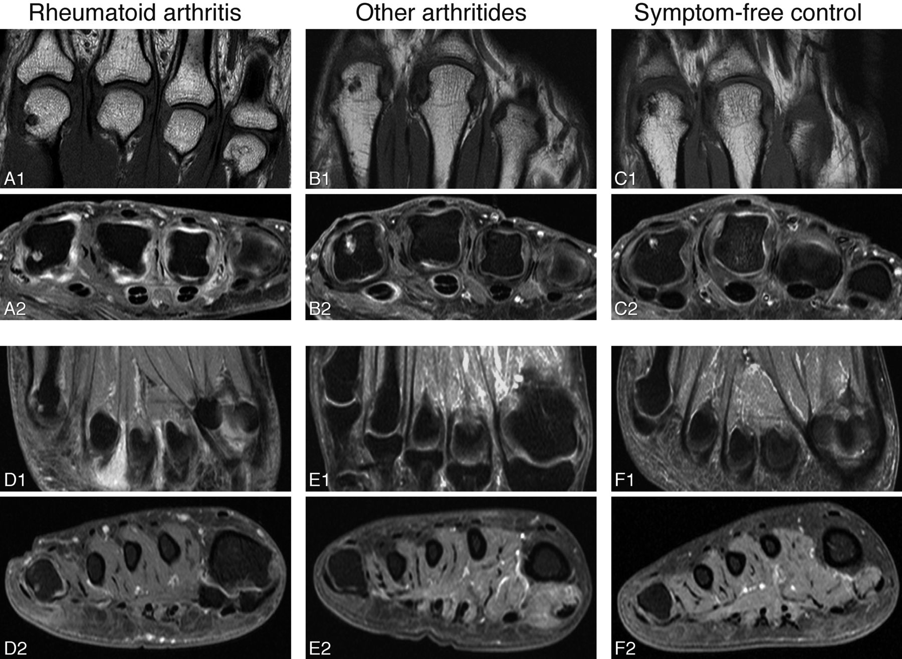
Then the location (the affected MCP or MTP joint) was assessed (table 4). In both patients with RA and controls, most erosions were located in the proximal part of the MCP and MTP joints: in patients with RA, 82%–95% of the erosions were located proximal in the joint, and in controls this was 81%–100% for the different age groups. As presented in table 4, overall the MCP and MTP bones that were frequently affected in patients with RA were also frequently affected in healthy controls. For instance, MCP2 and MCP3 were predilection sites for MRI-detected erosions in RA, but also in controls. However there were also some differences: erosions in MTP5 were more frequently present in patients with RA than in controls in most age groups (specificity 90%–98% for different age groups, table 2). In addition, erosions in MTP1 in the age group <40 almost exclusively occurred in RA (specificity 98%); the specificity was lower in older age groups (specificity 86% if aged 40–59 and 63% if aged ≥60). Examples of MRI-detected erosions are shown in figure 2.

Examples of erosions in patients with rheumatoid arthritis (RA), patients with other arthritides and symptom-free controls. MR images of patients with RA (A, D), patients with other arthritides (B, E) and symptom-free controls (C, F). Examples of erosions in MCP2 (A–C) and of a small erosion in MTP5 are shown (D). Erosions in MCP2 were observed in all different groups (A–C) while erosions in MTP5 were mainly observed in patients with RA (D). Patient B was diagnosed with polyarticular gout. Person C was aged 48 years. Coronal (A1, B1, C1, D1, E1, F1) and axial (A2, B2, C2, D2, E2, F2) images are shown. MRI sequences included coronal T1-weighted fast spin echo (FSE) sequences and axial T1-weighted FSE sequences with fat suppression after contrast enhancement. MCP, metacarpophalangeal joint; MTP, metatarsophalangeal joint.
Location of erosions in bones of the MCP and MTP joints of patients with RA and symptom-free controls, depicted per age category (18–39, 40–59, ≥60 years)
Erosions with the simultaneous presence of BME and/or synovitis are more frequent in patients with RA than in symptom-free controls
Then we questioned whether the combined presence of erosions with surrounding inflammation was specific for RA. At bone level, in patients with RA, 33% (95/285) of the total number of MCP and MTP bones with erosions only had erosions without synovitis and/or BME while in controls this was 77% (105/136, table 5). Similarly, when analysed on person level, 16% of the patients with RA only had erosions without inflammation and 40% had at least one erosion with inflammation in that same joint while in controls this was 30% and 12%, respectively (table 3). When analysing the different age groups, it appeared that within the age group <40 years, the simultaneous presence of erosions with inflammation was exclusively observed in patients with RA (specificity 100%). In the age group 40–59 years, the specificity was 91% and it was lower in persons aged ≥60 (specificity 71%), since in this age group erosions with inflammation were also observed in healthy controls (table 2). Thus, the presence of erosions with inflammation was specific for RA, but only if aged <60.
Frequencies of erosions in combination with inflammation in MCP and MTP bones of symptom-free controls and patients with RA; analysis on bone level
Altogether, the presence of grade ≥2 erosions and MTP5 erosions was specific for RA in all age groups, erosions with inflammation were specific for RA if aged <60 and MTP1 erosions if aged <40. Although these erosion characteristics were highly specific for RA, only 29% of all patients with RA had ≥1 erosion(s) with ≥1 of these characteristics.
Erosions in MTP5 and grade ≥2 erosions in all age groups and erosions in MTP1 if aged <40 remain specific for RA when compared with patients with other arthritides
Thus far, different erosion characteristics were compared between patients with RA and controls revealing some RA-specific characteristics. The next question is whether these characteristics are truly RA specific or are also present in other arthritides. Therefore, all analyses were repeated with patients with other arthritides as reference group. The total erosion scores of both patient groups were not significantly different (beta 0.92; 95% CI 0.84 to 1.01, figure 1B). Comparison of the different erosion characteristics showed that the presence of grade ≥2 erosions was RA specific in all age groups (specificity 100% if aged <40% and 96% if aged 40–59 and ≥60, table 2, online supplementary table 3). Also, MTP5 erosions were highly specific for RA in all age groups (specificity 100% if aged <40, 89% if aged 40–59, and 90% if aged ≥60, table 2, online supplementary table 4). The specificity of MTP1 erosions was 93% in patients aged <40, but at higher age specificity decreased to 66%. Erosions with inflammation were less specific for RA (specificity 49%–85% within different age groups) as these were also present in other arthritides. Thus, erosions with inflammation were not RA specific, but MTP5 erosions and grade ≥2 erosions were specific in all age groups and MTP1 erosions in patients aged <40. Twenty-one per cent of patients with RA had ≥1 erosion(s) with these characteristics (sensitivity 21%). Additionally, of all patients with erosions with one of these three finally identified features, 53% fulfilled the criteria for RA (positive predictive value (PPV) 53%), and of all patients without such erosions criteria were not fulfilled in 62% (negative predictive value (NPV) 62%).
MRI-detected erosions do not contribute to the identification of patients with UA that will progress to RA
Finally, the value of MRI-detected erosions was evaluated within patients with UA. Of the patients with UA, 15% (28/192) fulfilled the criteria for RA after 1 year. Of these patients, 11% had an RA-specific erosion at baseline, whereas 9% of the non-convertors had an RA-specific erosion (OR 1.3; 95% CI 0.3 to 4.8).
Discussion
Radiographic erosions specific for RA have been defined as the presence of ≥3 radiographic erosions on MCP, PIP, wrist or MTP joints and their presence is considered sufficiently specific to classify RA.11 MRI is a sensitive imaging modality that depicts cortical defects and therefore is suitable to detect erosive damage. Thus far, it was unknown which MRI-detected erosions on hand and foot joints are specific for RA. This large cross-sectional MRI study showed that on the group level, patients with RA had higher MRI-detected erosion scores in MCP and MTP joints than controls, but also that there was a large overlap on the individual level. Several erosion characteristics were studied in detail; this was done within three age strata as the total MRI erosion score was associated with age. Compared with controls from the general population, four characteristics were identified as RA specific: grade ≥2 erosions, MTP5 erosions, MTP1 erosions if aged <40 and erosions with local inflammation if aged <60. At least one of these characteristics is present in 29% of patients with RA.
Subsequently, patients with RA were compared with patients with early arthritis with other diagnoses, because studies comparing established cases and healthy controls will reveal the maximal contrast. Differences are often smaller when more clinically relevant patient groups are studied.21 22 Indeed, we observed that some erosion characteristics that were specific for RA when compared with controls were not specific when RA was compared with other arthritides. This was most prominent for the combined presence of erosions with inflammation. Nonetheless, some characteristics (grade ≥2 erosions, MTP5 erosions, MTP1 erosions in persons aged <40) were RA specific in both settings. Twenty-one per cent of patients with RA had ≥1 erosion(s) with ≥1 of these characteristics.
Although some erosion characteristics were identified as RA specific, an important overlap between patients with early RA and controls was observed. It has been recommended that novel imaging modalities, such as MRI, can be used to detect erosions early.1 The present data show that if all MRI-detected erosions (according to RAMRIS) would be considered as characteristic for RA or disease, this would yield many false-positive results.
We used the RAMRIS definition of erosions that basically evaluated the volume of the erosion in relation to the assessed bone. Others showed that small lesions on high-resolution peripheral quantitative CT were not entirely specific for RA and suggested that lesions >1.9 mm in diameter were highly specific.23 24 It remains to be determined if a phenotypic definition of MRI erosions, for instance, one that includes a description of the size of the cortical break, will be more discriminative; this is subject of further studies and is also considered within an ongoing EULAR task force.
Some of the findings on MRI-detected erosions are in line with previous findings on radiographic erosions. Radiographic erosions have been shown to occur more frequently at disease onset with higher age.25–31 MTP5 has been shown as a predilection site for RA-related erosions as well.32–34 We observed that the large majority of erosions (both in RA and in the other groups studied) were located in the proximal bone of the joint which is completely in line with previous findings.23 33 35
Erosive lesions in symptom-free controls have also been reported in other studies.12 The nature of these lesions is unclear. Because of the association with age, degenerative subchondral bone cysts may be one of the explanations. In addition, a very recent study, evaluating bone microstructure of MCP joints using high-resolution tomography and micro-CT, demonstrated that the number of so-called cortical microchannels (linking the synovial and bone marrow compartments) was higher in patients with RA than in healthy controls and was associated with erosions and age.36 It is intriguing to speculate that these channels have a causal role in erosion development, both in RA and controls. Another possibility is that mechanical strains are involved in erosion development, since erosions were frequently located in the foot (49% of the erosions in patients with RA and 38% in symptom-free controls). However, a pathophysiological explanation for the findings done in symptom-free persons is beyond the scope of this study.
The location of erosions within the bone was not studied here. This information could not be discerned using RAMRIS as this method evaluates the volume of the erosive lesion per bone. However, previous studies have shown that the majority of erosive MRI lesions in MCP joints occurred adjacent to the radial collateral ligaments, both in patients with RA and in healthy controls.37 38 Similar observations were done in a study in patients with RA and healthy controls on the location of erosions as detected on CT.23 The location of erosions in the symptom-free controls that were studied here has been reported previously,13 and showed that also in these persons erosions were present adjacent to the collateral ligaments and were not situated centrally in the bone. Because of these previous reports, showing no difference in location of erosions within the bone between patients with RA and controls, we anticipated that this characteristic will not result in further discrimination of RA-specific erosions from other erosions.
Cross-sectional analyses revealed that of all patients with an erosion that was identified as characteristic for RA 53% actually had RA (PPV). Likewise, 62% of all patients without such erosions did not have RA (NPV), whereas 38% did fulfil the criteria for RA. These data illustrate that the absence or presence of RA-specific erosions at disease presentation is of moderate value to identify patients who fulfil the criteria for RA at the same point in time.
Longitudinal analysis within patients with UA suggested that the presence of RA-specific erosions was also not predictive for the development of RA. However, this analysis was of limited power. Additionally, other outcomes, such as the start of disease-modifying antirheumatic drugs (DMARD), should be studied, since DMARD treatment might hamper progression to RA. Finally, it was not possible to study the different RA-specific erosion features separately due to the limited number of patients. Further studies are warranted.
We studied an early RA population. Thirty-six per cent of the patients were rheumatoid factor negative and 48% were anti-citrullinated protein antibody negative which is comparable to other early RA cohorts.39 40 Our population is somewhat different from patients with RA included in clinical trials where generally a selection of patients with RA is included.
A limitation of this study is that it was cross-sectional in nature and that imaging follow-up was not studied. Sensitivity of readers could have been a problem that could be equally present in the three groups. The presence of serial MRI data facilitates the differentiation of erosions from vascular channels and entheseal attachments, as these should not change during follow-up. Erosions in contrast could progress over time, although this progression may also have been hampered by up-to-date treatment strategies. Serial MRIs were not made but would have been beneficial to evaluate if some erosions were falsely identified as such. However, if MRI will be used for early identification of patients in clinical practice, single MRI measurements will be made.
In conclusion, MRI-detected erosions (according to the RAMRIS definition) in MCP and MTP joints are not confined to RA, but also present in other arthritides and in symptom-free persons from the general population. On the individual level, there was a large overlap. Some erosion characteristics were identified as specific for RA (grade ≥2 erosions, MTP5 erosions and MTP1 erosions if aged <40), though these occurred in a minority (21%) of the patients. Longitudinal MRI may improve specificity; however, this was not tested in this study. The present data imply that if single measurements with novel imaging modalities such as MRI are used for the early detection of structural damage in clinical practice, the risk of false-positive findings should be considered.
Acknowledgments
The authors thank G Kracht from the Department of Radiology of the Leiden University Medical Center for processing the MRIs.
References
Footnotes
Handling editor Tore K Kvien
Contributors DMB, WPN and AHMvdHvM contributed to the conception and study design. DMB analysed the data. DMB, WPN, HWvS, MR, RBML and AHMvdHvM contributed to interpretation of the data. DMB and AHMvdHvM wrote the first version of the manuscript and WPN, HWvS, MR and RBML revised it critically. DMB, WPN, HWvS, MR, RBML and AHMvdHvM read and approved the final manuscript.
Funding This project has received funding from the European Research Council (ERC) under the European Union’s Horizon 2020 Research and Innovation Programme (Starting Grant, agreement number 714312) and from the Netherlands Organization for Health Research and Development (Vidi Grant).
Competing interests None declared.
Patient consent Obtained.
Ethics approval The study on patients with early arthritis and the study on healthy controls were both approved by the local medical ethics committee ‘Commissie Medische Ethiek’.
Provenance and peer review Not commissioned; externally peer reviewed.
Data sharing statement No additional data available.
Presented at This paper is based on work that was previously presented at the 2017 ACR/ARHP Annual Meeting, 7 November 2017, San Diego, California, and was published as a conference abstract: DMB et al, Arthritis Rheumatol. 2017; 69 (suppl 10).