Article Text

Abstract
Objectives To study the epidemiology and mortality in patients with biopsy-proven giant cell arteritis (GCA) in southern Sweden.
Methods The study area was the County of Skåne. Patients with a positive temporal artery biopsy between 1997 and 2010 were identified using a regional register and a structured review of all histopathology reports. Standardised mortality ratios (SMR) were calculated using data for the Swedish population as the reference.
Results There were 840 patients with biopsy-proven GCA (626 women). The annual incidence rate per 100 000 inhabitants aged ≥50 years was 14.1 (95% CI 13.1 to 15.0); 7.7 (6.7 to 8.7) for men and 19.6 (18.1 to 21.1) for women, without seasonal variations. The incidence increased with age, with estimates of 2.0, 11.8, and 31.3 per 100 000 in the age groups 50–60, 61–70, 71–80 years, respectively (p<0.001). The age-standardised and sex-standardised incidence rate decreased from 15.9/100 000 in 1997–2001 to 13.3/100 000 in 2007–2010 (p=0.026). Two hundred and seventy-nine patients (207 women) died during the observation period. Mortality was significantly increased over the first 2 years after GCA diagnosis (SMR 1.52 (95% CI 1.20 to 1.85)), but not with longer follow-up. The estimated excess mortality was greater in women and in patients aged ≤70 years at diagnosis.
Conclusions In this large population-based study of biopsy-proven GCA from southern Sweden, the incidence of GCA may have decreased over time. Short-term mortality was increased, in particular among those diagnosed at ≤70 years of age, but long-term survival was not impaired.
- Epidemiology
- Giant Cell Arteritis
- Outcomes research
Statistics from Altmetric.com
Introduction
Giant cell (temporal) arteritis (GCA) is a primary systemic vasculitis of unknown aetiology, affecting large arteries, especially the aorta and its main branches.1 GCA has distinct epidemiological and clinical characteristics, and it is more common among women. The disease affects older adults, rarely occurs before the age of 50 years, and has an incidence that rises rapidly after 50 years of age. Common clinical presentations include a new-onset headache, scalp tenderness, fever and constitutional systemic features.2–4 The most feared complication is visual impairment or irreversible blindness due to involvement of the arteries supplying the optic nerves.5 ,6 The diagnosis of GCA depends on the clinical characteristics and is usually confirmed by obtaining a temporal artery biopsy (TAB) demonstrating vasculitis.
GCA is most common in populations of northern European ancestry. The incidence of biopsy-proven GCA in Gothenburg, Sweden, was reported to be 22 per 100 000 inhabitants aged ≥50 years.7 Similarly, the incidence of GCA in Rochester, Minnesota, USA, which has a large population of residents with Scandinavian ancestry, was 18.8 per 100 000.8 In other parts of the world, considerably lower incidence estimates have been reported, for example, 6.9/100 000 in Italy,9 9.5/100 000 in Israel,10 and 12.7/100 000 in the Otago region of New Zealand.11 Genetic and environmental factors have been implicated in the aetiology of GCA.12 A number of studies addressed the issue of mortality in patients with GCA; while the majority showed no increased mortality in comparison to the general population, a smaller number of studies revealed higher mortality rates.13–17
No updated epidemiologic data on GCA have been published from Sweden or other parts of northern Europe since the early 1990s. Incidence of GCA and survival could change over time due to changes in environmental exposure and lifestyle factors. Therefore, new epidemiologic studies of GCA using high-quality registries in well-defined populations are of importance.
This is a population-based study involving a larger geographical area than previous Swedish studies and covering a 14-year period. The aims of this study were (1) to estimate the annual incidence rate of biopsy-proven GCA in a large well-defined population in southern Sweden and (2) to study the survival of patients with biopsy-proven GCA in comparison with the general population.
Methods
The study area and population
The study area is Skåne, the southernmost county in Sweden, with a total population of 1 243 329 as on December 2010 (13.2% of the total Swedish population in 2.7% of the total area of Sweden).18 More than 95% of the population is Caucasian. The county includes urban and rural areas, and by tradition it has many people employed in agriculture, but also a large research and education sector.
The department of pathology in skåne
The department of clinical pathology in the county of Skåne operates at four hospitals (in Malmö, Lund, Helsingborg and Kristianstad), and provides histopathology services to all doctors, clinics and all the hospitals in Skåne. A single computerised system is used by all pathology units in Skåne, includes results for all pathology specimens examined, and allows computer assisted searches by topographic classification codes, histopathology diagnosis codes, clinical diagnosis, or free-text terms.
The identification of TABs and cases of biopsy-proven GCA
A search of the patient registry at the department of pathology in Skåne was performed using the following topographic codes: T41 for ‘artery’, T42750 for ‘temporal artery’, and T45 for ‘artery in the head’ for biopsies performed between 1997 and 2010 yielding a list of all individuals assigned at least one of the topographic codes at any time. Duplicates were removed. All histopathology reports were reviewed by one of the authors (AJM) to verify the diagnosis.
Patients were diagnosed as having GCA if the pathology report stated the diagnosis of ‘giant cell arteritis’, ‘temporal arteritis’, ‘granulomatous arteritis’, or unequivocally indicated infiltration of mononuclear cells into the arterial wall with or without giant cells. Patients with biopsy results showing ‘non-specific inflammation’, ‘periarteritis’, insufficient data to make a diagnosis, or non-specific inflammation considered by the pathologist secondary to atherosclerosis were excluded. Borderline cases were reviewed by two investigators (AJM and CT), and the final judgment was based on consensus.
Study subjects
All newly diagnosed persons with biopsy-proven GCA between 1997 and 2010 who were living in Skåne at the date of diagnosis were included. For survival analysis and to check for the residential address of the patients, the cohort was linked to the population register in Sweden; a comprehensive record run by the Swedish Tax Authority providing current information on who is living in Sweden and their residential address. The matching criteria were personal identification numbers which are unique for each person living in Sweden.
Statistical analysis
The differences between groups were compared using the non-parametric Mann-Whitney U test or the χ2 test. The Kaplan–Meier method was used to estimate survival rates. The analyses performed in this study were based on overall mortality (irrespective of cause of death). The expected number of deaths was calculated by period-specific person-years of follow-up with corresponding rates for the entire Swedish population, matched for age, sex, and year-specific mortality rates. The standardised mortality ratio (SMR) was obtained by dividing the observed number of deaths by the expected number. 95% CIs were calculated assuming a Poisson distribution of the observed cases. The denominator population used in the incidence estimates was the mean population aged ≥50 years during the period 1997–2010 (426 697). To study possible annual fluctuation or temporal changes in incidence rates, the incidence rate for each calendar year, and for three time periods (1997–2001, 2002–2006 and 2007–2010) were calculated separately, using the mean population aged ≥50 years during the relevant period as the denominator. The incidence estimates were standardised for age and sex, using the first time period (1997–2001) as the reference. Data regarding the season at diagnosis were analysed to study possible seasonal variations in incidence of biopsy-proven GCA. The seasons were defined as follow: winter (December–February), spring (March–May), summer (June–August) and autumn (September–November). To study the differences in mortality rates according to age at diagnosis, the cohort was divided into two groups based on age at diagnosis of <70 years and ≥70 years, respectively.
Statistical analysis was performed using the Statistical Package for the Social Sciences; SPSS for Windows, V.20.0 (IBM SPSS Statistics).
Results
Case identification
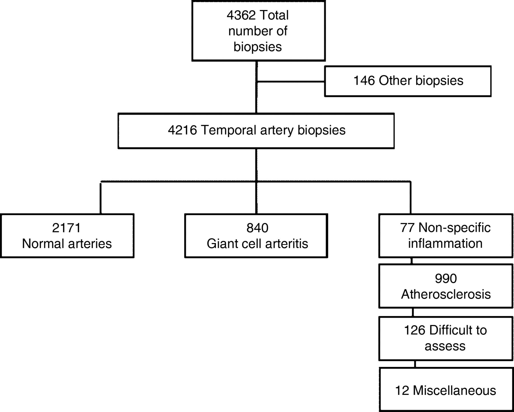
A total of 4216 TABs (2872 women) were identified (figure 1). The annual rate of performing a new TAB during the study period was 71/100 000 aged ≥50 years (95% CI 68 to 73); 90/100 000 among women and 48/100 000 among men. The annual incidence of TAB among those aged ≥50 years decreased from 76/100 000 in 1997–2001 to 70/100 000 in 2002–2006 and 65/100 000 in 2007–2010. The proportion with a positive TAB remained stable around 20%. Accordingly, there were 840 patients with a positive TAB for GCA (626 women (74.5%)); the woman to man ratio was 3 : 1. The median age at time of diagnosis was 75.9 years (IQR 69.9–81.2) for all patients; 75.7 (IQR 69.0–81.4) for men and 76.0 (IQR 70.2–81.2) for women.

The results of a search of the database of the department of pathology in Skåne 1997–2010 for cases of giant cell arteritis. The numbers refer to the number of biopsies or the results of histopathology examination of the temporal artery biopsies. Other biopsies: tissue biopsies from organs other than temporal artery that assigned codes of ‘artery’.
Incidence rates
The annual incidence rate of biopsy-proven GCA per 100 000 people aged ≥50 years was 14.1 (95% CI 13.1 to 15.0); 7.7 (6.7–8.7) for men and 19.6 (18.1–21.1) for women (table 1). The incidence of biopsy-proven GCA was significantly higher among women than men (p<0.001). The incidence rates significantly increased with age: 2.0, 11.8 and 31.3 per 100 000 people in the age groups 50–60, 61–70 and 71–80 years (p<0.001), respectively, while slightly decreased in the age group ≥81 years (28.3 per 100 000; p=0.239 vs the 71–80 year group), figure 2.
Annual incidence rate of biopsy-proven giant cell arteritis in southern Sweden

Age-specific incidence of biopsy-proven giant cell arteritis (GCA) in southern Sweden. Upper and lower limits represent the 95% CI.
The data were divided into three periods (1997–2001, 2002–2006 and 2007–2010) in order to identify possible changes in incidence rates. A total of 325 patients diagnosed during the first period with an incidence rate of 15.9 (95% CI 14.2 to 17.7). The population increased by 7% during period 2, and a total of 281 patients were diagnosed with biopsy-proven GCA for a corresponding age and sex-standardised incidence rate of 13.4 (95% CI 11.8 to 15.0); and 234 patients were diagnosed during period 3 with corresponding age and sex-standardised incidence of 13.3 (95% CI 11.7 to 14.9). The incidence rate in period 1 was significantly higher compared to period 2 (p=0.037) and period 3 (p=0.026). There was no statistically significant difference in the incidence rate between periods 2 and 3. The incidence rates per year are shown in figure 3⇓. There were some fluctuations in the incidence between 1997 and 2002, whereas after 2002, the incidence estimates were quite stable.

Incidence rates per year of biopsy-proven giant-cell arteritis (GCA) in southern Sweden.

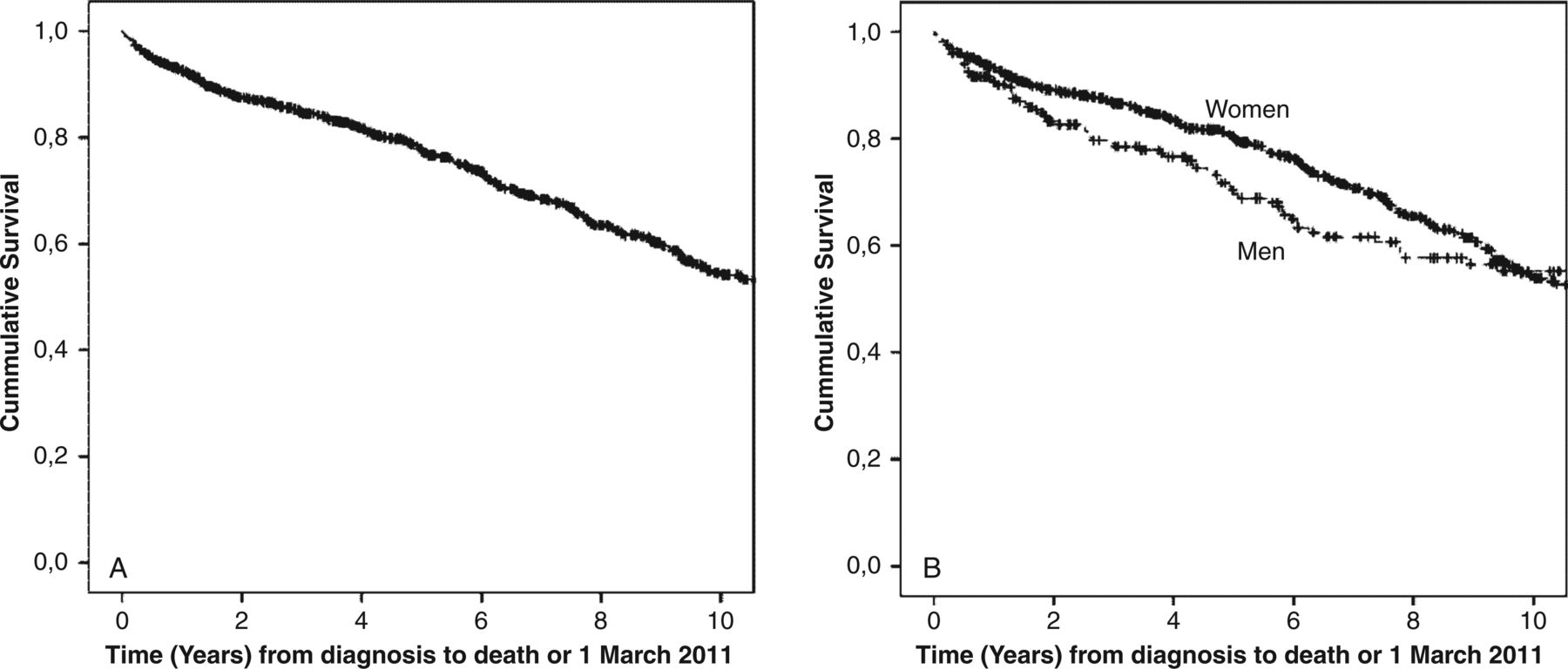
Survival curves according to Kaplan–Meier analysis in patients with biopsy-proven giant cell arteritis (GCA) in southern Sweden. (A) Overall survival in 840 patients. (B) Survival in women (n=626) versus men (n=214).
Seasonal variations for diagnosis of GCA
The number of patients with biopsy-proven GCA during the study period as grouped by season was 190 patients (22.6%) in winter, 232 patients (27.6%) in spring, 228 patients (27.1%) in summer, and 190 patients (22.6%) in autumn. The monthly distribution of the diagnosis of biopsy-proved GCA did not differ significantly (data not shown).
Survival rates
The median time of follow-up for all patients from diagnosis to 1 March 2011 or death was 62.0 months (IQR 25.0–106.1). Two hundred and seventy-nine patients (207 women) died during this follow-up period. Thirty patients (21 women) died within 3 months from diagnosis, and 47 (30 women) died within 6 months. For all patients the absolute survival rate was 92.5% at 1 year, 77.6% at 5 years, and 54.2% at 10 years. The corresponding rates for men were 90.6%, 69.5% and 55.2%, respectively, and for women, 93.2%, 80.2% and 53.8%, respectively (p=0.481 for comparison between men and women) (figure 4).
Standardised mortality ratio
At last follow-up, the SMR was 0.91 (95% CI 0.80 to 1.02) for all patients; 0.82 (0.63 to 1.01) for men and 0.95 (0.82 to 1.08) for women (table 2). Overall mortality was significantly increased after 1 year (SMR 2.17 (95% CI 1.48 to 2.86); p<0.001) and after 2 years (SMR 1.52 (95% CI 1.20 to 1.85); p<0.001), but not with longer follow-up. In women, mortality was significantly increased during the first 2 years after GCA diagnosis (SMR 1.53 (95% CI 1.12 to 1.93); p=0.007). Patients aged ≤70 at diagnosis had a greater estimated excess mortality (SMR 4.68 (95% CI 0.93 to 8.42)) compared to those aged >70 (SMR 1.98 (95% CI 1.29 to 2.66)), although with wide CI due to the limited number of deaths in this subset (table 2).
Standardised mortality ratios (SMR) of patients with biopsy-proven giant cell arteritis compared with the general Swedish population
Discussion
This is a population-based study presenting the epidemiological characteristics of patients diagnosed with biopsy-proven GCA in a stable, large population in southern Sweden over a 14-year period. Our study supports previous reports on increasing incidence with age, and that the disease is much more common in women than in men.
The estimated incidence of biopsy-proven GCA in southern Sweden of 14.1 per 100 000 for people aged 50 years and older was comparable with previous estimates of 16/100 000 from the northwestern part of Skåne in 199119 and 17/100 000 from Gothenburg, Sweden in 1981,20 but lower than the rate of 22/100 000 reported in 1993 in a more recent study from Gothenburg.7 The method used in case retrieval was similar in the Gothenburg study and the present investigation, as both identified patients with biopsy-proven GCA from the records of the department of pathology. There are no major differences in the population distribution and geographical features between Gothenburg and Skåne. A number of studies have reported on the incidence rate of GCA in the world,8 ,21–25 but direct comparison with our study can only be made with studies that were restricted to biopsy-proven GCA. In these studies, the estimated incidence per 100 000 aged ≥50 years ranged from 9.5 in Israel,10 10.1 in Spain,26 and 12.7/100 000 in New Zealand,11 to 15.1 in Denmark21 and 17.4–26.2 in Finland.27
We found a significantly decreasing incidence over the study period, and these results were not changed after adjustment for age in our population. Therefore, this difference might reflect a true decrease in the incidence rate of biopsy-proven GCA with time, although changes in other factors, such as propensity to rely on TAB in the diagnostic work-up for suspected GCA, may have changed over time. Ultrasonography for diagnosis of GCA was introduced very recently, is used in a limited fashion in our region, and it is unlikely that such testing would affect incidence estimates through 2010.
There was a slight decrease in the incidence of TAB during the study period, whereas the proportion with a positive TAB was stable. This could reflect a change over time in the practice of ordering TAB.
The relevance of the observed decrease in incidence to the overall burden of GCA is unknown, since we investigated only biopsy-proven GCA. Furthermore, although there were fluctuations in the incidence during the first part of the observation period, the incidence appeared to be stable from 2002 onwards, indicating that GCA remains a relatively common condition in this population.
A previous study from Minnesota showed an increased incidence of GCA between 1950 and 1979; however, thereafter, the incidence remained stable through 1999.8 A study from the UK also reported stable incidence of GCA from 1990 to 2001, whereas there was a marked increase in the incidence of polymyalgia rheumatica during the same period.28 The percentage of positive biopsies in our area was 20%; similar to the recently reported rate of 19% in New Zealand11 and higher than the rates of 17% in Iceland,22 15% in Denmark21 and 13% in Gothenburg, Sweden.29
Differences in methodology used in these studies and different time periods of the studies might partially explain the differences in the evolution of incidence rates of GCA in these populations. Greater awareness among physicians about the disease and easier access to diagnostic testing may also have influenced the rate of diagnosis, although it is unlikely such factors would have sustained effect on incidence rates for several decades.
The aetiology of GCA is unknown, and there are studies that have tried to elucidate possible seasonal variation, seeking clues on the aetiology (infections vs allergic/environmental). In our study, no seasonal variation was found, a finding similar to a study from Spain.26 However, several other studies have found seasonal variation in the diagnosis of GCA to be present. In Denmark, an association has been found between onset of GCA and two peaks of Mycoplasma pneumonia infection,21 while in Jerusalem, a peak incidence was found in the months of May–June.10 In UK, and throughout the whole study period 1990–2001, there was a clear seasonal variation with more patients diagnosed during the warmer summer months.28 By contrast, researchers in Gothenburg, Sweden, found an increased incidence of GCA in winter and autumn months.7
In this study, the mortality in patients with GCA was comparable with the Swedish general population with a SMR of nearly 1.00 at 5 years follow-up. However, the mortality was higher early in the disease course, especially in patients younger than 70 years at time of diagnosis. Most previous studies showed no increase in mortality compared with the general population,13 including studies of patients with biopsy-proven GCA from Gothenburg14 and Australia.17 Increased mortality was previously reported in studies from Denmark16 and northern Sweden,30 in the latter, mainly due to cardiovascular disease. These discrepancies may be explained by methodological issues such as different criteria for case recruitment and different clinical settings, and by factors such as ethnicity and secular trends. The present results suggest that GCA has an impact on survival mainly during the first few years after diagnosis, possibly due to comorbidities related to active disease or intensive treatment. With longer follow-up of elderly patients, other non-disease-related factors may become more important, and the relative impact of GCA may decrease.
Strengths of this study include the population-based setting, with no referral or selection-biases, and that it included only patients with confirmed diagnosis by histopathology examination. To the best of our knowledge, it is the largest study reported on the epidemiology of biopsy-proven GCA so far.
A possible limitation explaining the lack of seasonal variation could be long lag-times between symptom onset and time of biopsy. To address this, analyses of a subsample (155 subjects) was performed, which suggests that the impact of such bias is limited, since the median delay between onset of symptoms to date of TAB was less than 1 month (unpublished data). This finding is consistent with previously reported data.31
In conclusion, the incidence of biopsy-proven GCA in southern Sweden is comparable with studies from other European countries but lower than previously reported from Sweden. We confirmed an increasing incidence with age, with higher rates in women. The reduced incidence over time during the study period indicates that secular trends and lifestyle factors may affect the occurrence of GCA. The increased short-term mortality among patients with GCA, in particular, in patients diagnosed before the age of 70 years, suggests that further improvements in the management of GCA may be needed, and that additional investigations are warranted, including studies on the link between GCA and all-cause mortality, the impact of specific disease manifestations, and the burden of treatment-related complications and comorbidities.
Acknowledgments
This study was supported by research grants from The Faculty of Medicine, Lund University, and The Swedish Rheumatism Association (Reumatikerförbundet).
References
Footnotes
Handling editor Tore K Kvien
Contributors All authors were involved in drafting the article or revising it critically, and approved the final version. AJM had full access to all the data in the study and takes responsibility for the integrity of the data and the accuracy of the data analysis. Study design: AJM, LJ, PAM and CT. Statistical analysis: AJM and JAN. Data collection: AJM.
Competing interests None.
Ethics approval The study was approved by the Regional Ethical Review Board in Lund, Sweden.
Provenance and peer review Not commissioned; externally peer reviewed.