Article Text

Abstract
Objectives: Raised levels of the cytokines interleukin (IL) 6 and IL10 have been reported in patients with systemic lupus erythematosus (SLE).
Objective: To determine if levels of IL6 and IL10 correlate with organ/system-specific disease activity in SLE, using the British Isles Lupus Assessment Group (BILAG) Disease Activity Index.
Methods: Levels of IL6 and IL10 in serum samples from 171 patients with SLE and 50 normal controls were determined by enzyme linked immunosorbent assay (ELISA). Levels of cytokines in individual patients with SLE were compared with the presence or absence of active disease in eight organ/systems using the BILAG index.
Results: Levels of IL6 were significantly higher (p = 0.005) in patients with active compared with inactive haematological disease, as scored by the BILAG index. Further analysis showed that this association was dependent on an inverse correlation (p = 0.002, r = −0.26) between IL6 levels and haemoglobin levels in patients with SLE. In contrast, IL10 levels did not correlate with individual organ/system disease activity.
Conclusions: Raised levels of IL6 in SLE may influence the development of anaemia in this disease. These findings are in agreement with an increasing number of studies, which support physiological links between IL6 and anaemia. Importantly, with the exception of the haematological system, our studies do not provide evidence of any individual organ/system which would respond to therapeutic manipulation of either IL6 or IL10 levels.
- BILAG, British Isles Lupus Assessment Group
- ELISA, enzyme linked immunosorbent assay
- hsp, heat shock protein
- IL, interleukin
- PBMCs, peripheral blood mononuclear cells
- SLE, systemic lupus erythematosus
- BILAG
- anaemia
- cytokines
- systemic lupus erythematosus
Statistics from Altmetric.com
- BILAG, British Isles Lupus Assessment Group
- ELISA, enzyme linked immunosorbent assay
- hsp, heat shock protein
- IL, interleukin
- PBMCs, peripheral blood mononuclear cells
- SLE, systemic lupus erythematosus
The clinical expression of systemic lupus erythematosus (SLE) is the consequence of its complex immunopathology, involving B lymphocyte hyperactivity, the production of a wide spectrum of autoantibodies and the failure of T lymphocytes to suppress autoreactive B cell clones. Such immune disturbances may, in part, be explained by the dysregulation of cytokines, which have important roles in regulating the functions of cells within the normal immune system.
It is now well established that serum levels of the cytokines interleukin (IL) 61 and IL102 are raised in patients with SLE, being highest in those patients with active disease. IL6 is a multifunctional cytokine produced in response to inflammatory stimuli, including IL1 and tumour necrosis factor α, with pivotal roles in regulating the host immune response to infection. Thus IL6 has been found to be a potent stimulator of the differentiation and activation of lymphoid and myeloid cells3 and the production of acute phase proteins within the liver.4 IL6 is also a key regulator of various other cellular processes, including erythropoiesis,5,6 neuronal cell differentiation,7 and bone metabolism.8
IL10 may be considered to have largely anti-inflammatory properties on cells of the immune system, down regulating cell mediated immune responses and supporting antibody mediated immunity (for references see Moore et al9). In view of the functions of IL6 and IL10 in the normal immune system, a role for raised IL6 and IL10 levels in the pathogenesis of SLE has been suggested and is supported by various studies. Peripheral blood mononuclear cells (PBMCs) from patients with untreated SLE spontaneously release large amounts of IL610 and IL1011 in vitro. There is also evidence that disease severity in patients with SLE correlates with a raised ratio of IL10:interferon γ-secreting PBMCs.12 Furthermore, spontaneous immunoglobulin production (particularly anti-DNA antibodies) in cultured PBMCs from patients with SLE, is increased in response to either IL6 or IL10 and inhibited in response to a neutralising anti-IL61 or neutralising anti-IL10 antibody.13
Defects in the regulation of autoreactive B and T lymphocytes by apoptosis may also have a pathogenic role in SLE. It has been shown that IL10 increases expression of the anti-apoptotic protein Bcl-2 by germinal centre B lymphocytes, which may result in B cell longevity.14 Administration of anti-IL10 antibodies delays the onset of autoimmunity and production of autoantibodies in NZB/W F1 lupus prone mice.15 Furthermore, a monoclonal antibody to IL10 has been used with some success in treating six patients with SLE.16
The British Isles Lupus Assessment Group (BILAG) index is a computerised index based on the principle of the physician’s intention to treat, for measuring clinical disease activity in patients with SLE.17 Eight organ based systems are separately assessed. Patients attending the SLE clinic at the Centre for Rheumatology, University College London, are routinely assessed using the BILAG index.
Previous studies on correlations between disease activity and IL6 or IL10 levels in SLE did not differentiate between activity in different organs or systems. This differentiation is important in trying to identify which patients might respond best to anti-IL6 or anti-IL10 treatment. The objective of this paper is to compare levels of the cytokines IL6 and IL10 with organ-specific disease activity in patients with SLE, using the BILAG index.
PATIENTS AND METHODS
Patients
One hundred and seventy one consecutive patients with SLE (166 female, 5 male; mean age 37 years (range 18–68)) who fulfilled the criteria of the American College of Rheumatology for the classification of SLE18,19 were studied. No patients fulfilling these criteria were excluded. The ethnic breakdown of these patients was: white (68%), Afro-Caribbean (18%), South Asian (11%), Chinese (3%), and Other (1%). Of these 171 patients, 17 were taking no drugs to treat SLE at the time of the blood sample, 91 patients were taking hydroxychloroquine, 131 patients were taking immunosuppressive drugs. This included 127 patients taking prednisolone (mean (SD) dose 8.5 (5.4) mg/day), 53 patients taking azathioprine, and 11 patients taking methotrexate. Anti-dsDNA antibody levels were measured by enzyme linked immunosorbent assay (ELISA) and C3 levels by nephelometry as part of the routine clinical management of these patients.
Samples of venous blood from all patients were taken during morning outpatient attendance at the Centre for Rheumatology, University College London. The disease activity of these patients was assessed using the BILAG index.17 Eight organ/systems (constitutional, mucocutaneous, central nervous system, musculoskeletal, cardiovascular, vasculitis, renal, and haematological) are separately assessed and scored from A (the most active state) to E (never active in this system). We define active disease in an organ/system as A, B, or C. A global score was derived using the notation A = 9, B = 3, C = 1, D/E = 0.
Fifty normal control subjects (48 female, 2 male; mean age 28, range (22–42))—healthy volunteers consisting of hospital and laboratory staff—were studied. Samples of venous blood were taken in the morning. The ethnic breakdown of the normal subjects was: Caucasian (68%), Afro-Caribbean (18%), South Asian (12%), and Chinese (2%).
Anaemia in patients with SLE
Patients were categorised as presenting or not presenting with anaemia, where anaemia is defined as haemoglobin <125 g/l for women; <135 g/l for men.20
Measurement of IL6 and IL10 levels
Levels of IL6 in the serum of patients with SLE and normal control subjects were measured using the Quantikine high sensitivity human IL6 immunoassay (R & D Systems Europe, Abingdon, Oxon UK). The manufacturers report interassay precision as a coefficient of variation of 6.5–9.6%; intra-assay precision is reported as a coefficient of variation of 6.9–7.4%. Levels of IL10 in the serum of patients with SLE and normal control subjects were measured using the Biosource Cytoscreen human IL10 ultrasensitive immunoassay (Biosource International, Inc, California, USA). The manufacturers report interassay precision as a coefficient of variation of 5–7.8%; intra-assay precision is reported as a coefficient of variation of 2.6–4.5%.
Statistical analysis
Statistical analyses were performed using an SPSS computer software package (SPSS Science, Chicago, USA). Levels of the cytokines IL6 and IL10 were log10 normalised to reduce the effect of outliers, as described by Altman.21 Levels of these cytokines are expressed in g×10−13/ml. Thus the log 1 level is equivalent to 1 pg/ml before log transformation. Differences between mean levels of cytokines in patients with active compared with inactive disease for each of the eight BILAG categories of organs/systems were assessed for statistical significance using Student’s t test. A Bonferroni correction was applied such that for eight independent tests a significant p value was required to be less than 0.05/8 (p<0.006). Pearson correlations were performed to investigate associations between IL6 or IL10 levels and levels of the laboratory measured variables which define the haematological BILAG grade (haemoglobin, platelets, and white blood cell count) in individual patients. A value of p<0.05 was considered to be significant. χ2 Analyses were performed for evaluation of an association between IL6 levels and anaemia. Where a χ2 test was performed, a Pearson p value is provided, which was considered to be significant for p<0.05.
RESULTS
Serum levels of IL6 and IL10 in patients with SLE
Levels of the cytokines IL6 and IL10 were measured in 171 serum samples from patients with SLE and 50 normal controls by ELISA. In agreement with previous reports,1,2 a clear rise in IL6 and IL10 levels was seen in the patients with SLE compared with normal controls (table 1).
IL6 and IL10 serum levels in patients with SLE and normal subjects
Correlation of IL6, IL10 levels with disease activity in patients with SLE
Figure 1A shows that mean levels of IL6 were significantly higher (p = 0.005) in patients with active haematological disease than in patients with inactive haematological disease, as scored by the BILAG index. Associations of IL6 levels in patients with SLE with active disease in other organ/systems (fig 1A) or with global BILAG disease activity scores (data not shown) were not significant.

Bar charts representing levels of (A) IL6 and (B) IL10 in patients with SLE graded by the BILAG index. Error bars represent 95% confidence intervals for the mean. CON, constitutional; MUC, mucocutaneous; CNS, central nervous system; MUSC, musculoskeletal; CV, cardiovascular; VASC, vasculitis; RENAL, renal; HAEM, haematological.
Figure 1B shows mean levels of IL10 in patients with SLE graded by the BILAG index. No statistically significant differences in mean IL10 levels between active and inactive BILAG disease activity grades were found. Correlations of IL10 levels in patients with SLE with their respective global BILAG disease activity scores were also not found to be significant (data not shown).
Further analyses were performed to determine if IL6 or IL10 levels correlated with other SLE disease associated variables. Thus, levels of these cytokines were compared with anti-DNA antibody levels and complement C3 levels. IL10 levels correlated significantly with anti-DNA antibody levels (r = 0.26, p = 0.006) and with complement C3 levels (r = −0.22, p = 0.006). No significant correlations between IL6 levels and these variables were seen and there was no confounding correlation observed between corticosteroid treatment and either IL6 or IL10 levels (data not shown).
In view of the possibility that the expression of IL6 and IL10 may, in part, be under genetic control, analyses of the correlations between IL6, IL10 levels and BILAG disease activity scores and other SLE disease associated variables were subsequently repeated but restricted to the white Caucasian patient cohort, which constituted 116 (68%) of the 171 patients studied. The results of these analyses paralleled those of the original study of 171 patients. Thus, we observed higher (p = 0.01) mean levels of IL6 in patients with active (mean (SD) log IL6 1.78 (0.66)) compared with inactive (mean (SD) log IL6 1.47 (0.59)) BILAG haematological disease. No correlations of IL6 with activity in any other system or of IL10 with activity in any system were seen. IL10 levels but not IL6 levels correlated with anti-DNA antibody levels (r = 0.37, p<0.0005) and with complement C3 levels (r = −0.26, p = 0.008).
Analysis of the correlation of IL6 levels with BILAG haematological disease
Having established that mean levels of IL6 are higher in patients with SLE with active disease than in patients with inactive BILAG haematological disease, we determined correlations between IL6 and the laboratory measured variables which define this BILAG grade (haemoglobin concentration, platelets, and white blood cell counts).
IL6 levels were found to correlate inversely (negative value for the Pearson correlation coefficient) with the level of haemoglobin (Pearson r = −0.19, p = 0.002). Correlations between IL6 levels and either platelet or white blood cell counts in individual patients, did not attain statistical significance There was also no significant correlation between IL6 or IL10 levels and haematological indices other than haemoglobin level, particularly erythrocyte mean cell volume, erythrocyte mean cell haemoglobin—data not shown.
Correlation of IL6 levels with anaemia in patients with SLE
To analyse the relationship between IL6 levels in SLE and anaemia, 164 of the 171 patients were studied; the seven patients who were excluded had a known cause of low haemoglobin separate from SLE—namely, iron deficiency, chronic renal failure. None of the 164 patients studied had haemolytic anaemia and thus they could all be considered as having anaemia due to chronic disease.
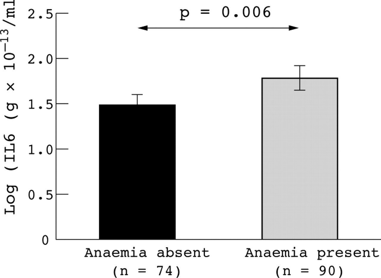
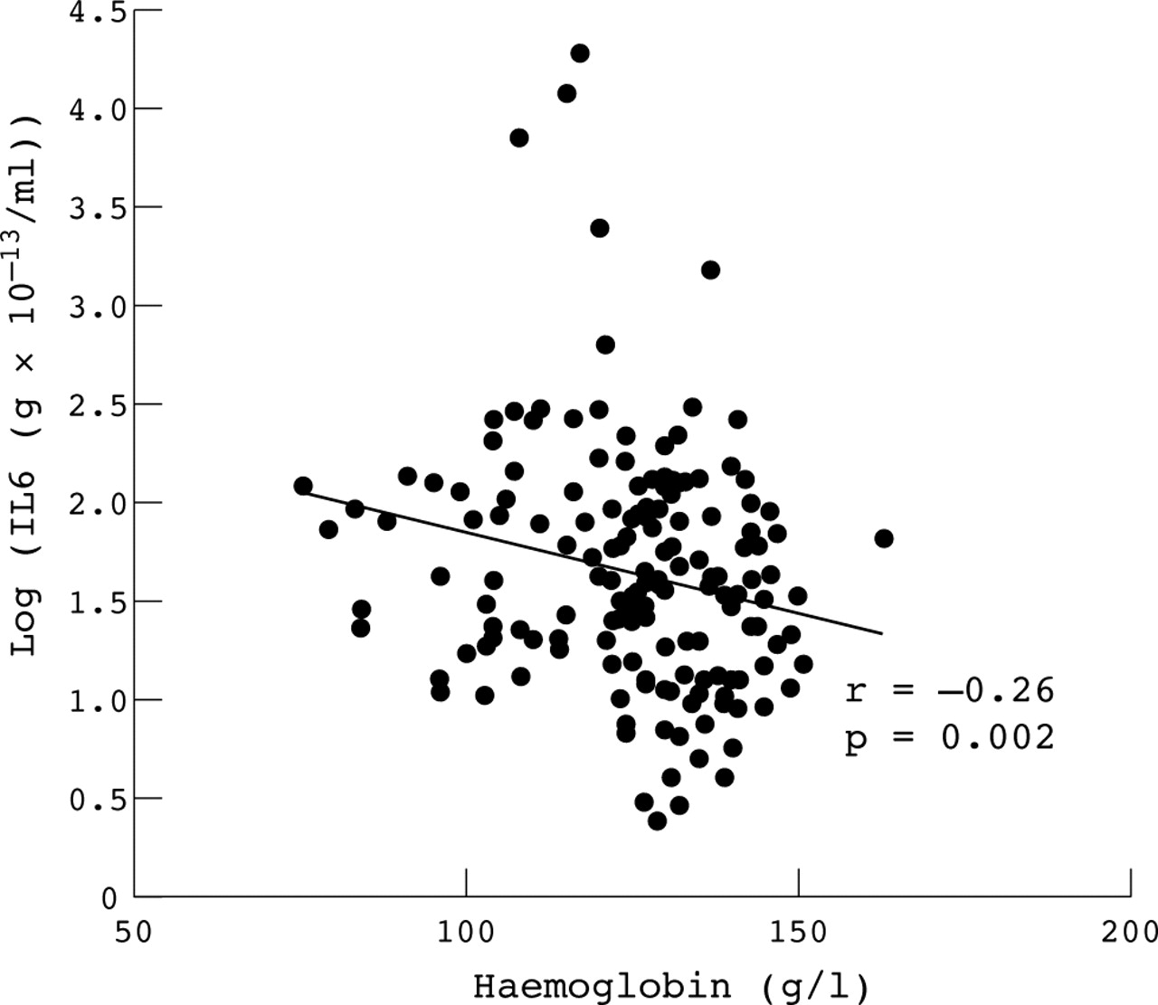
Figure 2 shows that there was an enhanced correlation between IL6 levels and haemoglobin levels in the 164 patients studied (r = −0.26, p = 0.002) compared with the original analysis in 171 patients (r = −0.19, p = 0.002). In agreement with this relationship, when we categorised patients as presenting or not presenting with anaemia and compared the mean IL6 level of each of these two groups, we observed significantly higher (p = 0.006) mean IL6 levels in those patients presenting with anaemia than in those patients without anaemia (fig 3).

Scattergraph comparing levels of IL6 and haemoglobin in individual patients with SLE. A trendline and associated Pearson correlation coefficient (r) and p value are shown.
Mean levels of IL6 in patients with or without anaemia. Error bars represent 95% confidence intervals for the mean. The p value represents significance of difference in mean values as determined by Student’s t test for equality of means.
Patients with SLE were then categorised as having normal or raised (above the normal 97.5th centile) IL6 levels and as presenting or not presenting with anaemia. A χ2 analysis was performed, which showed a significant correlation (p = 0.024) between raised levels of IL6 in patients with SLE and patient presentation with anaemia.
DISCUSSION
Although previous studies have suggested pathogenic roles for raised levels of IL6 and IL10 in SLE, any involvement of these cytokines with specific clinical manifestations has remained unclear. Here we compared levels of IL6 and IL10 in individual patients with SLE with organ/system disease activity using the BILAG index.17
An association between higher mean IL6 levels and active haematological disease was observed and was not paralleled by significant associations of IL6 with other clinical activity. Levels of IL6 were not found to correlate with global BILAG disease activity scores. In a previous study using the SLE Disease Activity Index (SLEDAI) and the SLE Activity Measure (SLAM), a lack of correlation between levels of IL6 and overall disease activity, was reported.22 However, in other studies, which measured levels of IL6 both before and after disease exacerbations,23 or at various stages of disease activity,24 levels of IL6 correlated well with overall disease activity. Possibly, therefore, any relationship between IL6 and disease activity in SLE may be most appropriately assessed by an evaluation of changes in these variables in the same patients over time.
The absence of a correlation between levels of IL10 and active clinical subsets in SLE or overall disease activity scores (global BILAG scores) is in agreement with a previous study.22 We observed a significant correlation between IL10 levels and both complement C3 levels and anti-DNA antibodies—which has been previously reported25 as supporting a role for IL10 in active disease in SLE. Serum IL10 levels have been shown to correlate with overall disease activity when serial measurements are taken in the same patients over time.24 Thus, as for IL6, any relationship between IL10 and disease activity, may be most appropriately assessed in patients by following significant changes in either variable over time.
Further analyses were performed to determine the relationship between IL6 and active haematological disease. It was established that this statistical association was dependent on a linear (negative coefficient) correlation of IL6 levels with haemoglobin levels in SLE, whereas no correlation was found between IL6 and platelets or white blood cell levels, which together with haemoglobin define the BILAG grade of haematological disease activity. When mean levels of IL6 were compared between patients categorised as presenting or not presenting with anaemia, levels of IL6 were clearly higher in the anaemic patients. Furthermore, a correlation between raised IL6 levels and anaemia was found to be significant by χ2 analysis. Hence our studies suggest for the first time a link between raised IL6 levels in SLE and anaemia.
In this large study we were unable to find any association between IL6 or IL10 and individual organ/system activity in SLE, apart from the inverse relationship between IL6 levels and haemoglobin concentrations described above. Thus, while various studies strongly suggest pathogenic consequences of raised levels of IL6 and IL10 in SLE, our studies do not provide evidence of any individual organ/system which would respond to therapeutic manipulation of either IL6 or IL10 levels. However, because previous studies in individual patients with SLE have demonstrated correlations between IL6/10 levels and global disease activity over time,23,24 it will be of interest to repeat these analyses as part of a longitudinal study.
A correlation between IL6 and haemoglobin levels has been reported in the geriatric syndrome of frailty. In this study, there was an inverse correlation between IL6 and haemoglobin levels, which was potentially related to the increased chronic inflammatory state marked by an increase in serum IL6 and unlikely to be due to iron deficiency.26 In experimental animals and patients, administration of IL6 has correlated with the development of anaemia. Treatment of patients with cancer with IL6 led to a rapid decrease in haemoglobin concentrations,27 and administration of IL6 to rhesus monkeys correlated with a decline in packed cell volume.28 Thus these studies are in agreement with our own observations and support a link between a chronic increase in IL6 and the development of anaemia.
Of particular interest to this report are the growing number of studies which support a functional link between the dysregulation of proinflammatory cytokines (including IL6, IL1, interferon γ, and tumour necrosis factor α) and the development of anaemia in various chronic inflammatory disease states and age associated syndromes. These studies have focused on specific roles for these cytokines (particularly IL6) both in the inhibition of erythropoiesis5,6 and in altered iron transport/metabolism.29 For example, IL6 has been shown to inhibit erythroid progenitor cell proliferation and terminal differentiation and it is likely that this is mediated by IL6 suppression of erythropoietin, which regulates this process.30,31 Furthermore, chronic iron deficiency has been correlated with raised IL6 production by lymphocytes, and this is likely to be mediated in part by IL6-induced hepatic iron storage.29
Interestingly, we have previously reported that IL6 levels in SLE correlate with heat shock protein (hsp) 90 levels32 and that changes in hsp90 levels in SLE correlate with changes in active haematological disease,33 as defined by the BILAG index. Hence, our studies support a hypothetical model in which IL6 levels influence levels of haemoglobin, where changes in hsp90 levels serve as a marker of significant changes in IL6 levels.
In summary, we investigated the relationship between IL6 and IL10 levels with activity of SLE in various organs and systems using the BILAG index. We found no correlations with activity in any individual organs/systems except a link between haematological activity and IL6. However, on further analysis, this apparent correlation was explained by an inverse relationship between IL6 levels and haemoglobin levels, which is not specific to lupus. Therefore our study was unable to identify a particular form of lupus activity which would respond well to therapeutic manipulation of IL6 or IL10 levels. Further studies are required, however, to understand more completely the mechanisms by which IL6 might influence the development of anaemia in SLE, both at the cellular and the molecular level.