Article Text

Abstract
Objective: To compare the clinical assessment of overall inflammatory activity in patients with rheumatoid arthritis (RA) with grey scale and power Doppler (PD) ultrasonography (US).
Methods: Ninety four consecutive patients with RA were included. Demographic and clinical data, C reactive protein (CRP) level, and erythrocyte sedimentation rate (ESR) were recorded for each patient. The presence of tenderness, swelling, and a subjective swelling score from 1 to 3 were independently assessed by two rheumatologists, who reached a consensus in 60 joints examined in each patient. All patients underwent a US examination by a third blinded rheumatologist, using PD. US joint effusion, synovitis, and PD signal were graded from 1 to 3 in the 60 joints. Joint count and joint index for effusion, synovitis, and PD signal were recorded. A 28 joint count for clinical and US variables was calculated. Interobserver reliability of the US examination was evaluated by a fourth blinded rheumatologist.
Results: US showed significantly more joints with effusion (mean 15.2) and synovitis (mean 14.6) than clinical examination (mean 11.5, p<0.05). A significant correlation was found between joint count and joint index for swelling, US effusion, synovitis, and PD signal. The 28 joint count for effusion, synovitis, and PD signal correlated highly with the corresponding 60 joint counts. US findings correlated better with CRP and ESR than clinical measures. Interobserver reliability was better for US findings than for clinical assessment.
Conclusion: US is a sensitive method for assessing joint inflammatory activity in RA, complementary to clinical evaluation.
- CRP, C reactive protein
- ESR, erythrocyte sedimentation rate
- HAQ, Health Assessment Questionnaire
- MCP, metacarpophalangeal
- MRI, magnetic resonance imaging
- PD, power Doppler
- PIP, proximal interphalangeal
- PRF, pulse repetition frequency
- RA, rheumatoid arthritis
- SJC, swollen joint count
- SJI, swollen joint index
- TJC, tender joint count
- US, ultrasonography
- VASOA, visual analogue scale for patient overall assessment of disease activity
- VASP, global pain intensity visual analogue scale
- rheumatoid arthritis
- ultrasonography
- power Doppler
Statistics from Altmetric.com
- CRP, C reactive protein
- ESR, erythrocyte sedimentation rate
- HAQ, Health Assessment Questionnaire
- MCP, metacarpophalangeal
- MRI, magnetic resonance imaging
- PD, power Doppler
- PIP, proximal interphalangeal
- PRF, pulse repetition frequency
- RA, rheumatoid arthritis
- SJC, swollen joint count
- SJI, swollen joint index
- TJC, tender joint count
- US, ultrasonography
- VASOA, visual analogue scale for patient overall assessment of disease activity
- VASP, global pain intensity visual analogue scale
Rheumatoid arthritis (RA) is a chronic inflammatory disease characterised by the development of synovitis, which damages cartilage, bone, ligaments, and tendons. Assessment of inflammatory activity is essential in daily practice to enable therapeutic decisions and to evaluate disease outcome and response to treatment.1
Traditionally, the degree of disease activity has been evaluated by measuring subjective clinical variables, laboratory measures, and radiographic findings.2–4 However, clinical evaluation of joint pain and swelling have not been sufficiently reliable,5 and conventional plain radiography depicts indirect signs of cartilage loss and bony erosions due to previous destructive synovial inflammatory activity.
High frequency ultrasonography (US) has greatly improved musculoskeletal imaging in rheumatology.6 Several studies have demonstrated that high frequency US is accurate for detecting joint effusion7,8,9,10,11,12,13,14,15,16 and synovitis,7,8,9,10,11,15,17,18,19, compared with magnetic resonance imaging (MRI)10,12,14,17 and direct arthroscopic visualisation.20–22 US is more sensitive and reproducible than clinical evaluation in assessing joint inflammation.23–26
Power Doppler (PD) US is a new technique of colour Doppler that improves the sensitivity to detect flow from small vessels and low velocity flow at the microvascular level.27,28 PD US detects indirect signs of increased vascularisation associated with soft tissue musculoskeletal inflammatory and infectious diseases,28,29 and enthesitis in spondyloarthropathies.30 The PD signal correlates highly with local clinical evaluation of joint inflammatory activity in the knee, metacarpophalangeal (MCP) and interphalangeal joints of patients with RA and other inflammatory arthropathies.31,32 Recent studies have shown that PD synovial vascularity correlates highly with histologically proved knee pannus33 and with the degree of synovial vascularisation of the knee34 and hip.35
This study aimed at comparing grey scale US and PD US with clinical and biological findings in the determinination of global inflammatory activity assessed in 60 joints of patients with RA. To the best of our knowledge, this is the first US and clinical study that has examined so many joints in each patient with RA.
PATIENTS AND METHODS
Ninety four consecutive patients (20 male, 74 female) who fulfilled the 1987 American Rheumatism Association criteria for RA36 attending the outpatient rheumatology clinic were included. Mean (SD) age was 57.6 (14.3) years (range 23–88) and mean (SD) disease duration was 69.3 (58.2) months (range 5–280). Patients who had had traumatic, septic, or microcrystalline arthritis, previous joint surgery, or isotopic synovectomy within the past 12 months before the study were excluded.
The following data were recorded for each patient: age, sex, disease duration, drugs received for RA at entry, rheumatoid factor (measured by nephelometry, normal level 0–20 IU/ml), and previous joint surgery or isotopic synovectomy. C reactive protein (CRP) level (measured by nephelometry, normal range 0–8 mg/l) and erythrocyte sedimentation rate (ESR; measured by the Westergren method, VESMATIC 60, version 2.05; Menarini Laboratory, Barcelona, Spain) were recorded from each patient’s routine laboratory test performed within 1 week of the study. Informed consent was obtained from all patients before the clinical and US evaluation.
Clinical assessment
The clinical evaluation was performed independently and sequentially by two blinded rheumatologists. One week before the study they carried out a consensus joint examination in patients with RA (not included in the study) for 20 hours. The following bilateral joints were assessed for tenderness and swelling: glenohumeral, acromioclavicular, sternoclavicular, elbow, wrist, MCP, proximal interphalangeal (PIP) of hands, knee, ankle, (tibiotalar), subtalar, mid-tarsal, metatarsophalangeal, and PIP of feet (total in 94 patients 5640 joints). Hip joints were assessed for tenderness and pain on passive motion. Hip swelling was indirectly considered if pain on passive motion was detected by physical examination. A subjective score from 1 to 3 was assigned for all swollen joints except for the hip (1 = mild; 2 moderate; 3 = marked). Immediately after physical examination, GB and FG compared their findings. If there were discrepancies for the presence or absence of joint tenderness and swelling or the swollen joint scores, they carried out a third examination together to reach consensus. These last results were compared with US findings. Individual physical examinations were used for estimating clinical interobserver agreement for tenderness, swelling and swelling scores. Tender joint count (TJC), swollen joint count (SJC), and a 60 swollen joint index (SJI; sum of the swelling score from each joint) were recorded for each patient.
The following clinical variables were also recorded: a global pain intensity visual analogue scale (VASP; 0–100 mm), a visual analogue scale for patient overall assessment of disease activity (VASOA; 0–100 mm), and a self assessment Spanish version of the Health Assessment Questionnaire (HAQ).37
US examination
All patients underwent a US examination within 30 minutes of the clinical evaluation by a single rheumatologist experienced in US (EN) who was unaware of the clinical findings. US examination was performed with two commercially available ultrasound real time scanners (Logiq 400CL, General Electric Medical Systems, Korea (scanner 1) and Logiq 700, General Electric Medical Systems, Waukesha, WI, USA (scanner 2)) using multifrequency linear array transducers, 6–13 MHz and 5–10 MHz, respectively, operating at 6.6 and 5 MHz of frequency for Doppler imaging, respectively, according to the manufacturer’s criteria. The first 69 patients were examined with scanner 1 and the last 25 patients with scanner 2. The presence of joint effusion and synovitis was systematically evaluated by US in each of the 60 joints clinically examined. Table 1 describes the US scanning method. The presence of effusion and/or synovitis was diagnosed in each joint according to the criteria listed in table 1.7–9,12–14,21,38–40 Distances were measured using electronic callipers. Effusion and synovitis were identified and distinguished according to the following definitions. Effusion was defined as hypoechoic or anechoic compressible intra-articular material, within synovial recesses. Synovitis was defined as echogenic non-compressible intra-articular tissue, within synovial recesses. Joint effusion and synovitis were subjectively graded from 1 to 3 (1 = mild; 2 = moderate; 3 = marked).
Ultrasonographic scanning of joints and criteria of effusion/synovitis
Synovial vascularisation was assessed by PD US in each of the 60 joints. PD imaging was performed by selecting a region of interest that included the bony margins, articular space, and a variable view of surrounding tissues (depending on the joint size). PD variables were adjusted to the lowest permissible pulse repetition frequency (PRF) to maximise sensitivity. This setting resulted in a PRF between 500 and 1000 Hz depending on the joint scanned. Low wall filters were used. The dynamic range was 20–40 dB. Colour gain was set just below the level at which colour noise appeared underlying bone (no flow should be visualised at the bony surface). This setting resulted in gains of from 18 to 30 dB. An image with maximal colour activity was selected for analysis. Flow was additionally demonstrated in two planes and confirmed by pulsed wave Doppler spectrum to exclude artefacts.
The intra-articular PD signal was subjectively graded on a semiquantitative scale from 0 to 3 (0 = absence, no intra-articular flow; 1 = mild, single vessel signal; 2 = moderate, confluent vessels; 3 = marked, vessel signals in more than half of the intra-articular area). In each patient we recorded joint count for US effusion (USJCE), joint count for synovitis (USJCS), joint count for PD signal (USJCPD), and a 60 joint index for effusion (USJIE), synovitis (USJIS), and PD signal (USJIPD) (sum of the effusion, synovitis, and PD signal scores, respectively, obtained from each joint). In addition, we calculated for each patient a 28 joint count (10 PIP joints of hands, 10 MCP joints, 2 wrist, 2 elbow, 2 shoulder, 2 knee joints)41 for tenderness (TJC28), swelling (SJC28), US effusion (USJCE28), US synovitis (USJCS28), and US PD signal (USJCPD28).
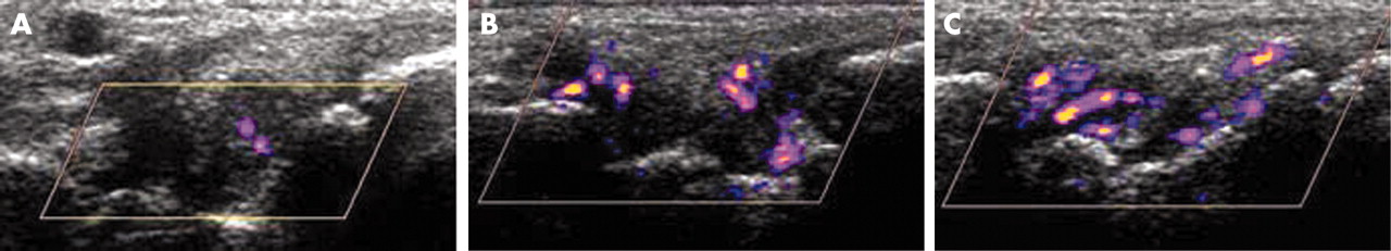
Inter- and intraobserver reliability of the US examination was evaluated by recording standard images of the 30 right joints from the first 22 patients evaluated with scanner 1 and the first 22 patients examined with scanner 2 in a digital archiving computer system. The saved images were blindly read by the same rheumatologist who performed US examination (EN) 3 months after the initial scanning, and by a fourth rheumatologist (JU) trained in US. These images did not show measurements. Before the study the investigators reached a consensus about the US scales. Figure 1 shows some examples of the US findings.
Longitudinal sonographic image of the wrist joint with moderate effusion, moderate synovitis, and mild (A), moderate (B), and marked (C) colour signal.
Statistical analysis
A χ2 test was applied for comparing cumulative variables, and Student’s t test and Pearson’s correlation were used for continuous variables. Any value of p<0.05 was considered significant.
Interobserver agreement between the clinical investigators and the US investigators and intraobserver US agreement were calculated using an unweighted κ test. The κ value measures agreement between pairs of observers, eliminating random concordance. A κ value <0.40 was considered poor, 0.40–0.60 moderate, 0.60–0.80 good, and 0.80–1 excellent.
RESULTS
Patients’ characteristics
Rheumatoid factor was positive in 73/94 (78%) patients. Therapeutic regimens included non-steroidal anti-inflammatory drugs in 60 (64%) patients, corticosteroids in 56 (60%), methotrexate in 61 (65%), leflunomide in 18 (19%), sulfasalazine in 10 (11%), antimalarial drugs in 10 (11%), gold salts in 9 (10%), infliximab in 8 (9%), etanercept in 4 (4%), ciclosporin in 2 (2%), and azathioprine in 2 (2%) patients. Two patients had a unilateral hip prosthesis, three patients a unilateral knee prosthesis, and one patient a bilateral hip and knee prosthesis. All surgical procedures had been performed more than 5 years before the study.
No significant differences were found between patients examined with scanners 1 and 2 for mean age, mean duration of symptoms, mean VASP and VASOA scores, mean joint count for tenderness and swelling, mean SJI, mean HAQ score and mean ESR and CRP values (table 2).
Demographic, clinical, and laboratory characteristics of patients evaluated with scanners 1 and 2
All joints, including nine prosthetic joints, could be easily assessed by US. The US examination took 20–30 minutes for each patient, not including documentation.
Clinical and ultrasonographic joint involvement
US detected more joints with effusion and synovitis than clinical examination. Mean (SD) USJCE was 15.2 (9.3), mean USJCS 14.6 (9.4), and mean SJC 11.5 (7.4) (p = 0.003 for mean USJCE v mean SJC and p = 0.01 for mean USJCS v mean SJC, respectively).
Correlation between clinical, ultrasonographic, and laboratory variables
VASP correlated highly with VASOA and HAQ (r>0.6, p<0.01). HAQ, VASP, and VASOA correlated weakly with ESR and CRP (r<0.4, p<0.05).
Table 3 shows the correlations between clinical, laboratory, and US variables. TJC, SJC, and SJI correlated poorly with HAQ, VASP, and VASOA (p<0.01). TJC did not correlate with ESR or CRP (p>0.05). SJC and SJI correlated poorly with ESR and moderately with CRP (p<0.01).
Correlation between clinical, ultrasonographic, and laboratory variables
US joint count and index for effusion, synovitis, and PD signal correlated highly with CRP (r>0.6, p<0.01). US variables correlated moderately with ESR (p<0.01), weakly with VASP, VASOA (p<0.05), and did not correlate with HAQ (p>0.05). Disease duration did not correlate with any clinical, US, or laboratory variable (p>0.05) (data not shown).
TJC and SJC correlated weakly (r = 0.49, p<0.01). TJC did not correlate with joint count for US effusion, US synovitis, and PD signal (p>0.05). SJC correlated with US joint counts (r = 0.58, 0.57, and 0.56, respectively, p<0.01). Correlations between SJI and US index for effusion, synovitis, and PD signal were better (r = 0.65, 0.64, and 0.58, respectively, p<0.01). A high correlation (r>0.80, p<0.01) was found between all the US variables.
TJC and SJC correlated highly with TJC28 (r = 0.95, p<0.01) and SJC28 (r = 0.96, p<0.01). In the same way, there was a high correlation between USJCE and USJCE28 (r = 0.92, p<0.01), USJCS and USJCS28 (r = 0.92, p<0.01), and between USJCPD and USJCPD28 (r = 0.94, p<0.01). USJCE28, USJCS28, and USJCPD28 showed similar correlations with clinical and laboratory measures as with the corresponding 60 joint counts (table 3).
For the 69 patients examined with scanner 1, US findings showed a similar correlation with clinical and laboratory measures as those obtained in the overall group (table 4). There was a high correlation between US parameters and CRP levels (p<0.01).
Correlation between clinical, ultrasonographic, and laboratory variables in patients evaluated with scanner 1
Clinical and ultrasonographic reliability
Clinical interobserver agreement ranged from poor to excellent (table 5). Overall interobserver agreement for tenderness was better than for swelling and for the swelling index. There was a high level of intraobserver and interobserver agreement, from moderate to excellent, for US effusion, synovitis, and PD signal (table 6).
Clinical interobserver agreement
Ultrasonographic intraobserver and interobserver agreement
DISCUSSION
We found that US was clearly more sensitive than physical examination in detecting joint swelling. In addition, US findings correlated better with CRP and ESR than clinical joint swelling. In keeping with our results, other studies have demonstrated that US detects subclinical synovitis.42,43 Because therapeutic decisions depend considerably on clinical synovitis, the undetectable synovitis may explain the continued bone damage found in patients with clinically controlled RA. In the same way, the results of Jevtic et al confirmed that in joints with inflammatory active pannus detected by contrast enhanced MRI, progression of bone-destructive changes is expected.44
Hypervascularisation and angiogenesis of the synovial membrane are considered to be primary pathogenic mechanisms responsible for invasive and joint destructive behaviour of rheumatoid pannus.45–47 Dynamic contrast enhanced MRI findings have demonstrated a close correlation with histological signs of knee synovial inflammation in patients with RA.48,49 However MRI is expensive, time consuming, and not widely available for routine use in most countries. Recently, PD US has demonstrated a high sensitivity (88.8%) and specificity (97.9%) for the assessment of inflammatory activity in the MCP joints of patients with RA compared with dynamic contrast enhanced MRI.50 One advantage of US over MRI is that examination of all peripheral joints can be done as many times as required and prosthesis or implants do not interfere with US images. Last but not least, rheumatologists can be trained to perform US, removing the need for referral to a radiologist and thus saving time and money.
US variables correlated with clinical joint swelling, CRP, and ESR, correlated weakly with VAS scales, and did not correlate with clinical joint tenderness and HAQ. Previous studies comparing clinical and US assessment have also reported a stronger correlation between US and physical examination of joint swelling than between US findings and patient’s perception of joint tenderness.17,26 Furthermore, Qvistgaard et al found that the degree of synovial vascularisation in finger joints detected by colour Doppler correlated with ESR, and not with VAS for pain, VAS for patient overall assessment, and HAQ scores in patients with RA.26 In fact, joint tenderness, VAS scales, and HAQ scores indicate either disease activity or structural joint damage or deformity secondary to previous synovial inflammation not present when clinical evaluation is performed.
This study evaluated 60 joints in each patient with RA, whereas similar previous reports investigated only a small number of joints, such as knee, MCP, and PIP joints.31–33 We considered that 60 joint counts represented an overall assessment of disease inflammatory activity. However, reduced joint counts for tenderness and swelling, such as the 28 joint count41 are widely used in the evaluation of RA inflammatory activity in daily practice and in clinical trials. Our 28 joint count for US effusion, synovitis, and PD signal correlated well with the 60 joint count. Thus, the reduced joint count could be used in US evaluation in rheumatological practice. Although effusion and synovitis can also be seen in other periarticular locations of the examined joints, we performed a simplified US investigation in order to shorten scanning duration. The proposed US evaluation of 28 joints can be performed in 15 minutes in daily practice.
Recent studies have reported the validity50 and reliability51 of cheaper US machines for assessing rheumatoid synovial inflammation, though most reports have used expensive machines like our scanner 2.31–35 Analysis of the results from scanner 1 showed that they were comparable with the overall results. Therefore, a considerably more affordable machine like scanner 1 could also be used.
Some limitations of our study should be mentioned. The assessment of a single selected US image instead of a real time examination of the joints performed by the second rheumatologist obviously introduces bias into the study. However, this is the standard way to record US examination in daily practice and the images for a second reading were chosen by an experienced sonographer. Our data are in accordance with those of Szkudlarek et al, who recently reported a high interobserver agreement for the identification and semiquantitative measurement of effusion, synovitis, and PD signal in the small joints of patients with RA.51 In addition, the rheumatologist obtaining the sonograms could not be completely unaware of a patient’s joint signs and symptoms. To avoid as much bias as possible, the US examination was carried out in the dark.
A number of factors potentially limit the use of PD. Firstly, no examination protocols or standard settings for PD US machines exist. In addition, PD US is extremely sensitive to tissue movement, especially at low PRF, which can result in “flash” artefacts. However, we used pulsed Doppler spectra as proof of the presence of vessels when images were doubtful.
CONCLUSION
We propose that the combination of grey scale US and PD could be used as a sensitive and reliable non-invasive and widely available method complementary to standard clinical assessment for evaluating rheumatoid synovial inflammation in daily management and clinical trials. Moreover, images of the examinations performed can be kept. PD may become a cost effective alternative to gadolinium enhanced MRI. Longitudinal studies that correlate PD findings with long term clinical changes and radiographic erosive joint damage in patients with RA are highly warranted.
Acknowledgments
We thank the staff members of the Department of Rheumatology, Hospital de la Princesa, Madrid, Spain; Alvaro-Gracia JM, MD; Castañeda S, MD; García-Vadillo A, MD; García de Vicuña R, MD; González-Alvaro I, MD; López-Bote JP, MD; Humbría A, MD; Ortiz A, MD; Bascones M, MD and Tomero E, MD; for allowing us to study their patients. We also thank Mr R Cuadrado, Mr J Gálvez, Mr A Escribano, and Mr LA Ortega, from General Electric Medical Systems Corporation for their technical support.
This study was supported by a grant from Schering-Plough.
REFERENCES
Footnotes
-
The second and third authors contributed equally to the study.