Article Text

Abstract
Objectives Although MRI data supports a link between spinal inflammation and formation of new bone in ankylosing spondylitis, anti-tumour necrosis factor α therapies have not been shown to prevent new bone formation. The authors aimed to demonstrate that while acute lesions resolve completely, more advanced lesions, characterised by evidence of reparation, are associated with new bone formation.
Methods MRI scans were performed at baseline, 12 and 52 weeks in 76 ankylosing spondylitis patients recruited to a placebo-controlled trial of adalimumab therapy. New syndesmophytes were assessed on lateral radiographs of the cervical and lumbar spine at baseline and 104 weeks. Anonymised MRI scans were read independently by two readers who recorded the presence/absence of acute (type A) and advanced (type B) vertebral corner inflammatory lesions (CIL) and fat lesions. The authors used generalised linear latent and mixed models analysis to adjust for the extent of syndesmophytes/ankylosis at baseline.
Results New syndesmophytes developed significantly more frequently from type B CIL (16.7%) compared with type A CIL (2.9%) (p=0.002) or no CIL (2.5%) (p<0.0001). This was also observed for both baseline and new vertebral corner fat lesions evolving over 52 weeks (11.1% (p<0.001) and 6.8% (p=0.03), respectively). The association with type B CIL (OR (95% CI 3.88, 1.20 to –12.57) and fat (OR 95% CI 4.83, 2.38– to 9.80), p<0.0001) was significant after adjustment for the extent of syndesmophytes/ankylosis at baseline.
Conclusions Our data supports the hypothesis that new bone formation is more likely in advanced inflammatory lesions and proceeds through a process of fat metaplasia, supporting a window of opportunity for disease modification.
Statistics from Altmetric.com
Introduction
Doubts have been raised about the relationship between inflammation and the development of ankylosis in ankylosing spondylitis (AS) since the results of trials of anti-tumour necrosis factor α (anti-TNFα) therapy in AS showed a failure to prevent radiographic progression on despite amelioration of inflammation on MRI.1,–,4 Therefore, an alternative hypothesis has been proposed where an unknown pathogenic trigger simultaneously induces both an acute inflammatory reaction and activation of stromal progenitor cells that ultimately leads to endochondral new bone.5 ,6 But the process of inflammation and new bone formation proceed along essentially disconnected or ‘uncoupled’ pathways once each has been activated.
The advent of fat-suppressed MRI has allowed non-invasive prospective examination of the relationship between prior inflammation and the subsequent development of new bone in patients with AS. Several prospective studies have now shown that vertebral inflammation visible on MRI predicts the development of new syndesmophytes.7,–,11 However, these observations fail to account for the lack of effect of anti-TNFα therapy on radiographic progression. Is it possible to reconcile these apparently disparate observations?
An alternative hypothesis has been proposed where acute inflammatory lesions resolve without sequelae, while resolution of inflammation in more advanced inflammatory lesions is accompanied by reparative processes and new bone formation.12 Vertebral corner inflammatory lesions (CIL) are first visible on MRI as increased signal on short tau inversion recovery (STIR) sequences. As these lesions evolve over time, the inflammation recedes from the vertebral corner either because of erosion or tissue metaplasia to fat or development of new bone. This is then evident on STIR MRI as increased signal that has receded from the vertebral corner, while the T1-weighted (T1W) image shows either increased signal denoting fat infiltration, or reduced signal indicating bone sclerosis or erosion. This more advanced vertebral CIL has been termed as type B CIL, while the uncomplicated lesion has been termed as type A CIL13. For an individual patient, the overall development of new bone during anti-TNFα therapy may therefore depend on the balance between the number of early and more advanced inflammatory lesions. If correct, this might have important implications for treatment, that is, it would support the benefits of early intervention with effective anti-inflammatory therapy as a strategy for disease modification. Therefore, we tested the hypothesis that new bone formation was more likely to develop in advanced (type B CIL) as compared with acute inflammatory (type A CIL) lesions in patients receiving anti-TNFα therapy.
Patients and methods
Patients
All patients in this MRI evaluation participated in the Canadian AS study (M03-606), a randomised, multicentre, double-blind, placebo-controlled comparison of adalimumab with placebo for the treatment of patients with active AS which has been reported previously.4 Patients were adults (≥18 years of age) with a diagnosis of AS defined by the modified New York criteria14 who were not responsive to or were intolerant of ≥1 non-steroidal anti-inflammatory drug. The study was approved by an independent ethics committee at seven of the 11 study centres. Four study centres used a central independent ethics committee. The study was performed in accordance with the ethical principles that originate in the Declaration of Helsinki. Written informed consent was obtained from each patient before any study-related procedures occurred.
Study design
Patients were randomised in a 1:1 ratio to receive either adalimumab 40 mg every other week or placebo during the initial 24-week double-blind period. Patients who failed to achieve an ASsessment in Ankylosing Spondylitis International Working Group 20% response (ASAS20)15 at week 12, week 16 or week 20 could initiate open-label adalimumab 40 mg. The MRI scans were performed at baseline, week 12 and week 52 using T1W and STIR sequences.
Standardised MRI definitions for spinal lesions
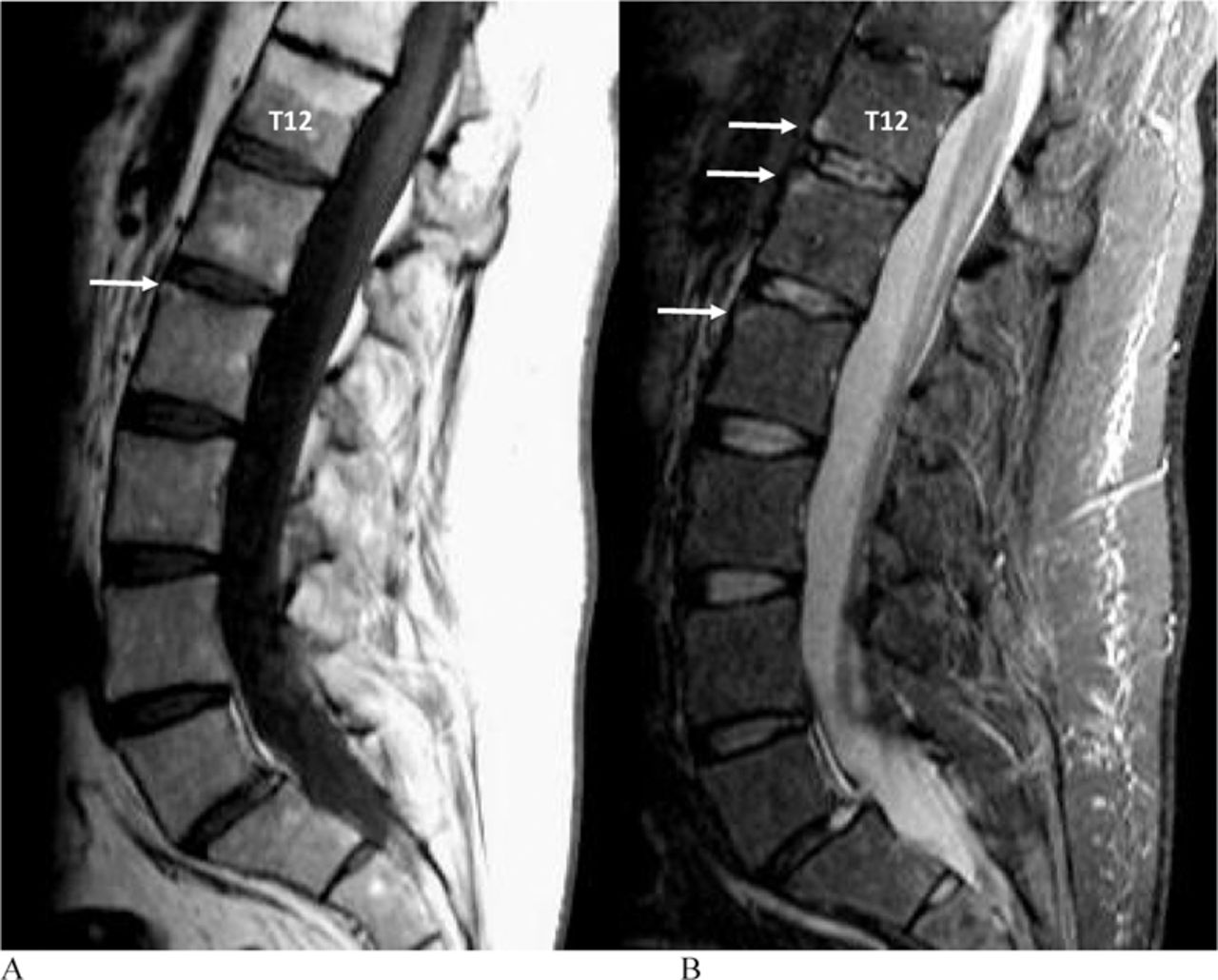
In reading the MRI images, we adhered to standardised definitions for acute and chronic spinal lesions developed by the Spondyloarthritis Research Consortium of Canada (SPARCC) and an international working group of rheumatologists and radiologists from Canada and Denmark.13 ,16 ,17 A vertebral CIL is defined as an increased STIR signal that is present in at least one sagittal slice which includes the spinal canal. A CIL may be further categorised as type B if the inflammatory signal on the STIR sequence has partially receded from the vertebral corner (figure 1). Increased signal intensity directly at the vertebral corner on T1W images indicates fat infiltration, while low intensity indicates erosion or new bone. In a type A CIL, the inflammatory signal on the STIR sequence extends completely to the vertebral corner.

T1 weighted image (A) and short tau inversion recovery sequence (B) MRI scans demonstrating a type A vertebral corner inflammatory lesion at T12 lower and type B corner inflammatory lesion at L1 upper and L2 upper. There is also a fat lesion at L2 upper.
Reading of Images
A unique MRI study number was randomly allocated to each patient thereby ensuring blinding to all patient demographics and treatment. Plain radiographs and MRIs were anonymised, assigned a random number and read independently. T1W and STIR sequences were read independently for assessment of vertebral CIL and fat inflammatory lesions, respectively. The anterior vertebral corners of the cervical (C2 lower to T1 upper) and lumbar (T12 lower to S1 upper) spine were examined for new syndesmophytes by comparing baseline and 2-year images on lateral radiographs of the cervical and lumbar spine by two readers with assessment according to consensus. The MRI images were read independently by readers who recorded the presence/absence of lesions at the anterior vertebral corners of the cervical (C2 lower to T1 upper) and lumbar (T12 lower to SI upper) spine.
Statistical analysis
All the primary analyses focused on concordant data between two readers as recommended previously.8 The primary analysis was a comparison of the proportion of new syndesmophytes developing at a vertebral corner with a type A versus a type B CIL.
In secondary analyses, we compared the proportion of new syndesmophytes developing from a persistent or resolved CIL and vertebral corners with fat infiltration versus those corners with neither CIL nor fat infiltration at any time-point.
A CIL was defined as being persistent if it was recorded as being present on each time-point. A CIL was defined as being completely resolved if it was recorded as being present at baseline and then absent at 52 weeks. Vertebral corners that already demonstrated syndesmophytes or ankylosis at baseline were excluded from this analysis. Fisher's exact test was used for comparisons of proportions.
Patient-level analyses comprised a comparison of type A versus type B CIL in patients who developed syndesmophytes versus those who did not develop syndesmophytes after 2 years, using unpaired t-tests.
It has been shown previously that the likelihood of finding a new syndesmophyte after 2 years' follow-up is higher in a patient who already has syndesmophytes and/or ankylosis at baseline.18 Consequently, we used generalised linear latent and mixed models (GLLAMM)19 that adjusted for within-patient dependence for the total number of vertebral corners with fat, inflammation and syndesmophytes/ankylosis. Multivariate adjustment was conducted at the level of the 24 anterior vertebral corners in the cervical and lumbar spine which formed the basis for this entire study.20
Results
Demographic and baseline characteristics for patients randomised to receive adalimumab (n=38) or placebo (n=44) have been described previously.4 These were similar between treatment groups and consistent with a typical AS population. At baseline and at week 12, all 44 patients in the placebo group and 38 in the adalimumab group had evaluable MRIs. At week 52, 42 placebo-treated patients and 38 adalimumab-treated patients had evaluable MRIs. MRI scans were assessed in 76 patients who had radiographic data at baseline and at 2 years. Data were unavailable for 64 vertebral corners in 22 patients primarily due to lack of radiographic visualisation of the C7 and T1 vertebral corners.
Baseline descriptive data for inflammatory and fat lesions
Individual spinal level
There were 220 (16.4%) CIL, 172 (12.8%) type A and 48 (3.6%) type B CIL out of 1345 vertebral corners without syndesmophytes or ankylosis at baseline that were evaluable by both MRI and radiography (table 1). Fat lesions were recorded in 179 (13.3%) of the vertebral corners of which 52 (3.9%) also had a concomitant inflammatory lesion. Fat lesions were recorded significantly more frequently at vertebral corners that also had a CIL (52/220 (23.6%)) compared with those without a CIL (127/1125 (11.3%)) (p<0.0001). This was particularly evident at vertebral corners with type B CIL (23/48 (47.9%)) compared with type A CIL (29/172 (16.9%)) (p<0.0001).
Number (%) of vertebral corners with inflammation and fat lesions on baseline MRI in 76 patients with ankylosing spondylitis
Patient level
The majority of patients (90.5%) had at least one CIL or fat lesion. Of the 74 patients who had available STIR MRI scans, 60 (81.1%) had at least one CIL on the baseline MRI (mean (SD) number 3.8 (3.6)), 54 (71.1%) had at least one type A CIL (mean (SD) number 2.9 (3.0)), 33 (43.4%) had at least one type B CIL (mean (SD) number 1.1 (1.8)), and 27 (36.5%) had both types of CIL. At least one fat lesion was observed in 54 (73%) of patients (mean (SD) number 4.1 (2.9)) and 47 (63.5%) had both CIL and fat lesions.
Radiography follow-up
The number (%) of patients who developed at least one new syndesmophyte after 2 years was 28 (36.8%), the mean (SD) number of new syndesmophytes per patient was 1.7 (1.0) and the total number (%) of new syndesmophytes was 48 (3.6%).
MRI follow-up
Resolution of CIL was recorded at 157 (71.4%) of vertebral corners and was not significantly different for type A versus type B CIL (128 (74.4%) and 29 (60.4%), respectively). Resolution of type A CIL was similar irrespective of the concomitant presence of fat lesions (23/29 (79.3%) and 105/143 (73.4%) with and without fat lesions, respectively). Resolution of type B CIL was less frequent at those vertebral corners that also had a fat lesion (11/23 (47.8%)) as compared with those who did not (18/25 (72%)). There were seven new CIL that developed from 12 to 52 weeks and all were type A. One patient developed three new CIL, while four patients each developed one new CIL. None of the new CILs was associated with the development of fat lesions or a new syndesmophyte.
A total of 59 new vertebral corner fat lesions had developed by week 52, of which 21 (16.7%) developed from a type A CIL that had resolved, four (9.8%) from a persistent type A CIL, six (24%) from a type B CIL that had resolved and one (6.7%) from a persistent type B CIL. The development of new fat lesions at vertebral corners with type A (25/143 (17.5%)) or type B (7/25 (28%)) CIL at baseline was significantly higher than those vertebral corners without CIL at baseline (27/998 (2.7%)) (p<0.0001 for both).
Association with radiographic progression
Spinal unit level data
The majority of new syndesmophytes (26/48 (54.2%)) occurred at those vertebral corners that had either a fat lesion and/or a CIL on baseline MRI (figure 2). New syndesmophytes developed significantly more frequently in those vertebral corners that had a CIL (5.9%) or fat lesion (11.2%) on baseline MRI as compared with those without either of these two lesions (1.8%) (p=0.002 and p<0.0001, respectively). New syndesmophytes were significantly associated with type B CIL (16.7%) versus type A CIL (2.9%) (p=0.002). Moreover, while the concomitant presence of both a CIL and a fat lesion at the same vertebral corner on baseline MRI was significantly associated with the development of new syndesmophytes (13.5%) (p<0.0001), all of these, save one, were type B CIL.

Percentage of patients with new syndesmophytes after 2 years on radiographs of anterior cervical and lumbar spine in a clinical trial cohort of 76 patients with ankylosing spondylitis receiving adalimumab stratified according to the presence or absence of vertebral corner inflammatory lesions (CIL), and fat lesions at the corresponding vertebral corner on the baseline MRI. *p<0.001. †p=0.002. ‡ 6 out of 7 vertebral corners had type B CIL.
New syndesmophytes developed significantly more frequently in vertebral corners with persistent CIL (11.1%) (p=0.0004), but this association was only evident for type B CIL (21.1%) (p=0.0005) (table 2). Similarly, the association with CIL that resolved was only observed for type B CIL (13.8%) (p=0.003). New syndesmophytes were observed at four of the 59 (6.8%) vertebral corners where new fat lesions were observed by week 52 which was a significantly greater proportion than those developing at vertebral corners with neither CIL nor fat lesions on baseline MRI (1.8%) (p=0.03).
Number (%) of new syndesmophytes after 2 years on radiographs of anterior cervical and lumbar spine in a clinical trial cohort of 76 patients with ankylosing spondylitis receiving adalimumab stratified according to the type of CIL and its persistence/resolution with treatment
Patient-level data
The number of CIL and fat lesions on baseline MRI was significantly higher in those patients that developed a new syndesmophyte after 2 years (table 3). This was observed irrespective of the type of CIL and whether the CIL resolved or persisted after 52 weeks of follow-up.
Comparison of the number (mean (SD) of CIL and fat lesions on baseline MRI and after 52 weeks follow-up on adalimumab therapy of 76 patients with ankylosing spondylitis according to whether patients did or did not develop a new syndesmophyte
Multilevel analysis
After adjusting for the total number of vertebral levels at baseline with syndesmophytes/ankylosis, the odds of a new syndesmophyte were significantly increased for a type B CIL (OR=3.88; 95% CI [1.20 –12.57], p=0.024) or a fat lesion (OR=4.83; 95% CI [2.38 to –9.80], p<0.0001) (table 4). After adjusting for within-patient variation for total number of vertebral corners with each of these predictors in the model that is, fat, type B CIL and syndesmophytes/ankylosis, the odds of a new syndesmophyte were significantly increased for a fat lesion (OR=4.0; 95% CI [1.74 –9.08], p=0.001) but not for type B CIL (OR=2.17; 95% CI [0.64 –7.40], p=0.21). The association with baseline extent of syndesmophytes/ankylosis was significant irrespective of the model.
Multivariate logistic regression analyses, in which baseline fat, inflammation and syndesmophytes/ankylosis were entered into a generalised linear latent and mixed model, to explore their association with the development of new syndesmophytes after 2 years in 76 patients with ankylosing spondylitis
Discussion
Our data supports the hypothesis that early inflammatory lesions may resolve without sequelae, while resolution of inflammation in more advanced inflammatory lesions, where reparative changes are already evident, is accompanied by new bone formation. First, we show that type B but not type A CIL are associated with the development of new syndesmophytes at the level of the patient, individual spinal units and when adjusted for the extent of baseline radiographic damage. Second, the development of new syndesmophytes from vertebral corners that have type B CIL at baseline occurs irrespective of whether the CILs persist or resolve. Third, we show that fat lesions are associated with the development of new syndesmophytes at the level of the patient, individual spinal unit and when adjusted for both the extent of baseline radiographic damage and the extent of inflammation. Of particular interest we observed the development of new syndesmophytes after 2 years at vertebral corners that developed new fat lesions from baseline to 52 weeks.
In patients with established disease, as exemplified by the baseline descriptive data in our cohort, almost half already have the more complex inflammatory lesions, and the majority have fat lesions. If the overall development of new bone during anti-TNF therapy depends on the balance between the number of early and more advanced inflammatory lesions, and if a substantial proportion of patients already have more complex inflammatory lesions, this might account for the lack of impact of adalimumab on radiographic progression at 2 years, as reported previously.3 However, the hypothesis also predicts that with longer-term follow-up there will be a declining rate of new bone formation because prolonged anti-TNFα therapy will not only induce resolution of type A lesions but also prevent development of new inflammatory lesions. Preliminary data supports this hypothesis as fewer syndesmophytes have been recorded in the later follow-up years of long-term therapy with infliximab compared with the first 4 years of followup21.
Our data also addresses current hypotheses directed at understanding the link between inflammation and ankylosis. It has been suggested that inflammation and ankylosis are uncoupled because one study reported a weak association between inflammation and new syndesmophytes,9 and because all reported studies7,–,9 have shown that new syndesmophytes occur at vertebral corners that appear normal on STIR MRI. A methodological approach that relies on inflammation in a vertebral unit9 that includes the entire bone marrow between two horizontal lines drawn across the middle of adjacent vertebrae, will result in weaker association than an approach based on direct evaluation of inflammation and new bone at the same anterior vertebral corner.7 ,8 In addition, all previous studies did not address lesions on T1W MRI, especially fat lesions. The majority of new syndesmophytes in our cohort developed from vertebral corners that had fat lesions and/or inflammation. Moreover, STIR MRI lacks sensitivity for inflammatory lesions.22 ,23 Consequently, our data is consistent with the hypothesis that inflammation and ankylosis are linked, and that an important intermediary in the sequence of events in human disease, but not in animal models reported to date, is the process of fat metaplasia. We further propose that while inflammation and ankylosis may be initially coupled this may become less evident as the lesion evolves into a fatty lesion. We did not assess the TIW sequence for vertebral corner erosions because these are often small and it is difficult to be certain that there is a breach of cortical bone because the signal is dark on both the STIR and T1W sequences. However, we have previously reported that vertebral corner erosions occur in a minority of patients (<20%)17 which casts doubt on a significant osteo-destructive process preceding the development of new bone as a reparative tissue response.23
Our finding that new syndesmophytes develop at the sites of both persistent and resolving type B CIL suggests that the process of new bone formation may eventually become autonomous from inflammation. While this observation appears at first sight to be at variance with previous reports demonstrating an association between resolution of new bone formation and new syndesmophytes,8 ,11 a significant consideration in the conduct of MRI studies is that it is often difficult to determine whether an inflammatory lesion has substantially resolved. This is particularly challenging for the assessment of type B CIL because the STIR signal can be heterogeneous and is often less pronounced or homogeneous than in type A CIL. However, it is relevant that combined MRI, and biomarker data, supports the hypothesis that resolution of inflammation is associated with new bone formation.24
In summary, our data shows that early inflammatory lesions resolve following anti-TNFα therapy and are not associated with the development of new syndesmophytes. While complex inflammatory lesions also resolve following anti-TNFα therapy, they are associated with the development of new syndesmophytes. Fat lesions, both established and newly evolving, are also associated with new bone formation. Our data supports a window-of-opportunity concept of disease modification for anti-inflammatory therapy in SpA, and a model of new bone formation that is dependent on the activation of inflammatory pathways followed by tissue metaplasia that includes fat. Further testing of these hypotheses is warranted in studies of early SpA.
Acknowledgments
WPM is a scientist of the Alberta Heritage Foundation for Medical Research.
References
Footnotes
-
Competing interests None.
-
Ethics approval The study was approved by an independent ethics committee at 7 of the 11 study centres; 4 study centres used a central independent ethics committee.
-
Provenance and peer review Not commissioned; externally peer reviewed.