Article Text

Abstract
Objectives To review and update the existing definition of a positive MRI for classification of axial spondyloarthritis (SpA).
Methods The Assessment in SpondyloArthritis International Society (ASAS) MRI working group conducted a consensus exercise to review the definition of a positive MRI for inclusion in the ASAS classification criteria of axial SpA. Existing definitions and new data relevant to the MRI diagnosis and classification of sacroiliitis and spondylitis in axial SpA, published since the ASAS definition first appeared in print in 2009, were reviewed and discussed. The precise wording of the existing definition was examined in detail and the data and a draft proposal were presented to and voted on by the ASAS membership.
Results The clear presence of bone marrow oedema on MRI in subchondral bone is still considered to be the defining observation that determines the presence of active sacroiliitis. Structural damage lesions seen on MRI may contribute to a decision by the observer that inflammatory lesions are genuinely due to SpA but are not required to meet the definition. The existing definition was clarified adding guidelines and images to assist in the application of the definition.
Conclusion The definition of a positive MRI for classification of axial SpA should continue to primarily depend on the imaging features of ‘active sacroiliitis’ until more data are available regarding MRI features of structural damage in the sacroiliac joint and MRI features in the spine and their utility when used for classification purposes.
- Magnetic Resonance Imaging
- Spondyloarthritis
- Ankylosing Spondylitis
Statistics from Altmetric.com
Introduction
Since the early 1990s MRI has been increasingly used to visualise inflammation in the sacroiliac (SI) joints and spine and it has become clear that inflammatory lesions can be visible on MRI before structural changes are detectable on radiography or CT.1 In 2009, the Assessment in SpondyloArthritis International Society (ASAS) published new criteria for axial spondyloarthritis (SpA) based on principles that incorporated demographic, clinical, laboratory and imaging components but now added an MRI definition so as to enable the identification of patients without evidence of structural change on radiography.2 A ‘positive MRI’ was defined in a publication which described and illustrated the variety of lesions that may be encountered on MRI of SI joints showing sacroiliitis and its differential diagnoses, and also defined the nature and extent of inflammation in the SI joints that would be necessary to meet the definition of ‘MRI positive for active sacroiliitis’.3 The definition relied on the observation of inflammation seen in subchondral bone and other observations were not required as part of the MRI definition.
As only 30%–50% of subjects with axial SpA are positive for active sacroiliitis on MRI,4–6 the question arose as to whether the wording of the current definition for a positive MRI is appropriate and whether structural change of the SI joint or findings on spine MRI should be incorporated into the ASAS definition of a positive MRI. The purpose of this consensus exercise was to examine and discuss whether data published in the last 5 years relevant to the diagnosis and classification of axial SpA are sufficient to merit a change in the MRI definition of a positive MRI and clarify any misunderstanding of the existing definition that may have become apparent since its first publication.
Methods
This manuscript has been developed on the basis of participation by 16 rheumatologists and 4 radiologists and 1 research fellow of the ASAS MRI working group with interest and experience in both SpA and MRI in a consensus exercise; presentation and discussion of evidence at a meeting on 5 September 2013 in Dusseldorf, Germany by the ASAS MRI working group; after refining the scope of the review, presentation during the annual assembly of ASAS on 17 January 2014 with voting on proposals open to all members; and consensus approval of the final manuscript by the members of the ASAS MRI working group.
Through the above process, the group was tasked with answering four questions related to MRI for inclusion in the ASAS classification criteria of axial SpA: (A) How does the current ASAS definition for a positive MRI perform? (B) Do we need to update the existing definition? (C) Do we need to add MRI features of structural changes of the SI joint to the definition? (D) Do we need to include features of SpA on MRI of the spine in the definition? At the consensus meeting, an updated systematic literature review was presented, followed by review of the definition of a positive MRI scan of the SI joint and the definition of a positive MRI scan of the spine. Next, new data (partly unpublished at that time but published and cited since) related to one or more of the study questions were presented by members of the group. Finally, the precise wording of the existing definition was examined in detail. During the 2014 annual assembly of ASAS a summary of the data and the draft proposal of the group was presented followed by voting open to all full ASAS members.
Results
There was consensus that there was no need to change the existing technical requirements necessary to reliably detect MRI features of inflammation or structural damage in bone marrow. As the presence of inflammation is the principal observation required by the current definition, this must be a focus for the MRI scan. The terms ‘bone marrow oedema’ (BMO) and ‘osteitis’ are considered to be equivalent in this context and the inflammatory and structural lesions have been previously described. The description for SI joint BMO is:
BMO is depicted as a hyperintense signal on short tau inversion recovery (STIR) images (or equivalent water-sensitive sequences) and usually as a hypointense signal on T1-weighted images (figure 1). The more intense the signal the more likely that it reflects active inflammation. A strong hyperintense signal is similar to that of cerebrospinal fluid. The sacral interforaminal bone marrow signal forms the reference for assignment of normal signal in the bone.
BMO is an indicator of active sacroiliitis but may be found in other diseases (figure 2) or as an incidental finding (figures 3 and 4).
Affected bone marrow areas are typically located periarticularly (subchondral bone marrow).
BMO may be associated with signs of structural damage such as sclerosis or erosion (figure 5).
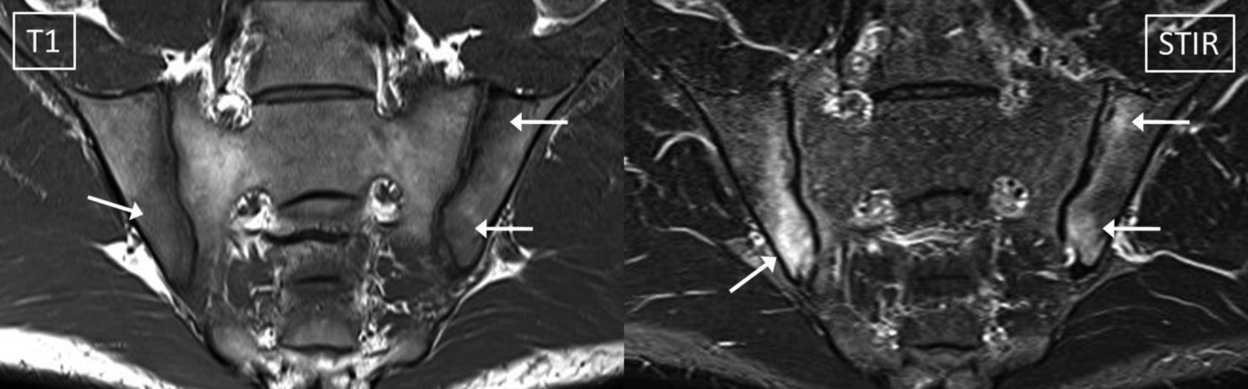
MRI sacroiliac (SI) joints—typical inflammatory sacroiliitis in non-radiographic axial spondyloarthritis. MRI of SI joints in a 26-year-old male with inflammatory low back pain of more than 3 months duration. C-reactive protein was 49.3 mg/L and HLA-B27 was positive. Pelvic radiograph was suspicious for spondyloarthritis but did not meet the definition for a positive radiograph according to the modified New York criteria—right SI joint grade 1 and left SI joint grade 0. The short tau inversion recovery (STIR) sequence shows abnormal increased signal (arrows) in the iliac bones bilaterally, typical for bone marrow oedema (BMO) due to inflammatory sacroiliitis. All the BMO are subchondral in location; the BMO is multifocal; each lesion is of a significant size; their margins are poorly defined; the right lower iliac lesion is larger and part of this lesion is intensely bright, similar in signal intensity to cerebrospinal fluid (not shown); and there are corresponding areas of diminished signal intensity on the T1-weighted sequence (arrows on T1). No erosion or other evidence of structural damage was visible on MRI or radiography. All these features are typical for inflammatory sacroiliitis.
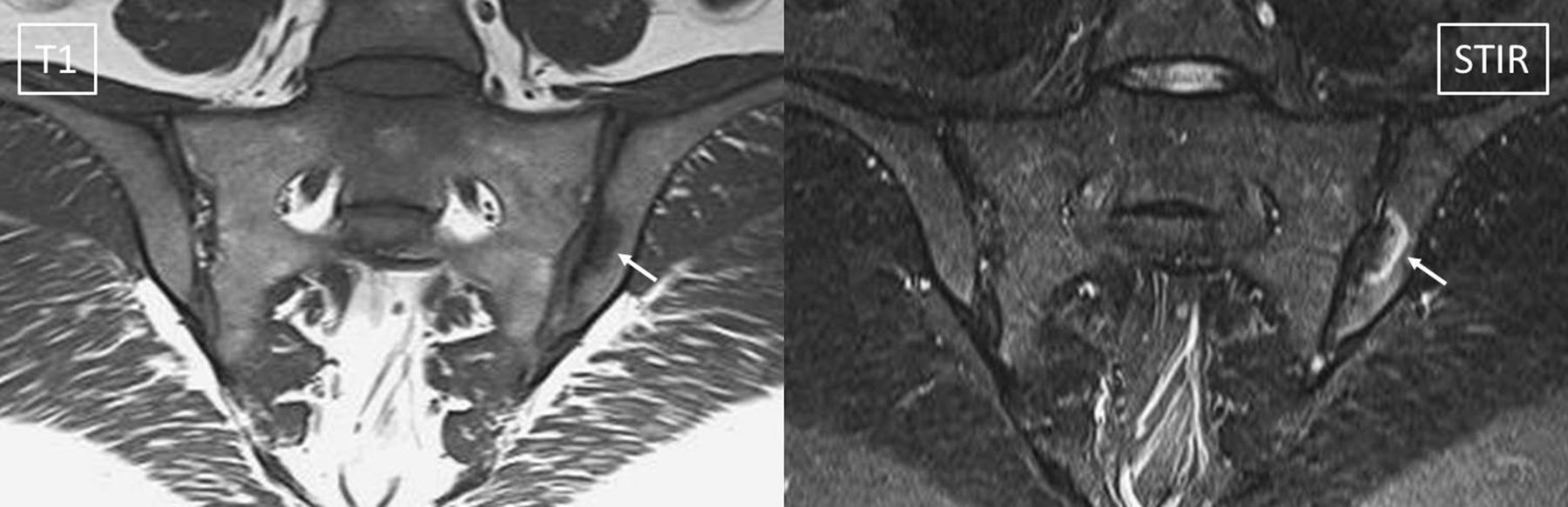
MRI sacroiliac (SI) joints—Osteitis Condensans Ilii (OCI). MRI of SI joints in a 28-year-old female with persistent low back and buttock pain of 4 years duration was performed 1 year after the second pregnancy. C-reactive protein was normal and HLA-B27 was positive. Pelvic radiograph revealed bilateral iliac sclerosis with joint space narrowing and minimal irregularity of the joint surface. The short tau inversion recovery (STIR) sequence shows abnormal increased signal (arrow) in the left iliac bone, with a non-specific appearance. The BMO has an arcuate contour surrounding an area of diminished signal intensity on the T1-weighted sequence (arrow on T1) that corresponded to radiographic sclerosis. More prominent sclerosis and less intense BMO were seen on multiple slices. The subchondral location of the finding may be seen in lesions related to either spondyloarthritis or mechanical causes. The very sharp definition of the borders of the abnormality does not help distinguish the aetiology. The T1 sequence did not show evidence of structural damage (erosion, fat metaplasia or ankylosis) except for sclerosis, which is a non-specific observation. The patient was followed up for 10 years and a diagnosis of OCI was subsequently confirmed.

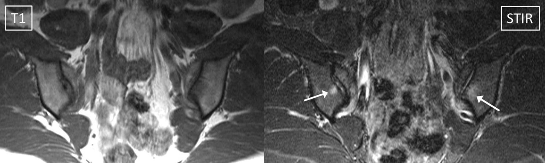
MRI sacroiliac (SI) joints—mechanical back pain. MRI of SI joints in a 31-year-old male with mechanical low back pain of more than 3 months duration. C-reactive protein was normal. On pelvic radiography, the SI joints were normal. The short tau inversion recovery (STIR) sequence shows abnormal increased signal (arrow) in the subchondral bone of the left ilium with a non-specific appearance. Although a BMO lesion is clearly present, it is small and on the T1-weighted sequence an even smaller focus of very low signal is seen in the same location paralleling the articular surface. There was no evidence of structural damage in the SI joints. These coronal images also show evidence of disc degeneration at L5/S1 with loss of height and signal intensity of the nucleus pulposis, bulging annulus and Modic type 1 reactive inflammation (bright on STIR) at the perimeter of the disc. The patient was followed up and the final diagnosis for the cause of the mechanical low back pain was disc degeneration. The cause of the left SI lesion is unproven but it most likely represents a small fatigue stress reaction in association with mild osteoarthritis of the SI joint. These MRI observations are frequently seen in weight-bearing joints as they degenerate.
MRI sacroiliac (SI) joints—healthy volunteer. MRI of SI joints in a 35-year-old female health services worker who was an asymptomatic volunteer enrolled as a control subject into an ethics-approved research project. There was no history of pregnancy. The subject was fit and healthy and did not participate in any endurance activities. Clinical evaluation confirmed the absence of any symptoms, signs or risk factors for spondyloarthritis (SpA). The volunteer has been followed for 10 years and remains asymptomatic. The short tau inversion recovery (STIR) sequence shows abnormal increased signal (arrows) in the subchondral bone of both SI joints. The findings were clearly visible on at least two slices bilaterally. The lesions on STIR are small (12 mm on left and 8 mm on right) and are located close to the anterior borders of the SI joints. Some brighter signals in the ‘joint space’ seen on these and other images are suspicious for cartilage degeneration. Minimal signal change is present on the T1-weighted sequence and no structural damage changes are present. The follow-up MRI performed 8 weeks later as part of the clinical trial was unchanged.
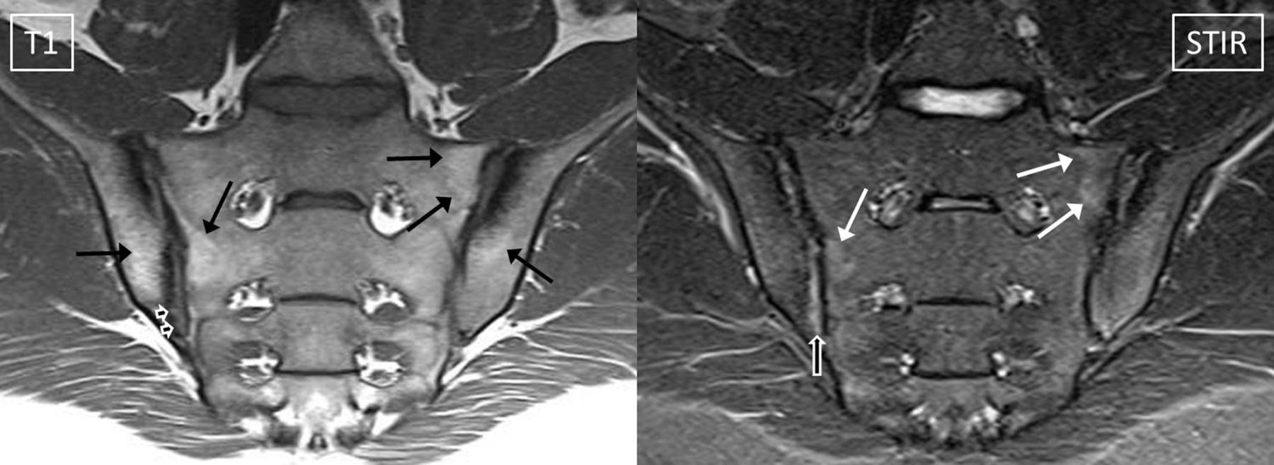
MRI sacroiliac (SI) joints—typical findings in non-radiographic axial spondyloarthritis (SpA) showing minimal inflammation and structural damage changes. MRI of SI joints in a 20-year-old male with inflammatory low back pain of more than 3 months duration. C-reactive protein was normal at the time of the MRI scan and HLA-B27 was positive. Pelvic radiograph was suspicious for SpA but did not meet the definition for a positive radiograph according to the modified New York criteria—right SI joint grade 2 and left SI joint grade 0. The short tau inversion recovery (STIR) sequence shows subtle increased signal (white arrows) in the subchondral bone of the right and left sacral alae suspicious for bone marrow oedema (BMO). However, the lesions are faint and heterogeneous and the right sacral lesion was only visible on one slice. If no other findings were present, it would be difficult to decide whether the BMO changes alone do or do not meet the criteria for a positive MRI. However, multiple other abnormalities are present on the MRI that materially influence the decision. On the T1-weighted sequence, subtle articular surface erosion is definitely seen at the caudal end of the right ilium (arrowheads); subtle foci of fat metaplasia are seen in all four bones (black arrows); and bilateral iliac subchondral sclerosis is present. Additionally, on the STIR sequence, abnormal increased signal overlying the area of articular surface erosion is typical for inflammation in the cartilage/joint space (compound arrow). Each finding individually is non-specific; however, in combination the appearance and distribution of all the findings are typical for inflammatory sacroiliitis. In conclusion, the MRI scan meets the Assessment in SpondyloArthritis International Society (ASAS) definition for a positive MRI because (1) BMO on STIR is present; (2) the inflammation is located in a typical anatomical area (subchondral bone) and (3) the MRI appearance is highly suggestive of SpA—in this case, because the findings of BMO are supported by MRI findings of structural damage (erosion, fat metaplasia and sclerosis) that are typical in appearance and distribution for SpA.
New data regarding the classification of patients with early axial SpA were presented from several cohorts that are currently under investigation. Using the DESIR cohort of subjects with inflammatory back pain (IBP) of less than 3 years duration before the age of 50, the reliability of classification of radiographs and MRI was compared between rheumatologists and radiologists of 25 local recruiting centres and central readers.5 ,7 The results indicate that the existing MRI definition could be applied across multiple centres with the expectation of acceptable reliability and at least with better reliability than the X-ray definition of sacroiliitis according to the modified New York criteria.
A detailed analysis of SI joint MRI scans from a pair of Canadian/Swiss inception cohorts of 157 consecutive subjects ≤50 years old with back pain that included age and sex-matched controls suggests that benefit might be gained from adding SI erosion to the definition.8 This was observed in both nr-axSpA patients with short symptom duration (mean 1.3 years) and those with longer duration (mean 10 years). However, the analysis did not take into consideration whether these subjects had or had not already met the ASAS classification for axial SpA by the clinical arm and so the incremental benefit to classification by adding erosion to the definition is not clear.
In the SPACE cohort of subjects with chronic back pain of less than 2 years duration starting before age 45, the effect of adding structural change to the definition of a positive SI joint MRI was analysed by each feature individually and in combination.9 In this cohort, there was no single lesion or combination of lesions that would confer a significant benefit to sensitivity of the ASAS MRI definition without a corresponding risk of losing specificity.
With regard to the spine, the Canadian/Swiss inception cohorts examined the incremental value of spine MRI and concluded that while sensitivity was enhanced by 16% with combined assessment of the spine in addition to the sacroiliac joint (SIJ), false-positive diagnoses of SpA were increased by a similar degree.10 Data were also analysed for the spine MRI in the SPACE cohort with the effect on classification of the subjects analysed by lesion type and also by the number of qualifying lesions present with a range of cut-off thresholds analysed for each lesion separately. For each type of lesion, the marginal benefit (sensitivity) for adding spine MRI to the definition comes at the price of both diminished specificity and additional financial cost.11
In summary, new data were presented indicating that:
For the SI joint, the current definition of a positive MRI (active sacroiliitis) performs satisfactorily for the classification of axial SpA according to the ASAS axial SpA criteria, and can be interpreted across many centres with substantial reader agreement.
Evaluation of structural features, especially erosions, may enhance confidence in the classification of axial SpA emphasising the importance of simultaneous assessment of T1W and fat-suppressed sequences, and the contextual interpretation of MRI. However, the effect on classification of the addition of any structural damage feature to the definition of a positive SI joint MRI is not yet clear, in part due to variations in MRI acquisition protocol and advancing MRI technology that compounded the complexities of achieving consensus for definitions for each MRI structural damage lesion and the setting of thresholds for any defined lesion or combination of lesions.
There is no consistent beneficial effect of adding features of SpA on spine MRI to the definition.
Following extensive discussion, the consensus opinion of the group was that ‘The definition of a positive MRI should not be changed at this time. The utility of the structural damage changes of the SI joints and the addition of features on MRI of the spine for classification purposes is not yet clear and this continues to be an important research agenda’. The available data (not all data from some references were available at the time) were then presented and discussed at the annual assembly of ASAS on 17 January 2014. The meeting concluded with voting open to all members and a proposal to not change the existing definition was unanimously approved.
Definition of sacroiliitis on MRI
After deciding to not change the definition of sacroiliitis on MRI for application in the ASAS classification criteria, it was agreed by consensus to provide some clarification of the existing definition and guidelines for the application of the definition. The presentation of the existing definition was reformatted accordingly (box 1) and guidelines for the application of the definition are now provided (box 2).
Definition of a positive MRI (active sacroiliitis) for the classification of axial spondyloarthritis (SpA) according to the Assessment in SpondyloArthritis International Society (ASAS) axial SpA criteria
Inflammation of the sacroiliac joints highly suggestive of SpA is required for the fulfilment of the imaging criterion ‘active sacroiliitis on MRI’ according to the ASAS classification criteria for axial SpA.
The requirements are listed below and guidelines for the application of the definition are provided in box 2.
REQUIRED MRI evidence of bone marrow inflammation must be present and the features required for the definition of active sacroiliitis on MRI are:
1. Bone marrow oedema (BMO) on a T2-weighted sequence sensitive for free water (such as short tau inversion recovery (STIR) or T2FS) or bone marrow contrast enhancement on a T1-weighted sequence (such as T1FS post-Gd).
2. Inflammation must be clearly present and located in a typical anatomical area (subchondral bone).
3. MRI appearance must be highly suggestive of SpA.
NOT REQUIRED Other findings related to sacroiliitis may be observed on MRI but are not required to fulfil the imaging criterion ‘active sacroiliitis on MRI’:
The sole presence of other inflammatory lesions such as synovitis, enthesitis or capsulitis without concomitant BMO is not sufficient for the definition of ‘active sacroiliitis on MRI’.
In the absence of MRI signs of BMO, the presence of structural lesions such as fat metaplasia, sclerosis, erosion or ankylosis does not meet the definition of ‘active sacroiliitis on MRI’.
Guidelines for the application of the definition of a positive MRI (active sacroiliitis) for the classification of axial spondyloarthritis (SpA)
MRI interpretation:
Bone marrow oedema (BMO) representing an inflammatory lesion that meets the above criterion will usually be easily seen on at least two consecutive slices of an MRI scan. Detection of inflammation on a single slice may be sufficient for the criterion ‘highly suggestive of SpA’ if there is more than one inflammatory lesion present. However, it is rare for an MRI scan of the sacroiliac joints with definite evidence of active sacroiliitis to demonstrate lesions on only a single image, and caution should be exercised in the interpretation of small lesions.
It is essential that the reader of the MRI scan simultaneously review sequences designed to identify inflammation and sequences that focus on depiction of structural damage.
If an inflammatory bone marrow lesion appears to be present but it is hard to determine whether the lesion meets the criterion ‘highly suggestive of SpA’, then the decision may be influenced by the presence of concomitant structural damage, especially erosion, and/or other signs of inflammation, which in themselves do not suffice to meet the criterion.
Context:
Evaluation of an MRI scan should be performed objectively. However, MRI findings are non-specific and the determination of the importance of the observations should never be made in isolation of the clinical context as demographic, clinical and laboratory information may outweigh the importance of the MRI findings.
The definition and guidelines are primarily for the classification of patients with SpA and will not be suitable for use in some clinical situations.
Discussion
The ASAS/OMERACT MRI working group previously decided by consensus that the presence of subchondral BMO (or osteitis) in the SI joints reflecting inflammation highly suggestive of SpA should be regarded as essential to meet the definition of ‘active sacroiliitis on MRI’ in cases of axial SpA when radiographic changes are absent or doubtful.3 The purpose of this consensus exercise was to examine whether new data published in the last 5 years regarding axial SpA are sufficient to merit a change in the MRI definition of a positive MRI and clarify any misunderstanding of the existing definition that may have become apparent since its first publication. After detailed consideration, the unanimous consensus of ASAS members was to retain the existing definition with a slight rewording that would help to emphasise the critical components of the definition.
The ASAS definition of ‘active sacroiliitis on MRI’ was the first definition of ‘a positive MRI’ to be widely used in clinical trials research. The development of the definition was based on published data and took other factors into consideration: universal agreement that, above all, MRI evidence of inflammation (which is radiographically occult) must be included in a definition of non-radiographic disease; easily applied MRI parameters; ability to apply the definition prospectively and retrospectively; and wording and illustration of the definition that was simple and intuitive. Ease of application of the criteria internationally is important and the minimum MRI technical requirements can be applied on any MRI platform and do not change with advancing technology. A wide range of newer sequences such as water excitation or chemical shift imaging may be used to detect bone marrow inflammation but do not change the principles of image acquisition or the definition of ‘active sacroiliitis’. Most centres use either STIR or T2FS as in most cases, T1-weighted contrast-enhanced sequences offer no additional benefit in either adults or children and contrast material is expensive and is therefore not recommended.12–15
The selection of an MRI definition is influenced by how it performs in a rigorous testing environment and by the ability to describe and illustrate the target lesion in terms that facilitate widespread application. This is another reason that the ASAS definition remains focused on the inflammatory lesion in the SI joint at this time and why structural damage lesions have not yet been added to the definition. Erosion of the joint surface may seem to be a logical choice for inclusion in a definition of sacroiliitis; however, currently, there is no international consensus as to how erosion should be defined on MRI or how it should be quantified. For example, it is not yet clear to what extent variation in MRI acquisition parameters, such as using a thin-slice high-resolution three-dimensional sequence, would alter the properties of a definition that includes structural damage. Features of axial SpA identifiable on spine MRI are still not included in the definition of a positive MRI because none of the candidate definitions would appear to confer a significant advantage over the current definition. The SI joints are involved before the spine in the majority of true axial SpA cases and the matter is complicated by the fact that degenerative changes in the spine are frequent and small foci of inflammation that are mechanical in origin may be indistinguishable from small inflammatory lesions due to SpA.10 ,16
The European Society of Skeletal Radiology (ESSR) recently reviewed the imaging appearances and the radiological features necessary for making a diagnosis of axial SpA and recommended that MRI is mandatory to look for early inflammatory lesions if axial SpA is suspected and radiographs are negative.17 The burden of disease required to meet a diagnosis of sacroiliitis described by the ESSR Arthritis Imaging Subcommittee is consistent with the ASAS definition for classification, which requires all lesions to be ‘highly suggestive of SpA’. It should be noted that the ESSR publication omits quotation of this critical component of the ASAS definition. In the case of minimal inflammatory changes, specific advice regarding the exact nature and extent of additional imaging features of SpA that would be required to heighten the suspicion of the observer is not provided and this remains an important focus for future research. As noted previously, doubtful cases of BMO should not be considered as positive, and this view is supported by data highlighting the prognostic role of more extensive BMO for the later development of radiographic sacroiliitis.18
The publication by ASAS of the first ‘definition of active sacroiliitis on magnetic resonance imaging (MRI) for classification of axial spondyloarthritis’ was an important step that has enabled many subsequent research projects. The existing definition is based on the observance of inflammatory lesions in typical locations related to the SI joint and can be applied by readers across the world with access to any MRI unit. The ASAS definition was reviewed by an expert group of rheumatologists and radiologists with experience in both SpA and MRI including systematic analysis of data from different cohorts. The results were presented by experts to the ASAS members. By a final vote of all members, the unanimous consensus was to retain the existing definition with a slight rewording that would help to emphasise the critical components of the definition. The contribution of MRI features of structural damage of the SI joint or spinal MRI features of SpA to a refined classification of axial SpA remains an important research agenda. However, the existing ASAS definition of a positive MRI continues to provide a solid basis for the application of MRI in the ASAS criteria for axial SpA.
References
Footnotes
Handling editor Tore K Kvien
Contributors All authors substantially contributed to the conception and/or design of the work, and the interpretation of data; drafting the work and/or revising it critically for important intellectual content; have final approval of the version published; and agree to be accountable for all aspects of the work.
Competing interests None declared.
Provenance and peer review Not commissioned; externally peer reviewed.