Article Text

Abstract
Objective: To correlate the number of chondrocytes in healthy and osteoarthritic human articular cartilage with age, and to evaluate the influence of donor age on total proteoglycan synthesis.
Methods: Chondrocytes were isolated from human articular cartilage derived from hip joints with and without osteoarthritic lesions. The cell number was normalised to cartilage sample wet weight. In addition, the influence of age on chondrocyte numbers was assessed histomorphometrically. Chondrocytes were grown as monolayer cultures for seven days in a chemically defined serum-free basal medium. Total proteoglycan synthesis was measured by [35S]sulphate incorporation into newly synthesised macromolecules.
Results: Chondrocyte numbers in healthy cartilage decreased significantly with advancing age (r = −0.69, p<0.0001). In contrast to healthy specimens, chondrocyte numbers were decreased in osteoarthritic cartilage irrespective of and unrelated to age, and differed markedly, by an average of 38%, from the cell numbers found in healthy individuals (p<0.0001). Regarding synthesis of matrix macromolecules, no dependence on patients’ age, either in healthy or in osteoarthritic specimens, could be observed.
Conclusions: Under the experimental conditions employed, chondrocytes from healthy and osteoarthritic joints synthesised comparable amounts of cartilage macromolecules, independent of age or underlying osteoarthritic disease. Thus the decrease in chondrocyte number in aging and osteoarthritic joints could be a crucial factor in limiting tissue replenishment.
- MMM, matrix macromolecules
- age
- cartilage
- chondrocyte
- osteoarthritis
Statistics from Altmetric.com
Aging is associated with progressive structural, functional, and metabolic alterations in a variety of tissues and systems. Many of these age related alterations have been implicated in subsequent impairment of physiological and physical function.1 Functional changes in the musculoskeletal system are among the most prevalent health problems of middle and old age.2 Osteoarthritis, the most common form of joint disease, is highly correlated with increasing age.3–5 Despite the strong relation between age and increased prevalence of degenerative cartilage changes, the underlying mechanisms whereby age is involved in the development or progression of osteoarthritis are so far unknown.6,7
Important age dependent changes in articular cartilage—which increase in prevalence, extent, and severity with advancing age8,9,10—concern matrix macromolecules (MMM) such as proteoglycans and collagens. Qualitative and quantitative variances in these components are observed, suggesting an alteration in the physiological cartilage composition and mechanical properties.2,9,11–17 Moreover, and possibly more important, an age related reduction in total proteoglycan synthesis after skeletal maturation has been reported.18–20
The role and influence of chondrocytes in this condition remain unclear. Several studies have suggested that the cell density of the whole thickness of the uncalcified articular cartilage declines sharply during the growth and maturation period of skeletal development, but remains relatively constant in adult life.21–24 On the other hand there is evidence that chondrocyte numbers decrease progressively in healthy articular cartilage as a function of age.25–28
Given the reduction in total proteoglycan synthesis with advancing age, we hypothesised that a decline in cell number was an important factor in limiting tissue maintenance. Thus we evaluated the cell numbers in healthy and osteoarthritic human articular cartilage in relation to increasing age, and investigated total MMM synthesis in healthy and osteoarthritic cartilage to determine a possible influence of age on proteoglycan production.
METHODS
Source of articular cartilage
Human articular cartilage from 41 patients (aged 37 to 87 years, mean age 62.9 years) without macroscopic osteoarthritic lesions were obtained from surgical specimens at the time of endoprosthetic replacement for acute transcervical femoral fractures and from organ donors.
The removal of cartilage from organ donors was approved by the ethics committee of the University of Vienna.
Human osteoarthritic cartilage samples were obtained from 30 individuals who underwent surgery for total hip endoprosthesis. The age of the patients ranged from 35 to 84 years with a mean age of 61.8 years.
Cell count and cell culture
Cartilage slices were aseptically dissected from the load bearing joint surfaces and finely minced. In osteoarthritis specimens, dissection of neocartilage at the joint margins was avoided. The wet weight of the samples obtained was then measured. Chondrocytes were released by overnight digestion in 0.2% collagenase B (Boehringer Mannheim, Mannheim, Germany) in a recently described, chemically defined, serum-free basal medium.29 Following digestion, an aliquot of the cell suspension was evaluated for chondrocyte number after Trypan blue staining in a Buerker–Tuerk chamber. The values are given in chondrocytes ×106/g wet weight. The fraction of dead chondrocytes was 5–10%, independent of advanced age or underlying osteoarthritic disease. In both osteoarthritic and unaffected cartilage samples, cell clusters formed about 2% of all chondrocytes and could easily be counted. To ensure completeness of tissue digestion, the suspension after collagenase incubation was subsequently filtered using a cell strainer (Falcon, Becton Dickinson Labware, Lincoln Park, New Jersey, USA), and the weight of the solid residues determined on a microgram scale. The percentage of solid residues after collagenase digestion was uniformly between 1.3% and 5.9% of the cartilage weight, with no difference between healthy and osteoarthritic samples (p = 0.46).
In an additional series of experiments, the influence of age on chondrocyte numbers was assessed histomorphometrically. We investigated histological samples from hip joints from 14 unaffected donors (aged 24 to 68 years, mean 43.5 years) and from 17 patients with osteoarthritis (aged 38 to 91 years, mean 70.4 years). Specimens of cartilage and subchondral bone were fixed in 7.5% formalin for 48 hours, decalcified with EDTA solution, and then paraffin embedded and stained with haematoxylin/eosin following standard protocols. Using the KS 300 version 3.0 software (Carl Zeiss Vision GmbH 1997) three randomly chosen full thickness cartilage areas were marked in each histological sample and the surface areas were calculated. The chondrocyte numbers were counted in the respective areas and the results are given as cell number/mm2.
For cell cultures, the chondrocyte filtrate was centrifuged at 500×g for 10 minutes. Pellets were resuspended in 1:1 mixture of Dulbecco’s modified Eagle’s medium (DMEM, 25 mM Hepes + 4500 mg/l glucose + pyridoxine, without sodium pyruvate; Life Technologies, Gaithersburg, Maryland, USA) and Ham’s F12 (Ham’s F-12 +L-glutamine; Life Technologies) containing 10% fetal bovine serum (PAA Laboratories, Linz, Austria) and antibiotics/antimycotics (100 U/ml penicillin G, 100 mg/ml streptomycin, and 0.25 µg/ml amphotericin B; Life Technologies). The isolated cells were grown as monolayer cultures in 24-well plates (Costar, Cambridge, Massachusetts, USA) in quadruplicate at a density of 1×10⁵ cells/cm2. At 90% confluence of the chondrocyte cultures (approximately 2×10⁵ cells/well), serum-containing medium was changed to basal medium and the cells were subsequently cultured for seven days. The medium was replaced every other day. Cultures were maintained at 37°C in humidified air and 5% CO2.
Biosynthesis of macromolecules
Proteoglycan synthesis rate for 17 healthy samples (from subjects aged 37 to 80 years, mean age 58.6 years) and 12 osteoarthritis samples (aged 35 to 84 years, mean age 58.6 years) was determined by [35S]sulphate incorporation into sulphated glycosaminoglycans, as previously described.30 Briefly, labelling of the cells with radioactivity was done by incubation with 20 µCi/ml of [35S]sulphate (carrier-free, Amersham, Buckinghamshire, UK) for six hours at 37°C in basal medium. Chondrocytes were extracted in guanidine-HCl buffer (4 M guanidine-HCl, 50 mM sodium acetate buffered at pH 7.2, in the presence of protease inhibitors). Unincorporated isotope was removed by using Sephadex G-25 gel chromatography (PD-10 columns; Pharmacia Biotech, Piscataway, New Jersey, USA). Values were obtained by liquid scintillation counting (model 1410 liquid scintillation counter, Wallac Oy, Turku, Finland) of aliquots from void volume fractions and normalised to protein content. Total cellular protein was determined by the Bradford method according to the manufacturer’s instructions (Bio-Rad Protein Assay; Bio Rad Laboratories GmbH, Munich, Germany).
Statistical analysis
Statistical analysis was done using Student’s t test. A normality test was carried out to determine whether the distribution of the samples was Gaussian. To examine relations between age and MMM synthesis rate, as well as between age and chondrocyte numbers, we used Pearson correlation calculations. Statistical significance was defined as a probability (p) value of <0.05.
RESULTS
Effect of age on chondrocyte numbers of healthy and osteoarthritis human articular cartilage
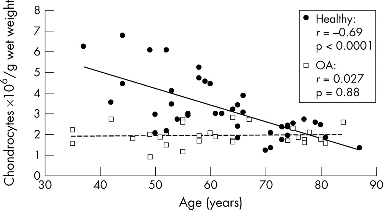
The evaluation of chondrocyte numbers after tissue digestion revealed an age dependent decrease of cells in the cartilage samples derived from joints without macroscopically visible defects (n = 41). In the patient age group <55 years (n = 12), a mean (SEM) of 4.2 (0.5)×106 cells/g wet weight of cartilage was found, whereas cartilage from patients >55 years old (n = 29) contained a mean of 2.7 (0.2)×106 chondrocytes/g wet weight—a significant (p<0.002) decrease of 35% (fig 1; r = −0.69, p<0.0001). To further evaluate whether this reduction in cartilage cellularity reflects a possible mechanism for the onset or progression of osteoarthritis, we compared the data from the healthy samples with the chondrocyte numbers in 30 tissue samples obtained from osteoarthritic joints. In contrast to the healthy specimens, the chondrocyte number in osteoarthritic cartilage had a mean (SEM) value of 1.9 (0.1)×106 cells/g wet weight, which differed markedly—by an average of 38%—from the number found in healthy cartilaginous tissue (p<0.0001). It is noteworthy that the decrease in chondrocyte numbers was already evident in younger patients with osteoarthritis and did not change further as a function of age (fig 1; r = 0.027, p = 0.88).

Changes in the total chondrocyte numbers of healthy and osteoarthritic articular cartilage with increasing age. Chondrocytes were enzymatically released from their extracellular matrix in 0.2% collagenase B. Directly after digestion cell numbers were assessed using light microscopy. The values are given as chondrocytes ×106/g wet weight. There was a decrease in chondrocyte numbers in healthy articular cartilage (black circles, black regression line) as a function of age (n = 41; r = −0.69, p<0.0001). Cellularity in osteoarthritic cartilage (white squares, dashed regression line) showed a significant reduction in cell number compared with healthy cartilage (p<0.0001), but no dependence on patients’ age (n = 30; r = 0.027, p = 0.88).
To support the above data, we evaluated the chondrocyte numbers/mm2 cartilage in histological sections from 14 healthy and 17 osteoarthritic patients. In accordance with the results from the cartilage digests, we found an age dependent decrease in cell numbers in healthy specimens (r = −0.89, p<0.0001) (fig 2). In osteoarthritic patients no such decrease could be seen (r = 0.12, p = 0.65). When we compared the healthy group with the osteoarthritic group we found a significant difference in cell numbers: healthy group, 91.6 (10.9) cells/mm2v osteoarthritis group, 48.6 (2.5) cells/mm2, p<0.007.

Chondrocyte numbers in histological sections from healthy and osteoarthritic cartilage. The values are given as chondrocytes/mm2 cartilage. Chondrocyte numbers in healthy articular cartilage (black circles, black regression line) decrease as a function of age (n = 14; r = −0.89, p<0.0001). In osteoarthritic cartilage (white squares, dashed regression line) no reduction in cell number could be seen (n = 17; r = 0.12, p<0.65). Analysis of the healthy v the osteoarthritic group showed a significant difference in cell numbers: healthy group, 91.6 (10.9) cells/mm2, v osteoarthritis group, 48.6 (2.5) cells/mm2 (mean (SEM)), p<0.007.
Effect of increasing age on biosynthetic activity in articular chondrocytes
To investigate whether the levels of biosynthetic activity in healthy and osteoarthritic human articular chondrocytes—as determined by synthesis of MMM—decreases with advancing age, we investigated cartilage specimens from osteoarthritic and non-osteoarthritic patients at different ages. The chondrocytes were cultured in serum-free basal medium for seven days. In cells from healthy individuals, [35S]sulphate incorporation rate ranged from 509 to 2689 cpm/mg protein, with an average of 1353 (161) cpm/mg protein (mean (SEM)). In osteoarthritis cultures, isotope uptake ranged from 118 to 2932 cpm/mg protein, with a mean of 1090 (249) cpm/mg protein. Although there was a mild trend towards decreasing MMM synthesis with increasing age, these differences were not significant, either among healthy samples (r = −0.23, p = 0.38) or among osteoarthritis samples (r = −0.19, p = 0.55) (fig 3A). Importantly, when we divided the healthy patient group into a “young” group (<55 years) and an “old” group (>55 years) and compared these groups with the osteoarthritis specimens (fig 3B), no significant difference could be observed (<55 years v osteoarthritis, p = 0.24; >55 years v osteoarthritis p = 0.8).

(A) Healthy (black circles, black regression line) and osteoarthritic (white squares, dashed regression line) human articular chondrocytes do not lose their biosynthetic capacity with age. Cells were cultured in serum-free basal medium without the addition of growth factors for seven days. The rate of proteoglycan synthesis was measured by [35S]sulphate incorporation into newly synthesised matrix proteoglycans present in the cell layer. Values were normalised to protein content and are given as counts per min/mg protein (cpm/mg protein). No age dependent decrease in total proteoglycan synthesis could be shown for healthy chondrocytes (r = −0.23, p = 0.38) or osteoarthritic chondrocytes (r = −0.19, p = 0.55). (B) Proteoglycan synthesis of postnatal human articular chondrocytes. Chondrocytes derived from healthy patients <55 years (white bars, n = 8), >55 years (light grey bars, n = 8), and osteoarthritis patients (dark grey bars, n = 13) were incubated in serum-free basal medium for seven days. On the final day of the incubation period, cell cultures were labelled with [35S]sulphate for six hours. The incorporated radiolabel in newly synthesised matrix macromolecules present in the cell layer was then measured, normalised to protein content, and given as cpm/mg protein. Values are mean (SEM). No difference between these groups could be determined (NS).
DISCUSSION
The ability of cells to maintain metabolic homeostasis is believed to decline with advancing age.6 Age represents a major risk factor for the occurrence of osteoarthritis.3 The question arises as to whether articular chondrocytes retain their capacity to uphold tissue homeostasis by synthesising matrix proteoglycans with advancing age. In the case of reduced biosynthetic activity, the maintenance of cartilage extracellular matrix would be impaired and subsequently lead to disruption of tissue integrity, as seen in osteoarthritis. Two possible underlying mechanisms for impaired proteoglycan synthesis in the aging articular cartilage could be a decrease in total cell number or a decline in proteoglycan synthesis rate.
Levels of biosynthetic activity may be decreased as a function of age or osteoarthritis, causing a breakdown of cartilage integrity. Under the experimental conditions employed in this study no such decrease—either with aging or with underlying osteoarthritis—was observed. On the other hand, there was a mild trend towards a decrease in macromolecular synthesis, and lack of statistical significance does not exclude the possibility that some patients with osteoarthritis may have a more severe impairment of proteoglycan biosynthesis. A possible limitation of the current study is that cell metabolic activity was assessed in monolayer cultures after initial expansion in serum-containing medium, thereby potentially overriding possible differences in metabolic activity between cells derived from osteoarthritic cartilage and controls. On the other hand, previous investigations did not show differences between explant cultures derived from osteoarthritic and healthy articular cartilage studied under serum-free conditions.31 Moreover, to minimise such potential effects, we exchanged the serum-containing to serum-free medium for seven days before assessing metabolic activity. Finally, in previous investigations we also did not observe dedifferentiation of cells under the experimental conditions employed.32,33 Nevertheless our data are in agreement with the reports by Lafeber and Brocklehurst, who previously described no difference in proteoglycan biosynthetic rate between healthy and osteoarthritic cartilage.31,34 A constant rate of proteoglycan biosynthesis in healthy articular chondrocytes between 30 and 95 years was also shown by Bayliss and colleagues.35 However, contrary findings were reported by Schafer et al,18 DeGroot et al,19 and Verbruggen et al,20 who all showed a significant reduction in proteoglycan synthesis as a result of aging. A possible explanation for these contradictory findings may be the use of different culture techniques. It must be assumed that all these results are valid, but studies employing in vivo rather than cell culture conditions may have to take mechanisms other than failure of chondrocyte metabolism into account.
As the cell counts in the monolayer cultures employed here were constant we assume that the age dependent reduction of biosynthetic activity is caused by an impaired cartilage cellularity, because it is well known that chondrocyte numbers decrease in healthy articular cartilage as a function of age.25–28 Furthermore it was reported that cell numbers in osteoarthritic cartilage declined in both fissured and intact joint surface areas compared with healthy specimens.28,36 Our present study confirms these data on healthy cartilage, showing a decrease in chondrocyte number with advancing age. In addition we found that cell numbers are markedly reduced in osteoarthritic patients independently of age, even in young patients. Thus the loss in MMM from articular cartilage—which represents an early event in osteoarthritis37—may be caused primarily by a decrease in cartilage cellularity. Moreover, with respect to the changes in matrix composition in old age and osteoarthritis,2,11,12,17 it is likely that a reduction in chondrocyte numbers plays a role in these condition, as the production of inappropriate non-cartilage-specific matrix constituents38,39 may be caused by an exhaustion or premature aging of the remaining cells.
The mechanisms leading to a loss of chondrocytes in aging and osteoarthritis are still unknown but it may result from a loss of responsiveness to anabolic growth factors40 or possibly from cell death.41,42 Whether the state of reduced tissue cellularity reflects cause or outcome of the osteoarthritic disease process is yet to be investigated.
Conclusions
We found an age related decrease in chondrocyte numbers in healthy cartilage, whereas cell counts in osteoarthritic tissue were reduced at all ages, even in younger patients. Chondrocytes released from cartilage samples synthesised similar amounts of MMM, regardless of their provenance—that is, whether they were from younger or older healthy cartilage or from osteoarthritic cartilage. These findings suggest that the reduction in cartilage cellularity may be an important factor in impairing tissue maintenance during aging and in osteoarthritis.