Review

Pathogenesis of Renal Disease in Systemic Lupus Erythematosus—The Role of Autoantibodies and Lymphocytes Subset Abnormalities

Nephrology Division, Department of Medicine, The University of Hong Kong, Hong Kong, China
*
Author to whom correspondence should be addressed.
Int. J. Mol. Sci. 2015, 16(4), 7917-7931; https://doi.org/10.3390/ijms16047917
Submission received: 11 January 2015 / Accepted: 2 March 2015 / Published: 9 April 2015

Abstract

:
Lupus nephritis (LN) is a common and severe organ manifestation of systemic lupus erythematosus (SLE), and is associated with significant patient morbidity and mortality. Autoantibodies and aberrations in lymphocyte subsets have putative roles in the pathogenesis of SLE and LN, and might reflect disease activity and are amenable to immunosuppressive treatments. Anti-DNA is one of the well-studied autoantibodies, which correlates with disease activity and has direct nephritogenic effects on resident renal cells and various glomerular components. Other important autoantibodies in the pathogenesis of LN include anti-C1q, anti-α-actinin and anti-nucleosome antibodies. Changes in naive and memory B cells and plasma cells have been observed in SLE and LN patients. These B cell subsets exert diverse effects during pathogenesis of LN such as production of autoantibodies, secretion of proinflammatory and anti-inflammatory cytokines and presentation of auto-antigens to effector cells. Aberration of T lymphocytes, especially the T-helper subsets, is also highly pertinent in the development of LN. In this context, important T helper subsets include Th1, Th2, Th9, Th17, TReg and follicular T-helper cells. The growing knowledge on these autoantibodies and lymphocyte subset abnormalities will enhance our understanding of SLE and LN, and hence help devise better strategies for disease monitoring and treatment.

1. Introduction

Systemic lupus erythematosus (SLE) is a chronic autoimmune disease with multi-systemic involvements. Lupus nephritis (LN) is a common and serious organ manifestation of SLE and is associated with substantial patient morbidity and mortality in certain racial groups such as Africo-Americans and Asians [1,2,3]. The pathogenesis of SLE and LN is highly complex. Genetic predispositions, proinflammatory and anti-inflammatory cytokines, autoantibodies, lymphocyte subset abnormalities as well as defects in the complement systems all have putative roles in the development of SLE. Among these factors, the contribution of autoantibodies and lymphocyte subset aberrations in the pathogenesis of renal disease in SLE cannot be over-emphasized. In this context, there is a close link between the production of autoantibodies and the abnormalities in lymphocyte subpopulations. In addition, changes in the level of autoantibodies and lymphocyte subsets can reflect disease activity in LN and are potential targets of immunosuppressive therapies. Hence, an enhanced understanding of these nephritogenic autoantibodies and lymphocyte subsets will help develop novel strategies for disease activity monitoring and treatment for LN.

2. The Role of Autoantibodies

The production of autoantibodies is one hallmark feature of SLE, and autoantibodies are frequently used as biomarkers for diagnosis and disease monitoring. While mounting evidence had suggested the pathogenic significance of conventional autoantibodies, new target auto-antigens and their relevant autoantibodies have been continually identified to help understand the pathogenesis of LN as well as to expand our diagnostic and disease monitoring tools. Various pathogenic mechanisms of these autoantibodies in LN have been proposed which include formation of circulating immune complexes which will deposit in renal tissues, binding of autoantibodies to “planted antigens” entrapped within the kidney parenchyma, and direct binding of autoantibodies to in-situ cross-reactive antigens on resident renal cells and extra-cellular matrix components. The following discussion highlights some of the autoantibodies that might have pathogenic significance in LN.

2.1. Anti-dsDNA Antibody

Anti-dsDNA antibody is an illustrative example of an autoantibody which has great significance in pathogenesis, diagnosis and disease activity monitoring in LN. The pathogenic role of anti-dsDNA is strongly supported by its close association with clinical disease activity [4], and its detection in eluates obtained from renal biopsy of LN patients [5,6,7]. Accumulating data have suggested that anti-dsDNA could directly bind to different resident renal cells and also the extracellular matrix components, and induce inflammation and cell function changes. In either pre-nephritic NZB/W F1 or BALB/c mice, the injection of anti-dsDNA antibodies would result in direct (cross-reactive) or indirect (chromatin-mediated) binding to mesangial cells [8,9,10]. Previous studies have also demonstrated that anti-dsDNA isolated from LN patients could bind to human mesangial cells and its binding activity correlated with disease activity [11]. An array of anti-dsDNA binding targets on mesangial cells has been proposed and they include annexin II, α-actinin, laminin or heparin sulphate [9,12,13,14]. In this context, the binding activity of anti-dsDNA to annexin II closely links with disease activity in human LN, and glomerular annexin II expression co-localizes with IgG and C3 deposits and correlates with severity of nephritis [9]. The relationship between anti-DNA and α-actinin is intriguing. Indeed, anti-α-actinin antibodies are detected in approximately one fifth of SLE patients [12]. One should also appreciate that more than 90% of patients with anti-dsDNA antibody had cross-reactivity to α-actinin [12]. Raised anti-α-actinin antibodies titres are detected prior to or at disease onset in LN patients when compared with active or inactive lupus patients who did not have evidence of nephritis [12]. Anti-α-actinin antibodies generated by Epstein–Barr virus transformation of lymphocytes isolated from SLE patients would cross-react with α-actinin and these cross-reacting antibodies could bind to mesangial cells and isolated glomeruli ex vivo [13]. Furthermore, alpha-actinin 4 and a splice variant of α-actinin 1 are both highly expressed in mesangial cells isolated from MRL/lpr mice and these observations suggested that upregulated α-actinin expression may affect the extent of immunoglobulin deposition in the pathogenesis of LN [14]. Nucleosomes are also important intra-renal targets of autoantibodies, and the loss of intra-renal nuclease would promote in-situ nucleosome accumulation and thus the development and binding of autoantibodies [15,16]. The presence of circulating chromatin fragments is important for glomerular mesangial matrix deposition of anti-dsDNA antibody-containing immune complexes in murine LN [10]. Also, the use of heparin to enhance degradation of nucleosomes could reduce their immunogenicity and prevent binding of nucleosome-IgG complexes in glomeruli of NZB/W F1 mice [17]. Anti-dsDNA isolated from LN patients could also bind to human proximal renal tubular epithelial cells (PTEC) and induce proinflammatory cytokine secretion and cell morphology alterations [18]. Affinity-purified autoantibodies to native DNA isolated from NZB/W F1 mice and two SLE patients with active LN exhibited cross-reactivity with the A and D SnRNP polypeptides and interaction with pig kidney cells [19]. Autoantibodies from one of these patients bind mostly to the cell surface and resulted in much significant complement-mediated cytolysis when compared to the patient whose autoantibodies were internalized [19]. It was also reported that murine anti-dsDNA antibody (mAb 3E10) had binding activity to human renal tubular cells, and residues required for binding DNA, but not HP8/HEVIN, were essential for antibody penetration [20]. Such observation implied that cellular penetration required the presence of DNA or antibody binding to a membrane component with close resemblance to DNA. These studies on how anti-dsDNA antibodies interact with PTEC have provided insight on the pathogenesis of tubulo-interstitial inflammation in LN. Circulating anti-endothelial cell antibodies have been detected in a substantial proportion of SLE patients, and correlates with disease activity [21,22]. Previous studies have reported that murine anti-dsDNA antibodies, in the presence of DNA, could bind to a 46 kDa plasma membrane protein on human umbilical vein endothelial cells (HUVEC), and the binding of DNA to HUVEC protein was augmented with IL-1α or TNF-α [23,24]. Also, anti-dsDNA binding to endothelial cells could stimulate release of IL-1 and IL-6, enhance von Willebrand factor cell surface expression, and upregulate IL-8, TGF-β and nitric oxide (NO) synthetase gene expression [25,26,27]. Previous studies have also shown that anti-dsDNA possessed in vitro and in vivo podocyte-binding activity [11,13,16]. Histopathological changes such as podocyte foot process effacement as well as subepithelial and subendothelial deposits were observed in BALB/c severe combined immunodeficiency (SCID) mice administered with a cell line that produced anti-dsDNA/α-actinin antibodies [13]. Although these findings implied α-actinin might be one candidate target of anti-dsDNA when they bound to endothelial cells, Mjelle et al. observed that anti-dsDNA with high affinity to α-actinin did not bind to such component in nephritic kidney sections, but rather to nucleosome-containing structures within the mesangial matrix or the glomerular basement membranes (GBM) [16]. The study by Krishnan et al. have also echoed that anti-dsDNA could bind directly to GBM and induce severe glomerulonephritis [28].

2.2. Anti-C1q Autoantibody and Other Autoantibodies

C1q is a component of C1 that belongs to the classical complement activation pathway. The binding of C1q to the Fc portion of IgG or IgM within immune complexes induces conformational changes in its collagen-like region to expose neoantigens, and facilitates the formation of autoantibodies against C1q [29,30,31]. Anti-C1q autoantibodies from active LN patients could prevent the clearance of apoptotic cells and interfere with the complement classical pathway activation in vitro [32]. Several studies have demonstrated that serum anti-C1q IgG titres correlated well with nephritic flares in SLE patients [33,34,35]. Although the exact pathogenic roles of anti-C1q in LN remained elusive, the good correlation with clinical activity and its association with complement activation pathways in LN strongly supported its role in development of LN. Other investigators have also reported on different autoantibodies and their pathogenic mechanisms in LN. One recent study suggested that circulating mesangial cell-binding IgG in proliferative LN patients correlated with serological activity and could predict renal flares [36]. Mesangial cell-binding IgG1, but not other subclasses, correlated with the amount of mesangial deposits on electron microscopy, and hence suggested a potential pathogenic significance of IgG1 in proliferative LN. Polyclonal IgG isolated from LN patients can decrease tyrosine phosphorylation of podocytic protein such as tubulin [37]. Other autoantibodies that have been reported to assume pathogenic potential in LN include anti-Ro, anti-Smith, anti-CRP, anti-serum amyloid protein and anti-ribosomal P protein antibodies [3,38,39]. The target antigens and the downstream cascade involved in the binding of these autoantibodies, however, remain to be elucidated.

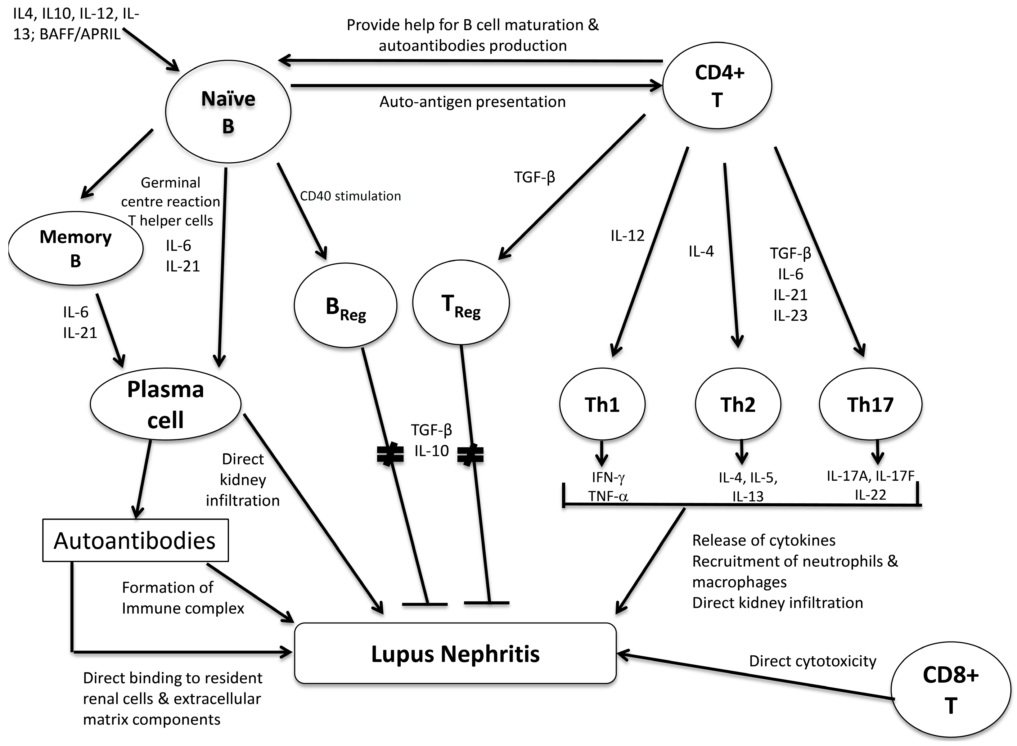
3. The Role of B Lymphocytes and Its Subsets

B lymphocytes have pleiotropic effects in the development of LN, including the generation of autoantibodies, secretion of proinflammatory and anti-inflammatory cytokines, auto-antigen presentation and direct infiltration to the kidneys (Figure 1). Antibody-secreting B cells and plasma cells were abundant in the kidneys of NZB/W F1 mice, and B cells that secrete nephritogenic anti-dsDNA could be isolated from nephritic MRL/lpr mice [40,41]. Interestingly, the frequency and autoantibody-secreting ability of the auto-reactive plasma cells within the kidney or bone marrow in NZB/W F1 mice were almost comparable, and the number of intra-renal plasma cells correlated with anti-dsDNA levels, and the activity and chronicity indices in LN [41]. Previous studies have reported that CXCL13 were highly expressed in NZB/W F mice with nephritis, and this would promote renal and other organ trafficking of B1 lymphocytes [42]. The pathogenic effect of B cells in LN is not limited to autoantibodies production. While genetically manipulated MRL/lpr mice that were incapable of antibody secretion still developed severe nephritis, B cell-deficient MRL/lpr mice were prevented from severe nephritis [43,44]. The improvement in renal manifestations in B cell-deficient mice might be explained by the failure of development of activated CD4+ and CD8+ T cells. IL-10-producing B cells, also known as regulatory B cells, also serve to regulate immune response and prevent autoimmunity [45]. However, the characterization of these IL-10-producing B cells and their roles in LN remains undefined. In human SLE, an increase in memory B cells and plasma cells coupled with a decrease of naive B cells are detected in the peripheral blood [46,47]. The antigen-experienced memory B cells have reduced regulation by FcγRIIb, and can be easily activated by the combination of toll-like receptor agonist or B cell activating factor (BAFF) [47,48]. Moreover, memory B cells have a low proliferation rate and are thus less susceptible to conventional immunosuppressive agents which are cell-cycle dependent, and hence a higher tendency to be involved in LN relapse [49]. In addition, SLE patients with active disease showed a marked expansion in plasma cells, and the frequency of circulating plasma cells correlated with disease activity scores, anti-dsDNA titre and serum immunoglobulin production [49,50]. In LN patients, local infiltration of B cells in the kidneys is associated with more severe renal disease and is augmented by BAFF [51]. The diverse and pivotal roles of B lymphocytes in the pathogenesis of SLE have aroused growing interest to use B cell-depleting therapy for the treatment of LN, but the human clinical trial data are less promising than one would have expected. In murine lupus models, the ablation of B cells via gene deletion or anti-CD20 administration prevents early death, inflammatory infiltrates and organ damage, while long-term administration of anti-CD20 eliminates autoantibody-secreting cells and is associated with reduction in survival niche for renal plasma cells [52,53]. However, recent randomized controlled trial data suggested that add-on rituximab (anti-CD20) to standard-of-care treatments did not improve clinical efficacy [54,55]. However, rituximab treatment might be useful in resistant LN or other refractory lupus manifestations [56]. Two randomized placebo-controlled phase III studies showed that belimumab (a BLys antagonist), when add-on to standard treatment, could enhance response rates in SLE patients without severe nephritis [57,58]. Post hoc analysis of patients with renal involvement from these studies suggested an efficacy signal of belimumab in LN, and hence further clinical trials are underway [59].

4. The Role of T lymphocytes and Its Subsets

T lymphocytes assume crucial roles in the development of LN such as provision of help to B cells to mature and produce autoantibodies, orchestration of B and T cell responses, release of proinflammatory and anti-inflammatory cytokines, and also the direct infiltration and cytotoxicity to renal parenchymal tissues (Figure 1). CD4+ helper T cells and CD8+ cytotoxic T cells are the two major subpopulations of T lymphocytes. Previous studies have revealed abundant mononuclear cells, CD4+ T cells and CD8+ T cells within the tubulo-interstitial lesions in LN patients [60]. However, depletion of CD8+ cytotoxic T cells in lupus-prone mice has produced conflicting data in SLE. β2-microglobulin deletion, which eliminated MHC Class I molecules and hence markedly impaired CD8+ T cell response, accelerated disease in NZB/W F1 mice [61]. Prolonged anti-CD8 administration also failed to alleviate disease in MRL/lpr or NZB/W F1 mice, in contrast to the favorable response to anti-CD4 treatment [62,63]. Studies on CD4+ helper T cells, however, have generated more important data regarding the development of SLE and LN. The following discussion highlights some of these important findings on CD4+ T cell subsets in the pathogenesis of LN.
Figure 1. The interaction between B and T lymphocyte subsets and their possible pathogenic roles in lupus nephritis.
Ijms 16 07917 g001

4.1. T-Helper Cells

4.1.1. Th1/Th2 Subsets

Th1 and Th2 responses represent two major T helper cell responses. In human LN, the Th1/Th2 balance exerts significant impact on the renal histopathological manifestations of LN. A predominant Th1 response was observed in both peripheral and renal tissues of patients with diffuse proliferative LN [64]. Okamoto et al. revealed that activated Th1 cells from kidneys of MRL/lpr mice with proliferative LN expressed high levels of T-cell receptor with IFN-γ, but not the cytokines relevant to Th2 or Th17 responses [65]. Urinary IL-12p70 elevation, which reflects a skewed Th1 response, is evident in diffuse proliferative but not membranous LN patients [66]. It was also demonstrated that peripheral IFN-γ/IL-4 ratio in diffuse proliferative LN was much higher than that in membranous LN, and was also correlated with the NIH activity index on renal biopsies [67,68]. MRL/lpr mice with deficiency of IFN-γ or its receptor had significantly milder proliferative LN changes [69,70]. In addition, IL-12-deficient MRL/lpr mice were associated with delay onset of diffuse proliferative LN and less severe renal histopathological changes [71]. Although these data have illustrated the importance of IFN-γ and Th1 response in SLE, it should be highlighted that IFN-γ might not be as important in the pathogenesis of human lupus as it is for the murine MRL/lpr model. On the other hand, Th2 response favored the development of membranous LN lesions in lupus-prone mice. MRL/lpr mice with WSX-1 defect exhibited pronounced Th2 but impaired Th1 responses, and developed renal lesions akin to human membranous LN [72]. As initial Th1 response is dependent on WSX-1, the loss of WSX-1 would polarize immunological functions towards Th2 response, and resulted in such phenotypic shift from diffuse proliferative to membranous LN in MRL/lpr mouse. These observations suggested the crucial role of Th2 response in the development of membranous LN.

4.1.2. Th17 and TReg

There is growing interest and data in Th17 and regulatory T cells (TReg) on their pathogenic roles in various autoimmune diseases including SLE. Th17 is a recently identified T helper cell subset characterized by the production of its signature cytokine IL-17, and has been implicated in causing inflammation and tissue injury in autoimmune disorders. In NZB/W F1 mice, there is significant renal infiltration of Th17 cells accompanied with abundant IL-17 production [73]. CD3+CD4CD8 T lymphocytes from MRL/lpr mice express copious amount of IL-17 and that as disease progresses, the expression of IL-17 and of IL-23 receptor in lymphocytes from these mice increases [74]. Cells extracted from MRL/lpr mice lymph nodes, upon treatment in vitro with IL-23, induced nephritis when transferred to non-autoimmune lymphocyte-deficient Rag-1(−/−) mice, and significant immunoglobulin and complement deposition were detected in these recipient mice [74]. Furthermore, IL-23 receptor deficiency or treatment with anti-IL23 monoclonal antibody ameliorates the development of LN in lupus-prone mice [75,76]. These data all suggested that an aberrant IL-17/IL23 axis is pivotal in the pathogenesis of murine LN. In human SLE, CD3+CD4C8 T cells produced high levels of IL-17 and IFN-γ, underwent expansion when stimulated in vitro with an anti-CD3 Ab in the presence of accessory cells, and were detected in the renal biopsies of LN patients [77]. SLE patients showed a significantly higher percentage of Th17 cells when compared to healthy controls, and these Th17 cells showed high expression levels of co-stimulatory molecules CD80 and CD134 [78]. Infiltration of Th17 cells with high CD134 expression was also detected in kidneys of LN patients [78]. The increased frequencies of Th17 cells was also coupled with significantly higher IL-17 and IL-23 levels when compared to healthy controls, and the number of Th17 cells correlates with disease activity scores (total and renal systemic lupus erythematosus disease activity index (SLEDAI)) and histopathological changes in the kidney [79]. Recent works by Zickert et al. have shown that high baseline IL-17 level was predictive of an unfavorable histopathological response, and non-responders (according to British Isles Lupus Activity Group (BILAG) scores) showed high circulating IL-23 levels [80]. These results suggested that a subset of LN patients exhibited Th17 phenotype that might affect response to treatment and could be evaluated as a biomarker for poor therapeutic response. Nevertheless, some recent findings have produced conflicting results regarding the role of Th17 in LN. For instance, Schmidt et al. revealed low frequency of infiltrating CD3+IL-17A+ cells in the kidneys of MRL/lpr and NZB/W F1 mice [81]. Also, IL-17A deficiency did not influence the renal or histological parameters in MRL/lpr mice with LN, nor did IL-17A antagonism alter the clinical course in nephritic NZB/W F1 mice [81]. While most data on Th17 cells still favors a pathogenic role in SLE and LN, the exact roles and pathogenic mechanisms of Th17 in LN needs to be further characterized. TReg cells (CD4+CD25+FOXP3+) play a protective role in autoimmune disorders. The treatment of anti-mouse thymocyte globulin followed by TGF-β1 in MRL/lpr mice induced TReg differentiation, and inhibited progression of proteinuria and improved renal histopathologies [82]. In murine LN, depletion of TReg in NZB/W F1 accelerated the onset of diffuse proliferative LN whereas their transfer into CD4/CD25 knock-out mice retarded the development of nephritis [53]. In human LN patients, a substantial shrinkage of CD4+CD25high and CD4+CD25+FoxP3+ TReg cells was observed in the peripheral blood, and such reduction in TReg number was accompanied with lower serum TGF-β1 levels [83] Moreover, the increase of peripheral Th17 cells was coupled with a decrease in TReg in LN patients, suggesting that the importance of Th17/TReg imbalance in the pathogenesis of renal disease in SLE [84].

4.1.3. Other T Helper Subsets (Follicular T Helper Cells, Th9 and Th22 Cells)

Follicular T helper cells (TFH) are antigen-experienced CD4+ T cells found at the B cell follicles of secondary lymphoid organs, and are characterized by the surface marker expression of CXCR5. They serve to form and maintain germinal cells, promote differentiation of naive B cells into memory B cells and plasma cells, and also to negatively select against auto-reactive B cells. Accumulating evidence has indicated the pivotal roles of TFH cells in the generation of pathogenic autoantibodies and tissue injury in SLE. The frequency of CD4+CXCR5+PD1+ TFH cells correlated with the number of plasma cells, anti-nuclear factor titer and disease activity in human SLE [85]. Furthermore, the number and percentage of CD4+CXCR5+PD1+ TFH cells diminished following treatment with corticosteroids in SLE patients [85]. Nevertheless, not all studies in lupus patients agreed on the increase in circulating TFH. Wong et al. reported a decrease in peripheral blood TFH cells although it was argued that such observation was due to a migration of these cells into the tissues [86]. The role of TFH in the formation and maintenance of germinal centres was relevant to the pathogenesis of tubulo-interstitial inflammation in LN. Ectopic germinal centres as well as aggregates of T and B cells have been found within the tubulo-interstitial infiltrates of the renal biopsies obtained from LN patients, and the presence of these ectopic germinal centres and lymphoid aggregates are strongly associated with renal tubular basement membrane immune complex deposition [87]. Moreover, the centroblasts and plasmablasts within these intrarenal germinal centres and lymphoid aggregates were capable of clonal expansion and somatic hypermutation [87]. Th22, characterized by the production of IL-22, is another newly identified T helper subset that is found to be significantly increased in activity SLE patients [88]. Although preliminary data suggested that Th22 was increased in SLE patients with skin manifestations but decreased in patients with isolated nephritis, the role of Th22 in specific organ involvement remained undefined [89]. Th9, which characteristically secretes IL-9, has both pro-inflammatory and anti-inflammatory actions, but the overall role is more skewed to pro-inflammatory in autoimmune diseases [90]. Taken together, the data regarding Th9 and Th22 on SLE are still relatively limited and their roles in the pathogenesis of LN remain unclear.

5. Conclusions

Production of autoantibodies and aberrations of lymphocyte subsets are two important pathogenic mechanisms of developing renal diseases in SLE patients. In this context, anti-dsDNA stands out to be a very important autoantibody as the monitoring of its titre is widely adopted in clinical practice and shows good association with clinical disease activities. Moreover, ample works have demonstrated that anti-dsDNA can bind to different resident renal components and mediate immunological injury to the kidneys. Future researches would be worthwhile to elucidate the target antigen of anti-dsDNA on resident renal cells and also the downstream events following these anti-dsDNA-antigen binding. As for lymphocyte subsets, current evidence suggested that the B cell repertoire might assume more pivotal roles and data from recent clinical trials on biologics that target the B cell repertoire or associated cytokines have lent further support to this notion. While imbalance of T cell subsets have been implicated in local renal injury and the different histological manifestations of LN, the exact mechanism of interaction between various T cell subsets and the role Treg still remain undefined. A better understanding of these pathogenic mechanisms will help develop novel approaches for disease activity monitoring and therapy.

Author Contributions

Desmond Y. H. Yap and Kar N. Lai contributed to preparation and writing of this review.

Conflicts of Interest

The authors declare no conflict of interest.

References

  1. Danchenko, N.; Satia, J.A.; Anthony, M.S. Epidemiology of systemic lupus erythematosus: A comparison of worldwide disease burden. Lupus 2006, 15, 308–318. [Google Scholar] [CrossRef] [PubMed]
  2. Osio-Salido, E.; Manapat-Reyes, H. Epidemiology of systemic lupus erythematosus in Asia. Lupus 2010, 19, 1365–1373. [Google Scholar] [CrossRef] [PubMed]
  3. Korbet, S.M.; Schwartz, M.M.; Evans, J.; Lewis, E.J. Severe lupus nephritis: Racial differences in presentation and outcome. J. Am. Soc. Nephrol. 2007, 18, 244–254. [Google Scholar] [CrossRef] [PubMed]
  4. Rovin, B.H.; Zhang, X. Biomarkers for lupus nephritis: The quest continues. Clin. J. Am. Soc. Nephrol. 2009, 4, 1858–1865. [Google Scholar] [CrossRef] [PubMed]
  5. Winfield, J.B.; Faiferman, I.; Koffler, D. Avidity of anti-DNA antibodies in serum and IgG glomerular eluates from patients with systemic lupus erythematosus. Association of high avidity antinative DNA antibody with glomerulonephritis. J. Clin. Investig. 1977, 59, 90–96. [Google Scholar] [CrossRef] [PubMed]
  6. Kalunian, K.C.; Panosian-Sahakian, N.; Ebling, F.M.; Cohen, A.H.; Louie, J.S.; Kaine, J.; Hahn, B.H. Idiotypic characteristics of immunoglobulins associated with systemic lupus erythematosus. Studies of antibodies deposited in glomeruli of humans. Arthritis Rheumatol. 1989, 32, 513–522. [Google Scholar] [CrossRef]
  7. Termaat, R.M.; Assmann, K.J.; van Son, J.P.; Dijkman, H.B.; Koene, R.A.; Berden, J.H. Antigen-specificity of antibodies bound to glomeruli of mice with systemic lupus erythematosus-like syndromes. Lab. Investig. 1993, 68, 164–173. [Google Scholar] [PubMed]
  8. Vlahakos, D.V.; Foster, M.H.; Adams, S.; Katz, M.; Ucci, A.A.; Barrett, K.J.; Datta, S.K.; Madaio, M.P. Anti-DNA antibodies form immune deposits at distinct glomerular and vascular sites. Kidney Int. 1992, 41, 1690–1700. [Google Scholar] [CrossRef]
  9. Yung, S.; Cheung, K.F.; Zhang, Q.; Chan, T.M. Anti-dsDNA antibodies bind to mesangial annexin II in lupus nephritis. J. Am. Soc. Nephrol. 2010, 21, 1912–1927. [Google Scholar] [CrossRef] [PubMed]
  10. Fenton, K.A.; Tommeras, B.; Marion, T.N.; Rekvig, O.P. Pure anti-dsDNA mAbs need chromatin structures to promote glomerular mesangial deposits in BALB/c mice. Autoimmunity 2010, 43, 179–188. [Google Scholar] [CrossRef] [PubMed]
  11. Chan, T.M.; Leung, J.K.; Ho, S.K.; Yung, S. Mesangial cell-binding anti-DNA antibodies in patients with systemic lupus erythematosus. J. Am. Soc. Nephrol. 2002, 13, 1219–1229. [Google Scholar] [CrossRef] [PubMed]
  12. Croquefer, S.; Renaudineau, Y.; Jousse, S.; Gueguen, P.; Ansart, S.; Saraux, A.; Youinou, P. The ananti-α-actinin test completes ananti-DNA determination in systemic lupus erythematosus. Ann. N. Y. Acad. Sci. 2005, 1050, 170–175. [Google Scholar] [CrossRef]
  13. Zhao, Z.; Weinstein, E.; Tuzova, M.; Davidson, A.; Mundel, P.; Marambio, P.; Putterman, C. Cross-reactivity of human lupus anti-DNA antibodies with α-actinin and nephritogenic potential. Arthritis Rheumatol. 2005, 52, 522–530. [Google Scholar] [CrossRef]
  14. Zhao, Z.; Deocharan, B.; Scherer, P.E.; Ozelius, L.J.; Putterman, C. Differential binding of cross-reactive anti-DNA antibodies to mesangial cells: The role of α-actinin. J. Immunol. 2006, 176, 7704–7714. [Google Scholar] [CrossRef] [PubMed]
  15. Mortensen, E.S.; Rekvig, O.P. Nephritogenic potential of anti-DNA antibodies against necrotic nucleosomes. J. Am. Soc. Nephrol. 2009, 20, 696–704. [Google Scholar] [CrossRef] [PubMed]
  16. Mjelle, J.E.; Rekvig, O.P.; van der Vlag, J.; Fenton, K.A. Nephritogenic antibodies bind in glomeruli through interaction with exposed chromatin fragments and not with renal cross-reactive antigens. Autoimmunity 2011, 44, 373–383. [Google Scholar] [CrossRef] [PubMed]
  17. Hedberg, A.; Fismen, S.; Fenton, K.A.; Fenton, C.; Osterud, B.; Mortensen, E.S.; Rekvig, O.P. Heparin exerts a dual effect on murine lupus nephritis by enhancing enzymatic chromatin degradation and preventing chromatin binding in glomerular membranes. Arthritis Rheumatol. 2011, 63, 1065–1075. [Google Scholar] [CrossRef]
  18. Yung, S.; Tsang, R.C.; Sun, Y.; Leung, J.K.; Chan, T.M. Effect of human anti-DNA antibodies on proximal renal tubular epithelial cell cytokine expression: Implications on tubulointerstitial inflammation in lupus nephritis. J. Am. Soc. Nephrol. 2005, 16, 3281–3294. [Google Scholar] [CrossRef] [PubMed]
  19. Koren, E.; Koscec, M.; Wolfson-Reichlin, M.; Ebling, F.M.; Tsao, B.; Hahn, B.H.; Reichlin, M. Murine and human antibodies to native DNA that cross-react with the A and D SnRNP polypeptides cause direct injury of cultured kidney cells. J. Immunol. 1995, 154, 4857–4864. [Google Scholar] [PubMed]
  20. Zack, D.J.; Stempniak, M.; Wong, A.L.; Taylor, C.; Weisbart, R.H. Mechanisms of cellular penetration and nuclear localization of an anti-double strand DNA autoantibody. J. Immunol. 1996, 157, 2082–2088. [Google Scholar] [PubMed]
  21. Chan, T.M.; Frampton, G.; Jayne, D.R.; Perry, G.J.; Lockwood, C.M.; Cameron, J.S. Clinical significance of anti-endothelial cell antibodies in systemic vasculitis: A longitudinal study comparing anti-endothelial cell antibodies and anti-neutrophil cytoplasm antibodies. Am. J. Kidney Dis. 1993, 22, 387–392. [Google Scholar] [CrossRef] [PubMed]
  22. Chan, T.M.; Cheng, I.K. A prospective study on anti-endothelial cell antibodies in patients with systemic lupus erythematosus. Clin. Immunol. Immunopathol. 1996, 78, 41–46. [Google Scholar] [CrossRef] [PubMed]
  23. Chan, T.M.; Frampton, G.; Cameron, J.S. Identification of DNA-binding proteins on human umbilical vein endothelial cell plasma membrane. Clin. Exp. Immunol. 1993, 91, 110–114. [Google Scholar] [CrossRef] [PubMed]
  24. Chan, T.M.; Frampton, G.; Staines, N.A.; Hobby, P.; Perry, G.J.; Cameron, J.S. Different mechanisms by which anti-DNA MoAbs bind to human endothelial cells and glomerular mesangial cells. Clin. Exp. Immunol. 1992, 88, 68–74. [Google Scholar] [CrossRef] [PubMed]
  25. Lai, K.N.; Leung, J.C.; Lai, B.K.; Li, P.K.; Lai, C.K. Anti-DNA autoantibodies stimulate the release of interleukin-1 and interleukin-6 from endothelial cells. J. Pathol. 1996, 178, 451–457. [Google Scholar] [CrossRef] [PubMed]
  26. Lai, K.N.; Leung, J.C.; Lai, K.B.; Lai, F.M.; Wong, K.C. Increased release of von Willebrand factor antigen from endothelial cells by anti-DNA autoantibodies. Ann. Rheum. Dis. 1996, 55, 57–62. [Google Scholar] [CrossRef] [PubMed]
  27. Lai, K.N.; Leung, J.C.; Lai, K.B.; Lai, C.K. Effect of anti-DNA autoantibodies on the gene expression of interleukin 8, transforming growth factor-β, and nitric oxide synthase in cultured endothelial cells. Scand. J. Rheumatol. 1997, 26, 461–467. [Google Scholar] [CrossRef] [PubMed]
  28. Krishnan, M.R.; Wang, C.; Marion, T.N. Anti-DNA autoantibodies initiate experimental lupus nephritis by binding directly to the glomerular basement membrane in mice. Kidney Int. 2012, 82, 184–192. [Google Scholar] [CrossRef] [PubMed]
  29. Heinz, H.P.; Rubin, K.; Laurell, A.B.; Loos, M. Common epitopes in Clq and collagen type II. Mol. Immunol. 1989, 26, 163–169. [Google Scholar] [CrossRef] [PubMed]
  30. Wener, M.H.; Mannik, M.; Schwartz, M.M.; Lewis, E.J. Relationship between renal pathology and the size of circulating immune complexes in patients with systemic lupus erythematosus. Medicine (Baltimore) 1987, 66, 85–97. [Google Scholar] [CrossRef]
  31. Uwatoko, S.; Mannik, M. Low-molecular weight C1q-binding immunoglobulin G in patients with systemic lupus erythematosus consists of autoantibodies to the collagen-like region of C1q. J. Clin. Investig. 1988, 82, 816–824. [Google Scholar] [CrossRef] [PubMed]
  32. Pang, Y.; Yang, X.W.; Song, Y.; Yu, F.; Zhao, M.H. Anti-C1q autoantibodies from active lupus nephritis patients could inhibit the clearance of apoptotic cells and complement classical pathway activation mediated by C1q in vitro. Immunobiology 2014, 219, 980–989. [Google Scholar] [CrossRef] [PubMed]
  33. Moroni, G.; Trendelenburg, M.; del Papa, N.; Quaglini, S.; Raschi, E.; Panzeri, P.; Testoni, C.; Tincani, A.; Banfi, G.; Balestrieri, G.; et al. Anti-C1q antibodies may help in diagnosing a renal flare in lupus nephritis. Am. J. Kidney Dis. 2001, 37, 490–498. [Google Scholar] [CrossRef] [PubMed]
  34. Siegert, C.E.; Daha, M.R.; Tseng, C.M.; Coremans, I.E.; van Es, L.A.; Breedveld, F.C. Predictive value of IgG autoantibodies against C1q for nephritis in systemic lupus erythematosus. Ann. Rheum. Dis. 1993, 52, 851–856. [Google Scholar] [CrossRef] [PubMed]
  35. Coremans, I.E.; Spronk, P.E.; Bootsma, H.; Daha, M.R.; van der Voort, E.A.; Kater, L.; Breedveld, F.C.; Kallenberg, C.G. Changes in antibodies to C1q predict renal relapses in systemic lupus erythematosus. Am. J. Kidney Dis. 1995, 26, 595–601. [Google Scholar] [CrossRef]
  36. Yap, D.Y.; Yung, S.; Zhang, Q.; Tang, C.; Chan, T.M. Mesangial cell-binding activity of serum immunoglobulin g in patients with lupus nephritis. PLoS ONE 2014, 9, e101987. [Google Scholar] [CrossRef] [PubMed] [Green Version]
  37. Manson, J.J.; Mills, K.; Jury, E.; Mason, L.; D’Cruz, D.P.; Ni, L.; Saleem, M.; Mathieson, P.; Isenberg, D.; Rahman, A. Pathogenic autoantibodies from patients with lupus nephritis cause reduced tyrosine phosphorylation of podocyte proteins, including tubulin. Lupus Sci. Med. 2014, 1, e000013. [Google Scholar] [CrossRef]
  38. Rahman, A.; Isenberg, D.A. Systemic lupus erythematosus. N. Engl. J. Med. 2008, 358, 929–939. [Google Scholar] [CrossRef] [PubMed]
  39. Doria, A.; Gatto, M. Nephritogenic-antinephritogenic antibody network in lupus glomerulonephritis. Lupus 2012, 21, 1492–1496. [Google Scholar] [CrossRef] [PubMed]
  40. Sekine, H.; Watanabe, H.; Gilkeson, G.S. Enrichment of anti-glomerular antigen antibody-producing cells in the kidneys of MRL/MpJ-Fas(lpr) mice. J. Immunol. 2004, 172, 3913–3921. [Google Scholar] [CrossRef] [PubMed]
  41. Espeli, M.; Bokers, S.; Giannico, G.; Dickinson, H.A.; Bards;ey, V.; Fogo, A.B.; Smith, K.G.C. Local renal autoantibody production in lupus nephritis. J. Am. Soc. Nephrol. 2011, 22, 296–305. [Google Scholar] [CrossRef] [PubMed]
  42. Ito, T.; Ishikawa, S.; Sato, T.; Akadegawa, K.; Yurino, H.; Kitabatake, M.; Hontsu, S.; Ezaki, T.; Kimura, H.; Matsushima, K. Defective B1 cell homing to the peritoneal cavity and preferential recruitment of B1 cells in the target organs in a murine model for systemic lupus erythematosus. J. Immunol. 2004, 172, 3628–3634. [Google Scholar] [CrossRef] [PubMed]
  43. Chan, O.T.; Hannum, L.G.; Haberman, A.M.; Madaio, M.P.; Shlomchik, M.J. A novel mouse with B cells but lacking serum antibody reveals an antibody-independent role for B cells in murine lupus. J. Exp. Med. 1999, 189, 1639–1648. [Google Scholar] [CrossRef] [PubMed]
  44. Chan, O.T.; Madaio, M.P.; Shlomchik, M.J. B cells are required for lupus nephritis in the polygenic, Fas-intact MRL model of systemic autoimmunity. J. Immunol. 1999, 163, 3592–3596. [Google Scholar] [PubMed]
  45. Harris, D.P.; Haynes, L.; Sayles, P.C.; Duso, D.K.; Eaton, S.M.; Lepak, N.M.; Johnson, L.L.; Swain, S.L.; Lund, F.E. Reciprocal regulation of polarized cytokine production by effector B and T cells. Nat. Immunol. 2000, 1, 475–482. [Google Scholar] [CrossRef] [PubMed]
  46. Anolik, J.H.; Barnard, J.; Cappione, A.; Pugh-Bernard, A.E.; Felgar, R.E.; Looney, R.J.; Sanz, I. Rituximab improves peripheral B cell abnormalities in human systemic lupus erythematosus. Arthritis Rheumatol. 2004, 50, 3580–3590. [Google Scholar] [CrossRef]
  47. Dorner, T.; Jacobi, A.M.; Lee, J.; Lipsky, P.E. Abnormalities of B cell subsets in patients with systemic lupus erythematosus. J. Immunol. Methods 2011, 363, 187–197. [Google Scholar] [CrossRef] [PubMed]
  48. Tiller, T.; Tsuiji, M.; Yurasov, S.; Velinzon, K.; Nussenzweig, M.C.; Wardemann, H. Autoreactivity in human IgG+ memory B cells. Immunity 2007, 26, 205–213. [Google Scholar] [CrossRef] [PubMed]
  49. Odendahl, M.; Jacobi, A.; Hansen, A.; Feist, E.; Hiepe, F.; Burmester, G.R.; Lipsky, P.E.; Radbruch, A.; Dorner, T. Disturbed peripheral B lymphocyte homeostasis in systemic lupus erythematosus. J. Immunol. 2000, 165, 5970–5979. [Google Scholar] [CrossRef] [PubMed]
  50. Jacobi, A.M.; Odendahl, M.; Reiter, K.; Bruns, A.; Burmester, G.R.; Radbruch, A.; Valet, G.; Lipsky, P.E.; Dorner, T. Correlation between circulating CD27 high plasma cells and disease activity in patients with systemic lupus erythematosus. Arthritis Rheumatol. 2003, 48, 1332–1342. [Google Scholar] [CrossRef]
  51. Sun, C.Y.; Shen, Y.; Chen, X.W.; Yan, Y.C.; Wu, F.X.; Dai, M.; Li, T.; Yang, C.D. The characteristics and significance of locally infiltrating B cells in lupus nephritis and their association with local BAFF expression. Int. J. Rheumatol. 2013, 2013, 954292. [Google Scholar] [PubMed]
  52. Renaudineau, Y.; Pers, J.O.; Bendaoud, B.; Jamin, C.; Youinou, P. Dysfunctional B cells in systemic lupus erythematosus. Autoimmun. Rev. 2004, 3, 516–523. [Google Scholar] [CrossRef] [PubMed]
  53. Wang, W.; Rangel-Moreno, J.; Owen, T.; Barnard, J.; Nevarez, S.; Ichikawa, H.T.; Anolik, J.H. Long-term B cell depletion in murine lupus eliminates autoantibody-secreting cells and is associated with alterations in the kidney plasma cell niche. J. Immunol. 2014, 192, 3011–3020. [Google Scholar] [CrossRef] [PubMed]
  54. Merrill, J.T.; Neuwelt, C.M.; Wallace, D.J.; Shanahan, J.C.; Latinis, K.M.; Oates, J.C.; Utset, T.O.; Gordon, C.; Isenberg, D.A.; Hsieh, H.J.; et al. Efficacy and safety of rituximab in moderately-to-severely active systemic lupus erythematosus: The randomized, double-blind, phase II/III systemic lupus erythematosus evaluation of rituximab trial. Arthritis Rheumatol. 2010, 62, 222–233. [Google Scholar] [CrossRef]
  55. Rovin, B.H.; Furie, R.; Latinis, K.; Looney, R.J.; Fervenza, F.C.; Sanchez-Guerrero, J.; Maciuca, R.; Zhang, D.; Garg, J.P.; Brunetta, P.; et al. Efficacy and safety of rituximab in patients with active proliferative lupus nephritis: The lupus nephritis assessment with rituximab study. Arthritis Rheumatol. 2012, 64, 1215–1226. [Google Scholar] [CrossRef]
  56. Gunnarsson, I.; Jonsdottir, T. Rituximab treatment in lupus nephritis—Where do we stand? Lupus 2013, 22, 381–389. [Google Scholar] [CrossRef] [PubMed]
  57. Furie, R.; Petri, M.; Zamani, O.; Cervera, R.; Wallace, D.J.; Tegzová, D.; Sanchez-Guerrero, J.; Schwarting, A.; Merrill, J.T.; Chatham, W.W.; et al. A phase III, randomized, placebo-controlled study of belimumab, a monoclonal antibody that inhibits B lymphocyte stimulator, in patients with systemic lupus erythematosus. Arthritis Rheumatol. 2011, 63, 3918–3930. [Google Scholar] [CrossRef]
  58. Navarra, S.V.; Guzman, R.M.; Gallacher, A.E.; Hall, S.; Levy, R.A.; Jimenez, R.E.; Li, E.K.; Thomas, M.; Kim, H.Y.; León, M.G.; et al. Efficacy and safety of belimumab in patients with active systemic lupus erythematosus: a randomised, placebo-controlled, phase 3 trial. Lancet 2011, 377, 721–731. [Google Scholar] [CrossRef] [PubMed]
  59. Dooley, M.; Houssiau, F.; Aranow, C.; D’Cruz, D.P.; Askanase, A.; Roth, D.A.; Zhong, Z.J.; Cooper, S.; Freimuth, W.W.; Ginzler, E.M.; et al. Effect of belimumab treatment on renal outcomes: Results from the phase 3 belimumab clinical trials in patients with SLE. Lupus 2013, 22, 63–72. [Google Scholar] [CrossRef] [PubMed]
  60. D’Agati, V.D.; Appel, G.B.; Estes, D.; Knowles, D.M., 2nd; Pirani, C.L. Monoclonal antibody identification of infiltrating mononuclear leukocytes in lupus nephritis. Kidney Int. 1986, 30, 573–581. [Google Scholar] [CrossRef] [PubMed]
  61. Mozes, E.; Lovchik, J.; Zinger, H.; Singer, D.S. MHC class I expression regulates susceptibility to spontaneous autoimmune disease in (NZBxNZW)F1 mice. Lupus 2005, 14, 308–314. [Google Scholar] [CrossRef] [PubMed]
  62. Merino, R.; Fossati, L.; Iwamoto, M.; Takahashi, S.; Lemoine, R.; Ibnou-Zekri, N.; Pugliatti, L.; Merino, J.; Izui, S. Effect of long-term anti-CD4 or anti-CD8 treatment on the development of lpr CD4CD8 double negative T cells and of the autoimmune syndrome in MRL-lpr/lpr mice. J. Autoimmun. 1995, 8, 33–45. [Google Scholar] [CrossRef] [PubMed]
  63. Adachi, Y.; Inaba, M.; Sugihara, A.; Koshiji, M.; Sugiura, K.; Amoh, Y.; Mori, S.; Kamiya, T.; Genba, H.; Ikehara, S. Effects of administration of monoclonal antibodies (anti-CD4 or anti-CD8) on the development of autoimmune diseases in (NZW × BXSB)F1 mice. Immunobiology 1998, 198, 451–464. [Google Scholar] [CrossRef] [PubMed]
  64. Masutani, K.; Akahoshi, M.; Tsuruya, K.; Tokumoto, M.; Ninomiya, T.; Kohsaka, T.; Fukuda, K.; Kanai, H.; Nakashima, H.; Otsuka, T.; et al. Predominance of Th1 immune response in diffuse proliferative lupus nephritis. Arthritis Rheumatol. 2001, 44, 2097–2106. [Google Scholar] [CrossRef]
  65. Okamoto, A.; Fujio, K.; Tsuno, N.H.; Takahashi, K.; Yamamoto, K. Kidney-infiltrating CD4+ T-cell clones promote nephritis in lupus-prone mice. Kidney Int. 2012, 82, 969–979. [Google Scholar] [CrossRef] [PubMed]
  66. Tucci, M.; Lombardi, L.; Richards, H.B.; Dammacco, F.; Silvestris, F. Overexpression of interleukin-12 and T helper 1 predominance in lupus nephritis. Clin. Exp. Immunol. 2008, 154, 247–254. [Google Scholar] [CrossRef] [PubMed]
  67. Akahoshi, M.; Nakashima, H.; Tanaka, Y.; Kohsaka, T.; Nagano, S.; Ohgami, E.; Arinobu, Y.; Yamaoka, K.; Niiro, H.; Shinozaki, M.; et al. Th1/Th2 balance of peripheral T helper cells in systemic lupus erythematosus. Arthritis Rheumatol. 1999, 42, 1644–1648. [Google Scholar]
  68. Masutani, K.; Taniguchi, M.; Nakashima, H.; Yotsueda, H.; Kudoh, Y.; Tsuruya, K.; Tokumoto, M.; Fukuda, K.; Kanai, H.; Hirakata, H.; Iida, M. Up-regulated interleukin-4 production by peripheral T-helper cells in idiopathic membranous nephropathy. Nephrol. Dial. Transplant. 2004, 19, 580–586. [Google Scholar] [CrossRef] [PubMed]
  69. Schwarting, A.; Wada, T.; Kinoshita, K.; Tesch, G.; Kelley, V.R. IFN-γ receptor signaling is essential for the initiation, acceleration, and destruction of autoimmune kidney disease in MRL-Fas(lpr) mice. J. Immunol. 1998, 161, 494–503. [Google Scholar] [PubMed]
  70. Haas, C.; Ryffel, B.; Le Hir, M. IFN-γ is essential for the development of autoimmune glomerulonephritis in MRL/Ipr mice. J. Immunol. 1997, 158, 5484–5491. [Google Scholar] [PubMed]
  71. Kikawada, E.; Lenda, D.M.; Kelley, V.R. IL-12 deficiency in MRL-Fas(lpr) mice delays nephritis and intrarenal IFN-γ expression, and diminishes systemic pathology. J. Immunol. 2003, 170, 3915–3925. [Google Scholar] [CrossRef] [PubMed]
  72. Shimizu, S.; Sugiyama, N.; Masutani, K.; Sadanaga, A.; Miyazaki, Y.; Inoue, Y.; Akahoshi, M.; Katafuchi, R.; Hirakata, H.; Harada, M.; et al. Membranous glomerulonephritis development with Th2-type immune deviations in MRL/lpr mice deficient for IL-27 receptor (WSX-1). J. Immunol. 2005, 175, 7185–7192. [Google Scholar] [CrossRef] [PubMed]
  73. Fleming, S.D.; Monestier, M.; Tsokos, G.C. Accelerated ischemia/reperfusion-induced injury in autoimmunity-prone mice. J. Immunol. 2004, 173, 4230–4235. [Google Scholar] [CrossRef] [PubMed]
  74. Zhang, Z.; Kyttaris, V.C.; Tsokos, G.C. The role of IL-23/IL-17 axis in lupus nephritis. J. Immunol. 2009, 183, 3160–3169. [Google Scholar] [CrossRef] [PubMed]
  75. Kyttaris, V.C.; Zhang, Z.; Kuchroo, V.K.; Oukka, M.; Tsokos, G.C. Cutting edge: IL-23 receptor deficiency prevents the development of lupus nephritis in C57BL/6-lpr/lpr mice. J. Immunol. 2010, 184, 4605–4609. [Google Scholar] [CrossRef] [PubMed]
  76. Kyttaris, V.C.; Kampagianni, O.; Tsokos, G.C. Treatment with anti-interleukin 23 antibody ameliorates disease in lupus-prone mice. Biomed. Res. Int. 2013, 2013, 861028. [Google Scholar] [CrossRef] [PubMed]
  77. Crispin, J.C.; Oukka, M.; Bayliss, G.; Cohen, R.A.; van Beek, C.A.; Stillman, I.E.; Kyttaris, V.C.; Juang, Y.-T.; Tsokos, G.C. Expanded double negative T cells in patients with systemic lupus erythematosus produce IL-17 and infiltrate the kidneys. J. Immunol. 2008, 181, 8761–8766. [Google Scholar] [CrossRef] [PubMed]
  78. Dolff, S.; Quandt, D.; Wilde, B.; Feldkamp, T.; Hua, F.; Cai, X.; Specker, C.; Kribben, A.; Kallenberg, C.G.; Witzke, O. Increased expression of costimulatory markers CD134 and CD80 on interleukin-17 producing T cells in patients with systemic lupus erythematosus. Arthritis Res. Ther. 2010, 12, R150. [Google Scholar] [CrossRef] [PubMed]
  79. Chen, D.Y.; Chen, Y.M.; Wen, M.C.; Hsieh, T.Y.; Hung, W.T.; Lan, J.L. The potential role of Th17 cells and Th17-related cytokines in the pathogenesis of lupus nephritis. Lupus 2012, 21, 1385–1396. [Google Scholar] [CrossRef] [PubMed]
  80. Zickert, A.; Amouddruz, P.; Sundstrom, Y.; Ronnelid, J.; Malmstrom, V.; Gunnarsson, I. Il-17 and IL-23 in lupus nephritis—Association to histopathology and response to treatment. BMC Immunol. 2015, 16, 7. [Google Scholar] [CrossRef]
  81. Schmidt, T.; Paust, H.J.; Krebs, C.F.; Turner, J.E.; Kaffke, A.; Bennstein, S.B.; Koyro, T.; Peters, A.; Velden, J.; Hünemörder, S.; et al. Function of the Th17/interleukin-17A immune response in murine lupus nephritis. Arthritis Rheumatol. 2015, 67, 475–487. [Google Scholar] [CrossRef] [PubMed]
  82. Kaplan, J.; Woodworth, L.; Smith, K.; Coco, J.; Vitsky, A.; McPherson, J. Therapeutic benefit of treatment with anti-thymocyte globulin and latent TGF-β1 in the MRL/lpr lupus mouse model. Lupus 2008, 17, 822–831. [Google Scholar] [CrossRef] [PubMed]
  83. Xing, Q.; Su, H.; Cui, J.; Wang, B. Role of Treg cells and TGF-β1 in patients with systemic lupus erythematosus: A possible relation with lupus nephritis. Immunol. Investig. 2012, 41, 15–27. [Google Scholar] [CrossRef]
  84. Xing, Q.; Wang, B.; Su, H.; Cui, J.; Li, J. Elevated Th17 cells are accompanied by FoxP3+ Treg cells decrease in patients with lupus nephritis. Rheumatol. Int. 2012, 32, 949–958. [Google Scholar] [CrossRef] [PubMed]
  85. Feng, X.; Wang, D.; Chen, J.; Lu, L.; Hua, B.; Li, X.; Tsao, B.P.; Sun, L. Inhibition of aberrant circulating Tfh cell proportions by corticosteroids in patients with systemic lupus erythematosus. PLoS ONE 2012, 7, e51982. [Google Scholar] [CrossRef] [PubMed]
  86. Wong, C.K.; Wong, P.T.; Tam, L.S.; Li, E.K.; Chen, D.P.; Lam, C.W. Elevated production of B cell chemokine CXCL13 is correlated with systemic lupus erythematosus disease activity. J. Clin. Immunol. 2010, 30, 45–52. [Google Scholar] [CrossRef] [PubMed]
  87. Chang, A.; Henderson, S.G.; Brandt, D.; Liu, N.; Guttikonda, R.; Hsieh, C.; Kaverina, N.; Utset, T.O.; Meehan, S.M.; Quigg, R.J.; et al. In situ B cell-mediated immune responses and tubulointerstitial inflammation in human lupus nephritis. J. Immunol. 2011, 186, 1849–1860. [Google Scholar]
  88. Zhao, L.; Jiang, Z.; Jiang, Y.; Ma, N.; Wang, K.; Zhang, Y.; Feng, L. IL-22+CD4+ T-cells in patients with active systemic lupus erythematosus. Exp. Biol. Med. (Maywood) 2013, 238, 193–199. [Google Scholar] [CrossRef]
  89. Yang, X.Y.; Wang, H.Y.; Zhao, X.Y.; Wang, L.J.; Lv, Q.H.; Wang, Q.Q. Th22, but not Th17 might be a good index to predict the tissue involvement of systemic lupus erythematosus. J. Clin. Immunol. 2013, 33, 767–774. [Google Scholar] [CrossRef] [PubMed]
  90. Leng, R.X.; Pan, H.F.; Ye, D.Q.; Xu, Y. Potential roles of IL-9 in the pathogenesis of systemic lupus erythematosus. Am. J. Clin. Exp. Immunol. 2012, 1, 28–32. [Google Scholar] [PubMed]

