
Diagnosis of gout for patients who have initial symptoms of ankle swelling and pain is difficult because talus fractures easily after injury, and it may be difficult to identify tophaceous gout by imaging alone.
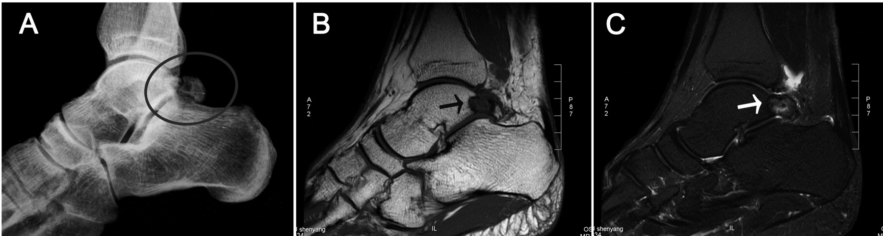
A 24-year-old man had persistent pain and swelling after spraining his right foot 1 year before presentation. Initial diagnosis was fracture of the right talus, based on plain radiograph (noncontinuous bone cortex on the back edge of right talus and a dissociative bone block, Figure 1A)1. After rest and antiinflammatory treatment, the pain and swelling were alleviated. Recently, the pain and swelling became much worse. Magnetic resonance images of the ankle showed a T1 hypointense and T2 hyperintense soft tissue mass. There was also a small irregular central area of low signal (Figure 1B, 1C). Plaster-like tissue mixed with a small amount of bone and soft tissue was noted during the surgery. The trigonal process of talus was heavily eroded. Pathological staining confirmed that the excised tissue was tophus. Blood uric acid and C-reactive protein levels after the surgery were 620 μmol/l and 18.7 mg/l, respectively. He was treated with the accepted gout maintenance regimen of nonsteroidal antiinflammatory drugs and colchicine after surgery2. One month after surgery, the pain and swelling were alleviated and ankle function was recovered.

A. Plain radiograph reveals fracture of the right talus, with noncontinuous bone cortex on the back edge of right talus and a dissociative bone block (circle). B and C. Magnetic resonance images of the ankle show a T1 hypointense and T2 hyperintense soft tissue mass. There was also a small irregular central area of low signal (arrow).
Doctors easily overlook the diagnosis of gout for patients who have ankle swelling and pain as initial symptoms, because talus fractures easily after injury. It may be difficult to identify tophaceous gout by imaging alone and therefore a preoperative assessment of ankle pain and swelling should include serum uric acid test and consideration of gout3.
REFERENCES
- 1.
- 2.
- 3.