
To the Editor:
Juvenile dermatomyositis (JDM) is an autoimmune vasculopathy involving primarily the proximal muscles and skin but can involve other organ systems, particularly the gastrointestinal (GI) tract1. In the presence of cutaneous vasculitic ulcerations, one needs to monitor for signs and symptoms suggestive of GI vasculopathy2, which can lead to bowel ischemia, necrosis, ulceration, and perforation1,2,3,4,5. The presence of gas in the bowel wall, also referred to as pneumatosis intestinalis (PI), is an ominous finding when it is due to a GI vasculopathy3. PI is a radiographic finding that is associated with both benign and life-threatening causes. The benign variant of PI has been described in relation to immunosuppressive therapy6 and to JDM itself7. We describe a child with JDM complicated by cutaneous ulcerations and benign PI.
An 8-year-old girl, following a 1-month history of rash and fatigue, was diagnosed with JDM based on the presence of Gottron sign, heliotrope rash, symmetric proximal muscle weakness, elevated serum muscle enzymes [creatine kinase (CK) 533 U/l (normal 44–189); lactate dehydrogenase 442 U/l (normal 142–261); aldolase 9.3 U/l (normal 0–7.6)] and findings consistent with myositis on magnetic resonance imaging of the proximal hip and shoulder girdle muscles. Antinuclear and anti-Jo1 antibodies were negative. The initial Childhood Myositis Assessment Scale8 (CMAS) was 44/52. Her nailfold capillaries were normal. Initial treatment included high-dose oral corticosteroids and methotrexate (MTX).
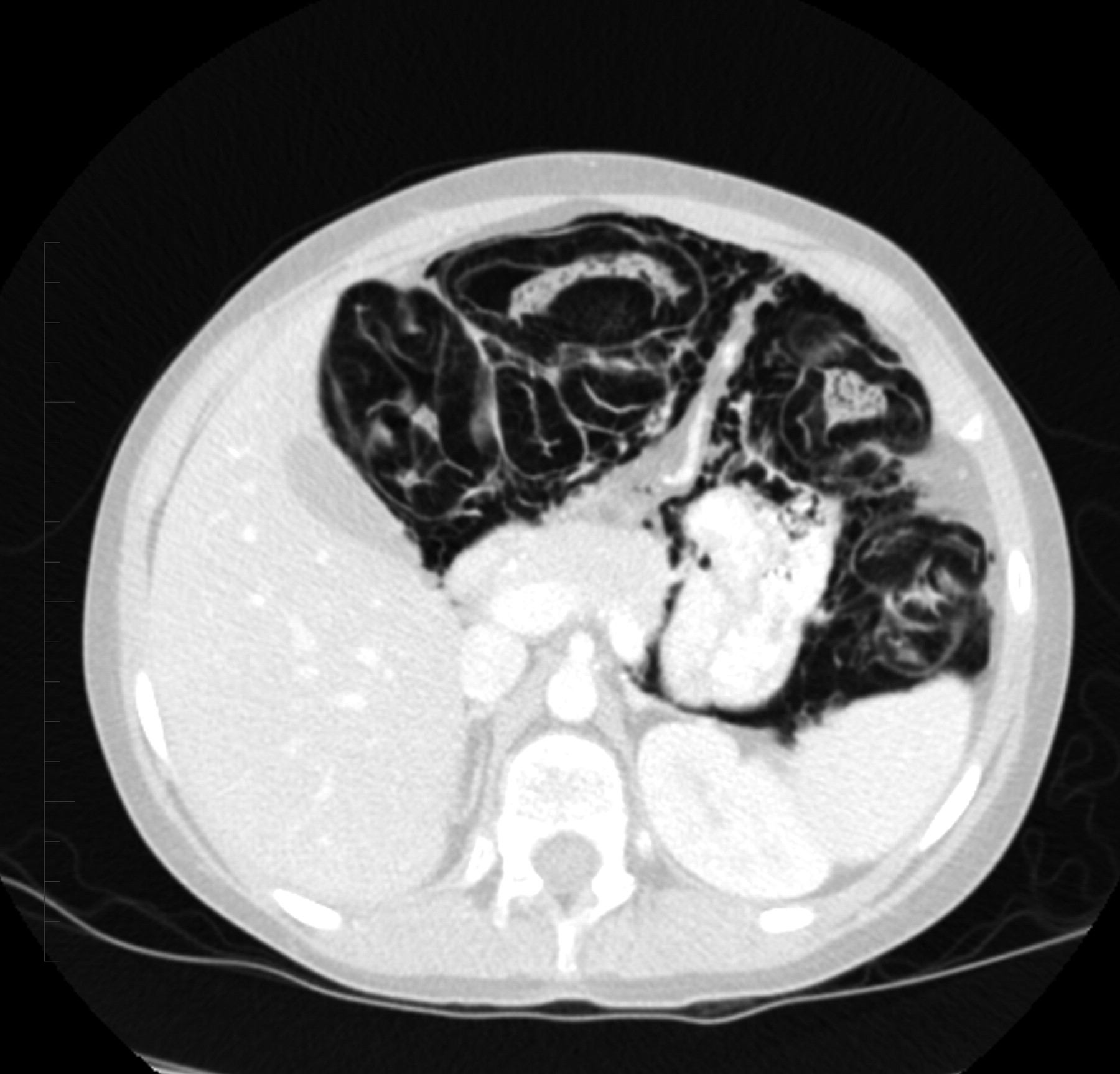
Three weeks after starting therapy, she presented with increased weakness, a vasculitic ulcer on the left upper eyelid and in the left nare associated with an increase in CK (1170 U/l); however, the other muscle enzymes remained unchanged. She was treated with intravenous pulse methylprednisolone once daily for 3 days. When reassessed a few days later for a cough, a chest radiograph showed free air under the right hemidiaphragm (Figure 1). She was asymptomatic. The eyelid and nare ulcers were healing, with no new cutaneous ulcerations. Her muscle strength (CMAS 46/52) had improved. Her abdomen was slightly distended but soft, non-tender with present bowel sounds. A pneumoperitoneum and extensive PI, primarily involving the large bowel, were confirmed by computed tomography (CT) of the abdomen (Figures 2 and 3). No associated signs of bowel wall thickening, fat stranding, or abdominal free fluid were present.

Free air under the right hemidiaphragm (arrow).

Infused CT scan of the abdomen, coronal view, showing extensive pneumatosis intestinalis in the large colon.
Infused CT scan of the abdomen, axial view, showing circumferential pneumatosis intestinalis of the transverse colon.
The patient was admitted to hospital for intravenous antibiotics and a short period of bowel rest. Additional investigations included a normal complete blood cell count, blood gas, lactic acid, fibrinogen, and coagulation profile. The muscle enzymes had improved with normalization of CK (52 U/l). D-dimers were negative; however, the von Willebrand factor antigen was elevated at 4.9 U/ml (normal 0.5–1.6). Anticardiolipin antibodies and lupus anticoagulant were negative. Family history was negative for vascular diseases. There was no escalation in immunosuppressive therapy. She remained asymptomatic and was discharged after 10 days without adverse sequelae. An abdominal radiograph performed 6 weeks later demonstrated persistence of PI with resolution of the pneumoperitoneum. The rest of her course was uncomplicated. Prednisone was discontinued 14 months from diagnosis and she continues on a tapering dose of MTX.
PI was first described in 17549. It is defined as the presence of gas within the bowel wall10 and is a radiographic finding that can be seen with many diagnostic entities11. The prevalence of PI is not known; however, this seems to be increasing with more frequent use of CT and immunosuppressive therapy10,12.
Benign causes of PI have been described in relation to systemic illness (collagen vascular disease, chronic pulmonary disease), medications, and GI procedures10,12. In 1973, Borns and Johnston suggested a link between PI and immunosuppressive therapy, particularly corticosteroids6. On pathologic specimens, air was discovered in the Peyer patches of the intestine13. This led to the hypothesis that steroids or stress cause lymphoid depletion and thus atrophy of the Peyer patches, resulting in alteration of the submucosal structural integrity, which permits intraluminal gas to dissect into the noninflamed bowel wall6,10,11,12.
The life-threatening causes of PI are those associated with bowel necrosis. In JDM, bowel necrosis occurs as a consequence of the vasculopathy1. A review in 1998 identified 6 patients in the literature with PI associated with JDM4; 4 had a benign clinical course similar to our patient, 1 had a life-threatening course, and 1 patient died4. In JDM, it is imperative to promptly identify a GI vasculopathy, as this can be fatal.
Cutaneous ulcerative disease in JDM may be associated with more widespread vasculitis and thus PI seen in this context would more commonly be secondary to bowel necrosis. Despite the cutaneous ulcerations in our patient, she did not have life-threatening PI. Pneumoperitoneum, as observed in our patient, can also occur in benign PI, which in this context is usually the consequence of the rupture of a subserosal cyst rather than a true perforation10,12. Worrisome features on imaging include signs of bowel wall inflammation or ischemia manifesting as bowel wall thickening, free fluid, or portal gas12, which were absent in our case.
The management of PI depends on the underlying etiology and most importantly on the clinical condition of the child. In the absence of sepsis or peritonitis, many children can be managed conservatively with antibiotic coverage and bowel rest10. The presence of PI alone is not an indication for surgical intervention or escalation of medical therapy. However, in children with a clinical deterioration, surgical intervention may be warranted10.
Our patient was asymptomatic and did not have the more worrisome features on imaging. Her presentation and clinical course, in our opinion, were consistent with benign PI, which we presumed was secondary to immunosuppressive therapy. However, we could not exclude the presence of a mild vasculopathy resulting in loss of mucosal integrity, given the elevated von Willebrand factor antigen. Both the benign and life-threatening variants of PI should be considered, particularly in patients at high risk for bowel necrosis.
Increased awareness of the benign form of PI in combination with clinical history, physical examination, and laboratory and radiographic evaluations will allow proper identification of this entity, which may prevent unnecessary escalation in medical therapy and/or surgical intervention.
REFERENCES
- 1.
- 2.
- 3.
- 4.
- 5.
- 6.
- 7.
- 8.
- 9.
- 10.
- 11.
- 12.
- 13.