
Main menu

  • Home
  • Content
    • First Release
    • Current
    • Archives
    • Collections
    • Audiovisual Rheum
    • 50th Volume Reprints
  • Resources
    • Guide for Authors
    • Submit Manuscript
    • Payment
    • Reviewers
    • Advertisers
    • Classified Ads
    • Reprints and Translations
    • Permissions
    • Meetings
    • FAQ
    • Policies
  • Subscribers
    • Subscription Information
    • Purchase Subscription
    • Your Account
    • Terms and Conditions
  • About Us
    • About Us
    • Editorial Board
    • Letter from the Editor
    • Duncan A. Gordon Award
    • Privacy/GDPR Policy
    • Accessibility
  • Contact Us
  • JRheum Supplements
  • Services

User menu

  • My Cart
  • Log In

Search

  • Advanced search
The Journal of Rheumatology
  • JRheum Supplements
  • Services
  • My Cart
  • Log In
The Journal of Rheumatology

Advanced Search

  • Home
  • Content
    • First Release
    • Current
    • Archives
    • Collections
    • Audiovisual Rheum
    • 50th Volume Reprints
  • Resources
    • Guide for Authors
    • Submit Manuscript
    • Payment
    • Reviewers
    • Advertisers
    • Classified Ads
    • Reprints and Translations
    • Permissions
    • Meetings
    • FAQ
    • Policies
  • Subscribers
    • Subscription Information
    • Purchase Subscription
    • Your Account
    • Terms and Conditions
  • About Us
    • About Us
    • Editorial Board
    • Letter from the Editor
    • Duncan A. Gordon Award
    • Privacy/GDPR Policy
    • Accessibility
  • Contact Us
  • Follow Jrheum on BlueSky
  • Follow jrheum on Twitter
  • Visit jrheum on Facebook
  • Follow jrheum on LinkedIn
  • Follow jrheum on YouTube
  • Follow jrheum on Instagram
  • Follow jrheum on RSS
Research ArticleImages in Rheumatology

Black-blood Magnetic Resonance Imaging in Giant Cell Arteritis

Jan Vynckier, Jelle Demeestere and Julie Lambert
The Journal of Rheumatology February 2021, 48 (2) 301-302; DOI: https://doi.org/10.3899/jrheum.190286
Jan Vynckier
Jan Vynckier, MD, Stroke Research Physician, Department of Neurology, Inselspital, Bern, Switzerland;
MD
Roles: Stroke Research Physician
  • Find this author on Google Scholar
  • Find this author on PubMed
  • Search for this author on this site
  • ORCID record for Jan Vynckier
  • For correspondence: janluc.vynckier{at}insel.ch
Jelle Demeestere
Jelle Demeestere, MD, Consultant Neurologist; Julie Lambert, MD, Consultant Neuroradiologist, Department of Neurology and Department of Radiology, University Hospitals Leuven, Leuven, Belgium.
MD
Roles: Consultant Neurologist
  • Find this author on Google Scholar
  • Find this author on PubMed
  • Search for this author on this site
  • ORCID record for Jelle Demeestere
Julie Lambert
Jelle Demeestere, MD, Consultant Neurologist; Julie Lambert, MD, Consultant Neuroradiologist, Department of Neurology and Department of Radiology, University Hospitals Leuven, Leuven, Belgium.
MD
Roles: Consultant Neuroradiologist
  • Find this author on Google Scholar
  • Find this author on PubMed
  • Search for this author on this site
  • ORCID record for Julie Lambert
  • Article
  • Figures & Data
  • Info & Metrics
  • References
  • PDF
PreviousNext
Loading

Giant cell arteritis (GCA) affects large extracranial supraaortic arteries. Ischemic stroke is a presenting feature in up to 10% and carries a mortality rate of 75%1.

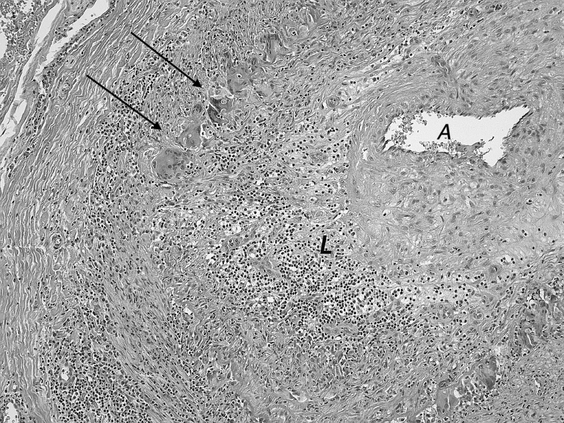
A 72-year-old woman was admitted with acute dizziness and jaw claudication. Clinical examination showed swollen temporal arteries, dysarthria, ataxia of the lower limbs, and left hemianopia. Erythrocyte sedimentation rate was elevated (48 mm/h). Brain magnetic resonance imaging (MRI) showed infarction of the right occipital lobe and the cerebellar hemispheres. High-resolution black-blood contrast-enhanced T1-weighted MRI showed vessel wall enhancement of the extradural vertebral and carotid arteries as well as the temporal and occipital arteries (Figure 1). A left temporal artery biopsy was performed, confirming giant cell formation and arterial wall inflammation with lymphocytic infiltrate (Figure 2). Despite maximal therapy with dual antiplatelet therapy and high-dose methylprednisolone, the patient died after having multiple strokes, 3 weeks after admission.

Figure 1.
  • Download figure
  • Open in new tab
  • Download powerpoint
Figure 1.

Axial black-blood MRI of the brain and extracranial arteries. There is contrast enhancement of the vessel wall of both temporal arteries (arrows). MRI: magnetic resonance imaging.

Figure 2.
  • Download figure
  • Open in new tab
  • Download powerpoint
Figure 2.

Histologic specimen with hematoxylin and eosin stain demonstrating a shrunken arterial lumen (A) with perivascular lymphocytic infiltrates (L) and typical giant cell formation (arrows).

The diagnosis of GCA remains challenging and often requires additional tests such as 18F-fluorodeoxyglucose positron emission tomography (FDG-PET) and biopsy. Black-blood MRI uses a prepulse for suppression of intraluminal gadolinium to create a high-contrast image of the blood vessel wall. This technique has been used for detection of vessel wall abnormalities, such as cervical dissection2 or central nervous system vasculitis and has been shown to detect arteritic anterior optic neuropathy in patients with GCA3. We report its use for diagnosis of GCA. Further studies are needed to investigate concordance between black-blood MRI and clinical symptoms, pathology, and FDG-PET.

  • Copyright © 2021 by the Journal of Rheumatology

REFERENCES

  1. 1.↵
    1. Samson M,
    2. Jacquin A,
    3. Audia S,
    4. Daubail B,
    5. Devilliers H,
    6. Petrella T, et al.
    Stroke associated with giant cell arteritis: a population-based study. J Neurol Neurosurg Psychiatry 2015;86:216-21.
    OpenUrlAbstract/FREE Full Text
  2. 2.↵
    1. Schwarz F,
    2. Strobl FF,
    3. Cyran CC,
    4. Helck AD,
    5. Hartmann M,
    6. Schindler A, et al.
    Reproducibility and differentiation of cervical arteriopathies using in vivo high-resolution black-blood MRI at 3 T. Neuroradiology 2016;58:569-76.
    OpenUrl
  3. 3.↵
    1. Sommer NN,
    2. Treitl KM,
    3. Coppenrath E,
    4. Kooijman H,
    5. Dechant C,
    6. Czihal M, et al.
    Three-dimensional high-resolution black-blood magnetic resonance imaging for detection of arteritic anterior ischemic optic neuropathy in patients with giant cell arteritis. Invest Radiol 2018;53:698-704.
    OpenUrlCrossRefPubMed
PreviousNext
Back to top

In this issue

The Journal of Rheumatology
Vol. 48, Issue 2
1 Feb 2021
  • Table of Contents
  • Table of Contents (PDF)
  • Index by Author
  • Editorial Board (PDF)
Print
Download PDF
Article Alerts
Sign In to Email Alerts with your Email Address
Email Article

Thank you for your interest in spreading the word about The Journal of Rheumatology.

NOTE: We only request your email address so that the person you are recommending the page to knows that you wanted them to see it, and that it is not junk mail. We do not capture any email address.

Enter multiple addresses on separate lines or separate them with commas.
Black-blood Magnetic Resonance Imaging in Giant Cell Arteritis
(Your Name) has forwarded a page to you from The Journal of Rheumatology
(Your Name) thought you would like to see this page from the The Journal of Rheumatology web site.
CAPTCHA
This question is for testing whether or not you are a human visitor and to prevent automated spam submissions.
Citation Tools
Black-blood Magnetic Resonance Imaging in Giant Cell Arteritis
Jan Vynckier, Jelle Demeestere, Julie Lambert
The Journal of Rheumatology Feb 2021, 48 (2) 301-302; DOI: 10.3899/jrheum.190286

Citation Manager Formats

  • BibTeX
  • Bookends
  • EasyBib
  • EndNote (tagged)
  • EndNote 8 (xml)
  • Medlars
  • Mendeley
  • Papers
  • RefWorks Tagged
  • Ref Manager
  • RIS
  • Zotero

 Request Permissions

Share
Black-blood Magnetic Resonance Imaging in Giant Cell Arteritis
Jan Vynckier, Jelle Demeestere, Julie Lambert
The Journal of Rheumatology Feb 2021, 48 (2) 301-302; DOI: 10.3899/jrheum.190286
del.icio.us logo Twitter logo Facebook logo  logo Mendeley logo
  • Tweet Widget
  •  logo
Bookmark this article

Jump to section

  • Article
    • REFERENCES
  • Figures & Data
  • Info & Metrics
  • References
  • PDF

Related Articles

Cited By...

More in this TOC Section

  • Classic Magnetic Resonance Imaging Features Unmask Progressive Multifocal Leukoencephalopathy in Rituximab-Treated Neuropsychiatric Systemic Lupus Erythematosus
  • Levamisole-Adulterated Cocaine-Induced Vasculitis Mimicking Acute Limb Ischemia
  • A Diagnostic Challenge: Palatal Nodules as the First Sign of IgG4-Related Disease
Show more Images in Rheumatology

Similar Articles

Content

  • First Release
  • Current
  • Archives
  • Collections
  • Audiovisual Rheum
  • COVID-19 and Rheumatology

Resources

  • Guide for Authors
  • Submit Manuscript
  • Author Payment
  • Reviewers
  • Advertisers
  • Classified Ads
  • Reprints and Translations
  • Permissions
  • Meetings
  • FAQ
  • Policies

Subscribers

  • Subscription Information
  • Purchase Subscription
  • Your Account
  • Terms and Conditions

More

  • About Us
  • Contact Us
  • My Alerts
  • My Folders
  • Privacy/GDPR Policy
  • RSS Feeds
The Journal of Rheumatology
The content of this site is intended for health care professionals.
Copyright © 2025 by The Journal of Rheumatology Publishing Co. Ltd.
Print ISSN: 0315-162X; Online ISSN: 1499-2752
Powered by HighWire