
Main menu

  • Home
  • Content
    • First Release
    • Current
    • Archives
    • Collections
    • Audiovisual Rheum
    • COVID-19 and Rheumatology
  • Resources
    • Guide for Authors
    • Submit Manuscript
    • Payment
    • Reviewers
    • Advertisers
    • Classified Ads
    • Reprints and Translations
    • Permissions
    • Meetings
    • FAQ
    • Policies
  • Subscribers
    • Subscription Information
    • Purchase Subscription
    • Your Account
    • Terms and Conditions
  • About Us
    • About Us
    • Editorial Board
    • Letter from the Editor
    • Duncan A. Gordon Award
    • Privacy/GDPR Policy
    • Accessibility
  • Contact Us
  • JRheum Supplements
  • Services

User menu

  • My Cart
  • Log In

Search

  • Advanced search
The Journal of Rheumatology
  • JRheum Supplements
  • Services
  • My Cart
  • Log In
The Journal of Rheumatology

Advanced Search

  • Home
  • Content
    • First Release
    • Current
    • Archives
    • Collections
    • Audiovisual Rheum
    • COVID-19 and Rheumatology
  • Resources
    • Guide for Authors
    • Submit Manuscript
    • Payment
    • Reviewers
    • Advertisers
    • Classified Ads
    • Reprints and Translations
    • Permissions
    • Meetings
    • FAQ
    • Policies
  • Subscribers
    • Subscription Information
    • Purchase Subscription
    • Your Account
    • Terms and Conditions
  • About Us
    • About Us
    • Editorial Board
    • Letter from the Editor
    • Duncan A. Gordon Award
    • Privacy/GDPR Policy
    • Accessibility
  • Contact Us
  • Follow jrheum on Twitter
  • Visit jrheum on Facebook
  • Follow jrheum on LinkedIn
  • Follow jrheum on YouTube
  • Follow jrheum on Instagram
  • Follow jrheum on RSS
Research ArticleOMERACT 2018: International Consensus Conference on Outcome Measures in Rheumatology, Terrigal, Australia, May 2018 Special Interest Groups, Part 2

The SPECTRA Collaboration OMERACT Working Group: Construct Validity of Joint Space Outcomes with High-resolution Peripheral Quantitative Computed Tomography

Sarah L. Manske, Scott C. Brunet, Stephanie Finzel, Kathryn S. Stok, Philip G. Conaghan, Steven K. Boyd and Cheryl Barnabe
The Journal of Rheumatology October 2019, 46 (10) 1369-1373; DOI: https://doi.org/10.3899/jrheum.180870
Sarah L. Manske
From the Department of Radiology, and Department of Medicine, McCaig Institute for Bone and Joint Health, Cumming School of Medicine, University of Calgary; Biomedical Engineering Graduate Program, Department of Radiology, Cumming School of Medicine, University of Calgary, Calgary, Alberta, Canada; Department of Rheumatology and Clinical Immunology, Medical Center University of Freiburg, Faculty of Medicine, University of Freiburg, Freiburg, Germany; Department of Biomedical Engineering, The University of Melbourne, Parkville, Australia; Leeds Institute for Rheumatic and Musculoskeletal Medicine, University of Leeds and UK National Institute for Health Research (NIHR) Leeds Biomedical Research Centre, Leeds, UK.
  • Find this author on Google Scholar
  • Find this author on PubMed
  • Search for this author on this site
  • ORCID record for Sarah L. Manske
  • For correspondence: smanske@ucalgary.ca
Scott C. Brunet
  • Find this author on Google Scholar
  • Find this author on PubMed
  • Search for this author on this site
  • ORCID record for Scott C. Brunet
Stephanie Finzel
  • Find this author on Google Scholar
  • Find this author on PubMed
  • Search for this author on this site
  • ORCID record for Stephanie Finzel
Kathryn S. Stok
  • Find this author on Google Scholar
  • Find this author on PubMed
  • Search for this author on this site
  • ORCID record for Kathryn S. Stok
Philip G. Conaghan
  • Find this author on Google Scholar
  • Find this author on PubMed
  • Search for this author on this site
  • ORCID record for Philip G. Conaghan
Steven K. Boyd
  • Find this author on Google Scholar
  • Find this author on PubMed
  • Search for this author on this site
  • ORCID record for Steven K. Boyd
Cheryl Barnabe
  • Find this author on Google Scholar
  • Find this author on PubMed
  • Search for this author on this site
  • ORCID record for Cheryl Barnabe
  • Article
  • Figures & Data
  • Info & Metrics
  • References
  • PDF
  • eLetters
PreviousNext
Loading

Abstract

Objective. We assessed construct validity of high-resolution peripheral quantitative computed tomography (HR-pQCT) joint space outcomes by comparison with radiographs in patients with rheumatoid arthritis.

Methods. In 43 patients, quantitative, volumetric, HR-pQCT measurements were compared with ordinal Sharp/van der Heijde scoring (SvdH) in the 2nd and 3rd metacarpophalangeal joints.

Results. Generalized estimating equations showed that joint space minimum, SD, and asymmetry by HR-pQCT were associated with SvdH scores (p < 0.05). There was a considerable range in HR-pQCT measurements at SvdH equal to 0.

Conclusion. HR-pQCT demonstrated construct validity outcomes and provides improved 3-D visualization of joint space.

Key Indexing Terms:
  • RHEUMATOID ARTHRITIS
  • RADIOGRAPHIC MICRO-COMPUTED TOMOGRAPHY
  • RADIOGRAPHY
  • METACARPOPHALANGEAL JOINT
  • OMERACT

Patients with rheumatoid arthritis (RA) develop joint damage as a result of an immune process involving synovium and periarticular bone, which manifests radiographically as joint space loss1,2. The importance of joint space narrowing on function has been demonstrated3. Conventional radiographs (CR) remain a feasible standard in clinical practice and research, but have known limitations in resolution, sensitivity, and responsiveness4. Radiograph scoring systems may be predisposed to floor and ceiling effects because significant progression is required for changes in ordinal scores, and scoring systems cannot demonstrate ongoing damage once the highest ordinal score has been achieved5,6,7.

State-of-the-art high-resolution peripheral quantitative computed tomography (HR-pQCT) is a sensitive tool used in the assessment of metacarpophalangeal (MCP) joints to describe joint space width (JSW), erosion number and size, as well as bone density and microarchitecture with high precision8,9,10,11. JSW outcomes are calculated volumetrically using an operator-independent algorithm12 and are minimally sensitive to positioning13. However, to date no comparisons have been made between HR-pQCT and radiography across the full range of joint damage scored on radiograph. To establish the construct validity of HR-pQCT–derived 3-D volumetric JSW outcomes, according to the OMERACT 2.0 filter14,15, a comparison with outcomes from CR is required. The purpose of our study was to compare HR-pQCT–derived outcomes of JSW with the radiographs evaluated using the joint space domain of the van der Heijde-modified total Sharp (SvdH) score system.

MATERIALS AND METHODS

Study design

Forty-three patients who met the 2010 American College of Rheumatology-European League Against Rheumatism Classification for RA16 were recruited for a larger study on the effects of biologic therapy on HR-pQCT outcomes. HR-pQCT scans were performed at the University of Calgary between 2014 and 2016 and CR were performed as standard of care and obtained from medical records. For this analysis, we paired an individual’s HR-pQCT scan and radiograph, conducted within 2 months of each other. Therapy was maintained for 3 months prior to HR-pQCT scans and did not change between radiography and HR-pQCT scans. All participants provided written informed consent prior to participation. Approval for all procedures was obtained by the Conjoint Health Research Ethics Board at the University of Calgary (REB 13-0743).

Conventional radiography

CR of the hands were scored using the SvdH score by a single experienced reader from Imaging Rheumatology International (Meersen, the Netherlands)17. Only the joint space domain from the second and third MCP of the dominant hand was used for this analysis, to match the joints scanned by HR-pQCT. Joint space narrowing was scored as 0 = normal, 1 = focal, 2 = generalized (> 50% of original joint space left), 3 = generalized (< 50% of the original joint space left or subluxation), and 4 = bony ankylosis or complete luxation.

HR-pQCT image acquisition

HR-pQCT scans of the second and third MCP joints of the participant’s dominant hand were acquired. The hand was secured in a custom positioning device (XtremeCTII, Scanco Medical) using an established protocol18. We obtained a reference radiograph in the coronal plane, and a reference line was placed at the distal cortical surface of the 2nd metacarpal head. Beginning 3 mm distal to the reference line, a total of 30.6 mm was acquired in three 10.2-mm sections or “stacks” with a nominal isotropic resolution of 60.7 µm using manufacturer standard settings (68 kVp, 1470 µA, 43 ms integration time).

HR-pQCT image processing and analysis

All image processing was conducted using Image Processing Language (Scanco Medical). The outer, periosteal surfaces of the 2nd and 3rd metacarpal and phalangeal bones were identified using an automated method9,19. To segment bone from soft tissue, a Gaussian filter (sigma = 0.5, support = 1) and fixed global threshold (12% of maximum greyscale value) were applied. Each scan was evaluated for motion in each stack using the manufacturer’s standard scoring system from 1–5. Scans that included a motion score of 4 or 5 were excluded.

Volumetric joint space was quantified using an algorithm developed by consensus from the Study grouP for eXtreme Computed Tomography in Rheumatoid Arthritis (SPECTRA)12. The 3-D JSW including mean (JSW.Mean, mm), maximum (JSW.Max, mm), minimum (JSW.Min, mm), SD (JSW.SD, mm), asymmetry [defined as JSW.Asymm = ratio of JSW.Max/JSW.Min, (1)] as well as volume (JSV, mm3) were calculated. Cuts through the sagittal and coronal planes (2-dimensional) were automatically generated for visualization. A rheumatologist with HR-pQCT expertise reviewed all joints and scored for degree of luxation (none, subluxation, luxation) and bone-on-bone contact (yes, no).

Statistical analyses

Data were visualized using strip charts to compare radiographic and HR-pQCT outcomes. Generalized estimating equations (GEE) were used to obtain an estimate of the ability of HR-pQCT outcomes to predict CR scoring, and account for the correlation between joints within a participant. All statistical analyses were performed using R (v3.3) and RStudio (v1.0.136).

RESULTS

Demographics

Five joints were excluded because of motion artifact in the HR-pQCT scans. One joint was excluded because of the inability of the algorithm to correctly define the joint space. The final dataset included 80 joints (40 2nd MCP and 40 3rd MCP) from 43 participants (34 female, 9 male). The participants had a mean age of 56 years (range 22–82 yrs, SD 13 yrs), and a mean disease duration of 12 years (range 0.5–39.1 yrs, SD 8.5 yrs). The mean time interval between radiograph and HR-pQCT acquisition was 1.5 weeks (range 0–9.1 weeks, SD 1.5 weeks).

JSW results

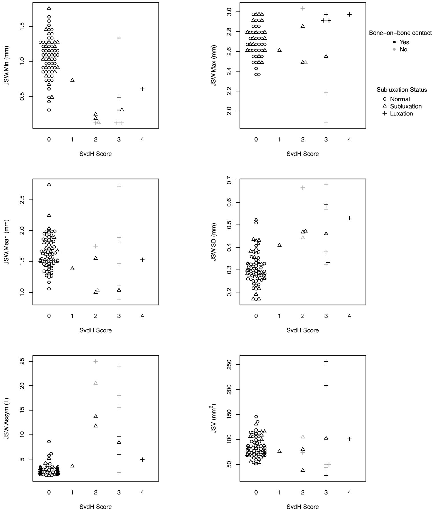
Distribution of SvdH scores for the 2nd and 3rd MCP is shown in Table 1. Figure 1 shows the distribution of HR-pQCT measurements by SvdH scores. More (sub)luxations were identified by HR-pQCT than radiograph. Bone-on-bone contact was observed in 5 joints on HR-pQCT, despite an absence of ankylosis observed on radiograph. GEE results indicated that decreased JSW.Min, increased JSW.SD, and increased JSW.Asymm were associated with increasing SvdH score (p < 0.05). However, no significant relationship was observed for JSW.Max, JSW.Mean, or JSV (p > 0.05). Visual examples of congruent and incongruent HR-pQCT and SvdH are shown in Figure 2.

Figure 1.
  • Download figure
  • Open in new tab
  • Download powerpoint
Figure 1.

Comparison between volumetric joint space outcomes derived from HR-pQCT with van der Heijde-modified Sharp joint space scores (SvdH) from conventional radiographs for the 2nd and 3rd MCP (n = 80 joints from 43 participants). MCP: metacarpophalangeal; JSW: joint space width; HR-pQCT: high-resolution peripheral quantitative computed tomography.

Figure 2.
  • Download figure
  • Open in new tab
  • Download powerpoint
Figure 2.

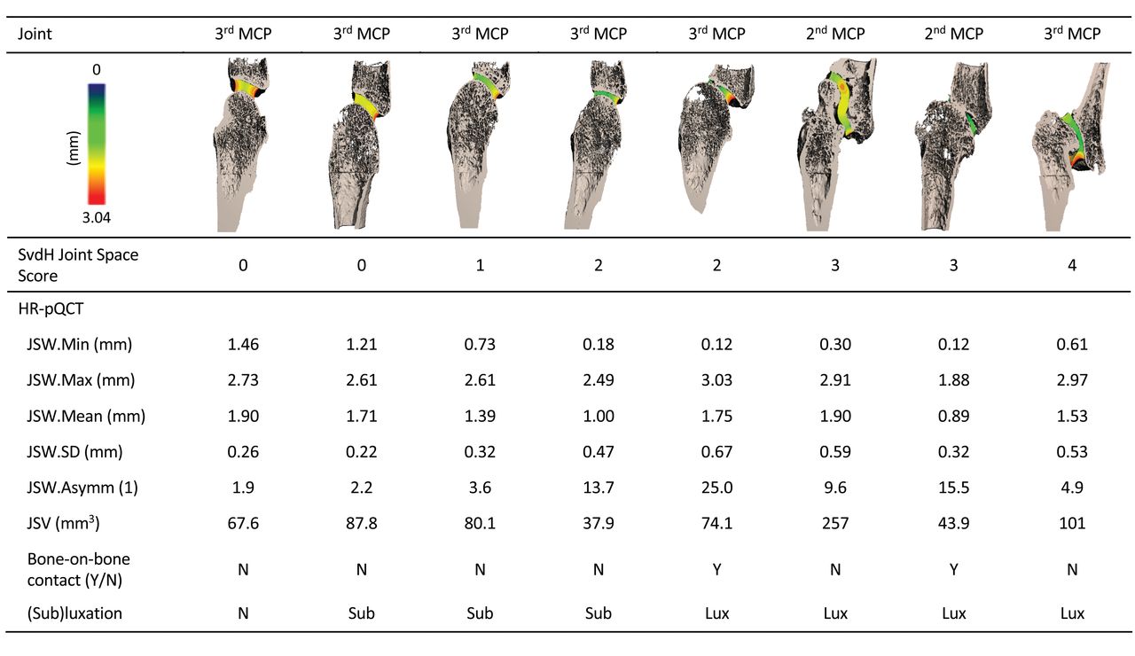
Examples of 3-D volumetric renderings of HR-pQCT scans and corresponding HR-pQCT outcomes across the range of van der Heijde-modified Sharp joint space scores (SvdH). HR-pQCT: high-resolution peripheral quantitative computed tomography; MCP: metacarpophalangeal; JSW: joint space width; JSV: joint space volume; sub: subluxation; lux: luxation.

View this table:
  • View inline
  • View popup
Table 1.

Frequency of joint space narrowing scored using the van der Heijde-modified Sharp scoring system on conventional radiographs (n = 80 joints from 43 participants).

DISCUSSION

Minimum JSW, SD, and asymmetry assessed by HR-pQCT were correlated with the joint space domain of a radiograph scoring system. However, large variability for SvdH scores across all quantitative outcomes confirms that the radiographic score is indeed an ordinal scale, and consequently progression would not be linear. The considerable number of subluxations, luxations, and bone-on-bone contact observed with HR-pQCT, and in previous studies using the current gold standard clinical CT20, suggests there may be a degree of subluxation and ankylosis that precedes detection of this damage on radiograph. Finally, it is difficult to interpret disease progression based on a single quantitative HR-pQCT outcome, such as volume, because there is a nonlinear association with radiographic score. The relationship between these pathological manifestations and clinical and patient-reported outcomes needs to be further investigated to understand how they relate to disease outcome.

HR-pQCT may provide advantages over radiography for monitoring disease progression, particularly in the context of clinical trials. HR-pQCT quantitative measurements appear less susceptible to floor and ceiling effects because there is a wide range of measurements in JSW.Min and JSW.SD when the ordinal score is assigned 0, and there is no maximum limit to SD and asymmetry outcomes. The quantitative design of the analysis and sensitivity of the technique will allow observation of increases or decreases in joint space. Further, automated image processing makes HR-pQCT outcomes less susceptible to reader interpretation.

A key limitation of HR-pQCT might be that fewer joints are accessible than in radiography; however, focusing on the 2nd and 3rd MCP of the dominant hand somewhat overcomes that limitation because those are the most commonly affected joints in RA. Although this cohort consisted of participants with severe disease activity, the most common SvdH score was 0. This is likely reflective of the general RA population, in which there is a need for more sensitive measures relevant to earlier stages of joint damage. Further, interpretation of volumetric joint space outcomes in (sub)luxed joints is a challenge, and whether progression in these highly damaged joints can be observed, and follows a similar quantitative pattern to less damaged joints, needs to be determined in a longitudinal analysis. While our study used second-generation HR-pQCT, the results should be directly applicable to first-generation HR-pQCT, because the algorithm accounts for changes in spatial resolution and we have previously observed minimal difference between measurements of the same joints on both scanner generations. Finally, because the algorithm identifies thickness of the joint space, joints with bone-on-bone contact typically correspond to a minimum width of 1 or 2 voxels rather than 0. There are plans to identify this bone-on-bone contact in the future; however, because the discrepancy between the current algorithm and the absolute minimum is smaller than our reproducibility error, this small discrepancy does not affect the results.

We have demonstrated aspects of construct validity for HR-pQCT against a well-established radiographic scoring system. Because HR-pQCT provides 3-D visualization of the joint that is not available using CR, its utility for assessing disease prior to evidence of radiographic joint space damage and with levels of significant joint space damage is promising. Further longitudinal studies are planned to understand the sensitivity of HR-pQCT outcomes to change over a wide range of joint damage and repair.

Acknowledgment

We thank Tessa Linkert for assistance with patient recruitment and data collection, Shaheer Chishti for assistance with data analysis, and Dr. Tak Fung for assistance with statistical analysis.

Footnotes

  • The study was funded by The Arthritis Society. PGC is supported in part by the UK NIHR Leeds Biomedical Research Centre.

  • Accepted for publication November 14, 2018.

REFERENCES

  1. 1.↵
    1. Schett G,
    2. Gravallese E
    . Bone erosion in rheumatoid arthritis: mechanisms, diagnosis and treatment. Nat Rev Rheumatol 2012;8:656–64.
    OpenUrlCrossRefPubMed
  2. 2.↵
    1. McInnes IB,
    2. Schett G
    . The pathogenesis of rheumatoid arthritis. N Engl J Med 2011;365:2205–19.
    OpenUrlCrossRefPubMed
  3. 3.↵
    1. Aletaha D,
    2. Funovits J,
    3. Smolen JS
    . Physical disability in rheumatoid arthritis is associated with cartilage damage rather than bone destruction. Ann Rheum Dis 2011;70:733–9.
    OpenUrlAbstract/FREE Full Text
  4. 4.↵
    1. Salaffi F,
    2. Gutierrez M,
    3. Carotti M
    . Ultrasound versus conventional radiography in the assessment of bone erosions in rheumatoid arthritis. Clin Exp Rheumatol 2014;32 Suppl 80:S85–90.
    OpenUrl
  5. 5.↵
    1. Scott DL,
    2. Laasonen L,
    3. Priolo F,
    4. Houssien DA,
    5. Bacarini L,
    6. Cerase A,
    7. et al.
    The radiological assessment of rheumatoid arthritis. Clin Exp Rheumatol 1997;15 Suppl 17:S53–61.
    OpenUrl
  6. 6.↵
    1. Ravindran V,
    2. Rachapalli S
    . An overview of commonly used radiographic scoring methods in rheumatoid arthritis clinical trials. Clin Rheumatol 2011;30:1–6.
    OpenUrlCrossRefPubMed
  7. 7.↵
    1. Yue J,
    2. Wu D,
    3. Tam LS
    . The role of imaging in early diagnosis and prevention of joint damage in inflammatory arthritis. Expert Rev Clin Immunol 2018;14:499–511.
    OpenUrl
  8. 8.↵
    1. Barnabe C,
    2. Buie H,
    3. Kan M,
    4. Szabo E,
    5. Barr SG,
    6. Martin L,
    7. et al.
    Reproducible metacarpal joint space width measurements using 3D analysis of images acquired with high-resolution peripheral quantitative computed tomography. Med Eng Phys 2013;35:1540–4.
    OpenUrlCrossRefPubMed
  9. 9.↵
    1. Burghardt AJ,
    2. Lee CH,
    3. Kuo D,
    4. Majumdar S,
    5. Imboden JB,
    6. Link TM,
    7. et al.
    Quantitative in vivo HR-pQCT imaging of 3D wrist and metacarpophalangeal joint space width in rheumatoid arthritis. Ann Biomed Eng 2013;41:2553–64.
    OpenUrlCrossRefPubMed
  10. 10.↵
    1. Töpfer D,
    2. Gerner B,
    3. Finzel S,
    4. Kraus S,
    5. Museyko O,
    6. Schett G,
    7. et al.
    Automated three-dimensional registration of high-resolution peripheral quantitative computed tomography data to quantify size and shape changes of arthritic bone erosions. Rheumatology 2015;54:2171–80.
    OpenUrlCrossRefPubMed
  11. 11.↵
    1. Kong S,
    2. Locrelle H,
    3. Amouzougan A,
    4. Denarie D,
    5. Collet P,
    6. Pallot-Prades B,
    7. et al.
    Remaining local subclinical joint inflammation is associated with deteriorated metacarpeal head bone microarchitecture in rheumatoid arthritis patients low disease activity. Joint Bone Spine 2018;85:569–72.
    OpenUrl
  12. 12.↵
    1. Stok KS,
    2. Finzel S,
    3. Burghardt AJ,
    4. Conaghan PG,
    5. Barnabe C;
    6. SPECTRA Collaboration
    . The SPECTRA collaboration OMERACT special interest group: current research and future directions. J Rheumatol 2017;44:1911–5.
    OpenUrlAbstract/FREE Full Text
  13. 13.↵
    1. Tom S,
    2. Frayne M,
    3. Manske SL,
    4. Burghardt AJ,
    5. Stok KS,
    6. Boyd SK,
    7. et al.
    Determining metacarpophalangeal flexion angle tolerance for reliable volumetric joint space measurements by high-resolution peripheral quantitative computed tomography. J Rheumatol 2016;43:1941–4.
    OpenUrlAbstract/FREE Full Text
  14. 14.↵
    1. Boers M,
    2. Kirwan JR,
    3. Wells G,
    4. Beaton D,
    5. Gossec L,
    6. d’Agostino MA,
    7. et al.
    Developing core outcome measurement sets for clinical trials: OMERACT filter 2.0. J Clin Epidemiol 2014;67:745–53.
    OpenUrlCrossRefPubMed
  15. 15.↵
    1. Boers M,
    2. Kirwan JR,
    3. Tugwell P,
    4. Beaton D,
    5. Bingham CO III,
    6. Conaghan PG,
    7. et al
    . The OMERACT Handbook. [Internet. Accessed May 17, 2017.] Available from: https://omeract.org/resources
  16. 16.↵
    1. Aletaha D,
    2. Neogi T,
    3. Silman AJ,
    4. Funovits J,
    5. Felson DT,
    6. Bingham CO 3rd,
    7. et al.
    2010 Rheumatoid arthritis classification criteria: an American College of Rheumatology/European League Against Rheumatism collaborative initiative. Arthritis Rheum 2010;62:2569–81.
    OpenUrlCrossRefPubMed
  17. 17.↵
    1. van der Heijde D
    . How to read radiographs according to the Sharp/van der Heijde method. J Rheumatol 1999;26:743–5.
    OpenUrlPubMed
  18. 18.↵
    1. Barnabe C,
    2. Feehan L;
    3. SPECTRA (Study GrouP for XTrEme-CT in RA)
    . High-resolution peripheral quantitative computed tomography imaging protocol for metacarpophalangeal joints in inflammatory arthritis: the SPECTRA collaboration. J Rheumatol 2012;39:1494–5.
    OpenUrlFREE Full Text
  19. 19.↵
    1. Burghardt AJ,
    2. Buie HR,
    3. Laib A,
    4. Majumdar S,
    5. Boyd SK
    . Reproducibility of direct quantitative measures of cortical bone microarchitecture of the distal radius and tibia by HR-pQCT. Bone 2010;47:519–28.
    OpenUrlCrossRefPubMed
  20. 20.↵
    1. Døhn UM,
    2. Conaghan PG,
    3. Eshed I,
    4. Boonen A,
    5. Bøyesen P,
    6. Peterfy CG,
    7. et al.
    The OMERACT-RAMRIS rheumatoid arthritis magnetic resonance imaging joint space narrowing score: intrareader and interreader reliability and agreement with computed tomography and conventional radiography. J Rheumatol 2014;41:392–7.
    OpenUrlAbstract/FREE Full Text
PreviousNext
Back to top

In this issue

The Journal of Rheumatology
Vol. 46, Issue 10
1 Oct 2019
  • Table of Contents
  • Table of Contents (PDF)
  • Index by Author
  • Editorial Board (PDF)
Print
Download PDF
Article Alerts
Sign In to Email Alerts with your Email Address
Email Article

Thank you for your interest in spreading the word about The Journal of Rheumatology.

NOTE: We only request your email address so that the person you are recommending the page to knows that you wanted them to see it, and that it is not junk mail. We do not capture any email address.

Enter multiple addresses on separate lines or separate them with commas.
The SPECTRA Collaboration OMERACT Working Group: Construct Validity of Joint Space Outcomes with High-resolution Peripheral Quantitative Computed Tomography
(Your Name) has forwarded a page to you from The Journal of Rheumatology
(Your Name) thought you would like to see this page from the The Journal of Rheumatology web site.
CAPTCHA
This question is for testing whether or not you are a human visitor and to prevent automated spam submissions.
Citation Tools
The SPECTRA Collaboration OMERACT Working Group: Construct Validity of Joint Space Outcomes with High-resolution Peripheral Quantitative Computed Tomography
Sarah L. Manske, Scott C. Brunet, Stephanie Finzel, Kathryn S. Stok, Philip G. Conaghan, Steven K. Boyd, Cheryl Barnabe
The Journal of Rheumatology Oct 2019, 46 (10) 1369-1373; DOI: 10.3899/jrheum.180870

Citation Manager Formats

  • BibTeX
  • Bookends
  • EasyBib
  • EndNote (tagged)
  • EndNote 8 (xml)
  • Medlars
  • Mendeley
  • Papers
  • RefWorks Tagged
  • Ref Manager
  • RIS
  • Zotero

 Request Permissions

Share
The SPECTRA Collaboration OMERACT Working Group: Construct Validity of Joint Space Outcomes with High-resolution Peripheral Quantitative Computed Tomography
Sarah L. Manske, Scott C. Brunet, Stephanie Finzel, Kathryn S. Stok, Philip G. Conaghan, Steven K. Boyd, Cheryl Barnabe
The Journal of Rheumatology Oct 2019, 46 (10) 1369-1373; DOI: 10.3899/jrheum.180870
del.icio.us logo Digg logo Reddit logo Twitter logo CiteULike logo Facebook logo Google logo Mendeley logo
  • Tweet Widget
  • Facebook Like
  • Google Plus One
Bookmark this article

Jump to section

  • Article
    • Abstract
    • MATERIALS AND METHODS
    • RESULTS
    • DISCUSSION
    • Acknowledgment
    • Footnotes
    • REFERENCES
  • Figures & Data
  • Info & Metrics
  • References
  • PDF
  • eLetters

Keywords

RHEUMATOID ARTHRITIS
RADIOGRAPHIC MICRO-COMPUTED TOMOGRAPHY
RADIOGRAPHY
METACARPOPHALANGEAL JOINT
OMERACT

Related Articles

Cited By...

More in this TOC Section

  • Pain Measurement in Rheumatic and Musculoskeletal Diseases: Where To Go from Here? Report from a Special Interest Group at OMERACT 2018
  • Adaptive Trial Designs in Rheumatology: Report from the OMERACT Special Interest Group
  • Musculoskeletal Ultrasound in Systemic Lupus Erythematosus: Systematic Literature Review by the Lupus Task Force of the OMERACT Ultrasound Working Group
Show more OMERACT 2018: International Consensus Conference on Outcome Measures in Rheumatology, Terrigal, Australia, May 2018 Special Interest Groups, Part 2

Similar Articles

Keywords

  • rheumatoid arthritis
  • RADIOGRAPHIC MICRO-COMPUTED TOMOGRAPHY
  • radiography
  • metacarpophalangeal joint
  • OMERACT

Content

  • First Release
  • Current
  • Archives
  • Collections
  • Audiovisual Rheum
  • COVID-19 and Rheumatology

Resources

  • Guide for Authors
  • Submit Manuscript
  • Author Payment
  • Reviewers
  • Advertisers
  • Classified Ads
  • Reprints and Translations
  • Permissions
  • Meetings
  • FAQ
  • Policies

Subscribers

  • Subscription Information
  • Purchase Subscription
  • Your Account
  • Terms and Conditions

More

  • About Us
  • Contact Us
  • My Alerts
  • My Folders
  • Privacy/GDPR Policy
  • RSS Feeds
The Journal of Rheumatology
The content of this site is intended for health care professionals.
Copyright © 2022 by The Journal of Rheumatology Publishing Co. Ltd.
Print ISSN: 0315-162X; Online ISSN: 1499-2752
Powered by HighWire