

Abstract
Objective. Inflammatory bowel disease (IBD) is generally reported to be associated with spondylarthropathies (SpA) in 5%–15% of cases. Systematic colonoscopic assessment by protocol demonstrated mucosal inflammation characteristic of Crohn disease (CD) in up to one-third of patients with SpA. Video capsule endoscopy (CE) is a superior diagnostic tool to detect small bowel mucosal disease. Our study compared the accuracy of CE to standard colonoscopy for detection of inflammatory bowel lesions in patients with SpA, and to describe predictors of small bowel inflammation (SBI) in this cohort.
Methods. Prospective cross-sectional study of adult patients followed for SpA. Patients were evaluated by CE and standard colonoscopy with biopsies. SBI was quantified using the Lewis Score. Additional screening tests included fecal calprotectin (FCP), C-reactive protein (CRP), and a diagnostic panel of serologic, inflammatory and genetic tests (SGI).
Results. There were 64 patients recruited (53% female, mean age 42 ± 13 yrs). Chronic gastrointestinal (GI) symptoms were present in 57%. CE revealed significant SBI in 27/64 (42.2%), compared to 7/64 (10.9%) by standard colonoscopy (p = 0.035). Elevated FCP was associated with small bowel CD (OR 4.5, 95% CI 1.01–19.9; p = 0.042). No correlation was observed with presence of GI symptoms, CRP, or SGI results. Finding CD led to a change in management in 65.2% of cases.
Conclusion. CE uncovered SBI consistent with CD in 42.2% of patients with SpA, with a significant incremental yield over colonoscopy of 31%. FCP levels were significantly correlated with CE results, while GI symptoms and SGI results were poor predictors of SBI.
Spondyloarthritis (SpA) is a group of inflammatory rheumatic diseases with common clinical, radiologic, and serologic features. Besides the prototype ankylosing spondylitis (AS), this group includes psoriatic arthritis, enteropathic arthritis associated with inflammatory bowel disease (IBD), reactive arthritis, and undifferentiated SpA1. Extraarticular manifestations and comorbidities are common, including anterior uveitis, psoriasis, and IBD. These should be assessed because they may affect treatment targets or followup measures2,3.
The association between IBD and SpA is well established. Both show familial clustering and may coexist. SpA was thought to affect < 10% of patients with IBD4,5, while radiologic evidence of sacroiliitis was detected in 18%. More recently, SpA prevalence rates ranging from 17% to 39% were reported in IBD6,7,8. A link between joint and gut inflammation has been proposed9,10.
Potential roles for HLA-B27, NOD2/CARD15, and IL-23R genes are implicated in predisposing to gut and/or joint inflammation11,12,13. Cytokines such as tumor necrosis factor (TNF)–α, interleukin (IL)-12, IL-23, and IL-17, are implicated in the inflammatory pathways for both14,15. The connection is further supported by overlapping treatment options for SpA and IBD. However, differences in therapeutic effectiveness have been observed in the gut and joints, affecting treatment choice if both disorders coexist10.
Overt symptoms or signs of accompanying intestinal inflammation are often absent. However, when colonoscopy was performed by protocol rather than symptom-based referral, a high prevalence (30%–44%) of ileal inflammation was observed16. SpA could be the sole clinical presentation of subclinical Crohn disease (CD), while in others, SpA with subclinical gut inflammation may progress to overt CD.
Conventional endoscopic and radiological techniques are limited in their capacity to investigate the small bowel, thus often unable to detect CD mucosal lesions17. Colonoscopy typically permits visualization of 20–30 cm of the terminal ileum (TI), leaving the remaining 4 meters of small bowel an unexplored space. Capsule endoscopy (CE) is superior to conventional techniques for the investigation of suspected small bowel inflammation (SBI)18,19. The primary objective of our study was to determine whether CE can more reliably reveal SBI due to CD in subjects with SpA versus colonoscopy. One pilot study was published supporting this concept20. SBI was demonstrated by CE in 6/20 patients, compared to only 1/20 by colonoscopy. Our hypothesis is that SBI due to CD are much more prevalent in patients with SpA than previously reported. Our secondary objectives were to compare the diagnostic yield of CE and colonoscopy in detecting SBI in SpA; to evaluate the clinical and laboratory variables including serological, genetic, and biomarker tests for predicting SBI in SpA; and to determine whether the additional diagnosis of CD resulted in a change in management.
MATERIALS AND METHODS
Our study population included consecutive adult patients aged 18–75 with SpA, diagnosed according to European Spondyloarthropathy Study Group (ESSG) or modified New York criteria. Our study received research ethics board (REB) approval from the McGill University Health Centre Research Ethics Office (study #GEN-08-053). All patients signed the REB-approved informed consent. Patients were classified into 2 groups: (a) subjects without GI symptoms or signs suggestive of IBD; and (b) patients with signs and/or symptoms suggestive of CD, including ≥ 1 of the following: diarrhea > 3 weeks, abdominal pain > 3 weeks, and extraintestinal manifestations suggestive of CD. The exclusion criteria included established diagnosis of IBD (CD and ulcerative colitis), psoriatic arthritis, reactive arthritis associated with an intestinal infection, known intestinal obstruction or obstructive symptoms (e.g., severe abdominal pain with nausea, vomiting, or abdominal distention), intestinal stricture identified by imaging, suspected stricture (followed by a patency capsule study that did not confirm SB patency), use of any nonsteroidal antiinflammatory drugs (NSAID) over the previous 4 weeks, treatment with any biological within the previous 6 months [except for etanercept (ETN)], diagnosis of celiac disease or positive celiac serology [antitransglutaminase (anti-TTG) or anti-endomysial antibody], and CE or colonoscopy within 1 year.
Study procedures
Articular symptoms were assessed using the Bath Ankylosing Spondylitis Functional Index (BASFI) and Bath Ankylosing Spondylitis Disease Activity Index (BASDAI). The Harvey-Bradshaw Index (HBI) was used to assess gastrointestinal (GI) symptoms. Anti-TTG and HLA-B27 were assessed if unknown. Fecal calprotectin was measured using Quantum Blue semiquantitative assay (Buhlmann). NOD2 mutations were assessed using a Sequenom array.
Capsule endoscopy
All studies used the PillCam™ SB2 or SB3 (Given Imaging). Preparation included a clear liquid diet the preceding day and a 12-h overnight fast. The study was reviewed using the RAPID software (Given Imaging). Mucosal inflammation was graded using the Lewis score (LS)21. LS is considered “positive” if > 135 for any tertile. For our study, we predefined mild SBI as LS ≥ 300 in any SB tertile, including ≥ 3 ulcers22. Moderate to severe SBI was defined as LS ≥ 790.
Colonoscopy
Patients underwent colonoscopy within 4 weeks after CE. Colonic, ileal, and rectal biopsies were routinely obtained. The endoscopist was blinded to CE results.
Serological and genetic testing
Patients were screened for IBD using the Prometheus Labs Serology, Genetics, and Inflammation (SGI) panel23.
Results of the biomarker panel were reported using a validated diagnostic algorithm as consistent with CD or ulcerative colitis (UC), or not consistent with IBD23. In addition, the results of each individual test included in the SGI panel were analyzed independently.
Management outcomes
Patients were seen in clinic 3 months after CE. They were questioned for a history of any adverse events, and changes in medication use were verified for those with a diagnosis of IBD.
Statistical analysis
We compared the characteristics of patients with and without findings of small bowel CD on CE. Categorical values were compared using chi-square test, and continuous variables with Student t test. The association with small bowel CD detected by CE was evaluated using univariate logistic regression. Sensitivity, specificity, and Spearman rank correlation values were determined. OR and CI were calculated when appropriate. Receiver-operated curve analysis was performed for prediction of small bowel CD by quantitative fecal calprotectin (FCP) values. P values < 0.05 were considered statistically significant. The analysis was performed using IBM SPSS statistical software Version 20.0.
RESULTS
There were 67 patients who underwent CE. Three subsequently refused to undergo colonoscopy and were withdrawn from analysis. The demographic and clinical characteristics of the patients are depicted in Table 1. As noted, ESSG and modified New York criteria were used to recruit patients for our study, which was initiated in 2012. Since then, Assessment of Spondyloarthritis international Society (ASAS) criteria were established and validated, allowing SpA to be further classified as axial (ax-) or peripheral (p-) SpA24. Longterm followup of the original ASAS cohort confirmed an excellent predictive validity for the ASAS axSpA and pSpA classification criteria24. In addition, patients fulfilling the “clinical arm” had disease characteristics in keeping with that of the “imaging arm.” Imaging criteria for ASAS were not uniformly carried out for earlier patients recruited. We thus retrospectively applied the clinical ASAS criteria for axSpA to our cohort, providing further information and clarification about the type of SpA recruited. Patients were thus reclassified as axSpA or pSpA (Table 1), blinded to whether CD was uncovered.
The clinical and demographic characteristics of the SpACE Capsule Study patients. Values are n (%) unless otherwise specified.
CE and colonoscopy findings
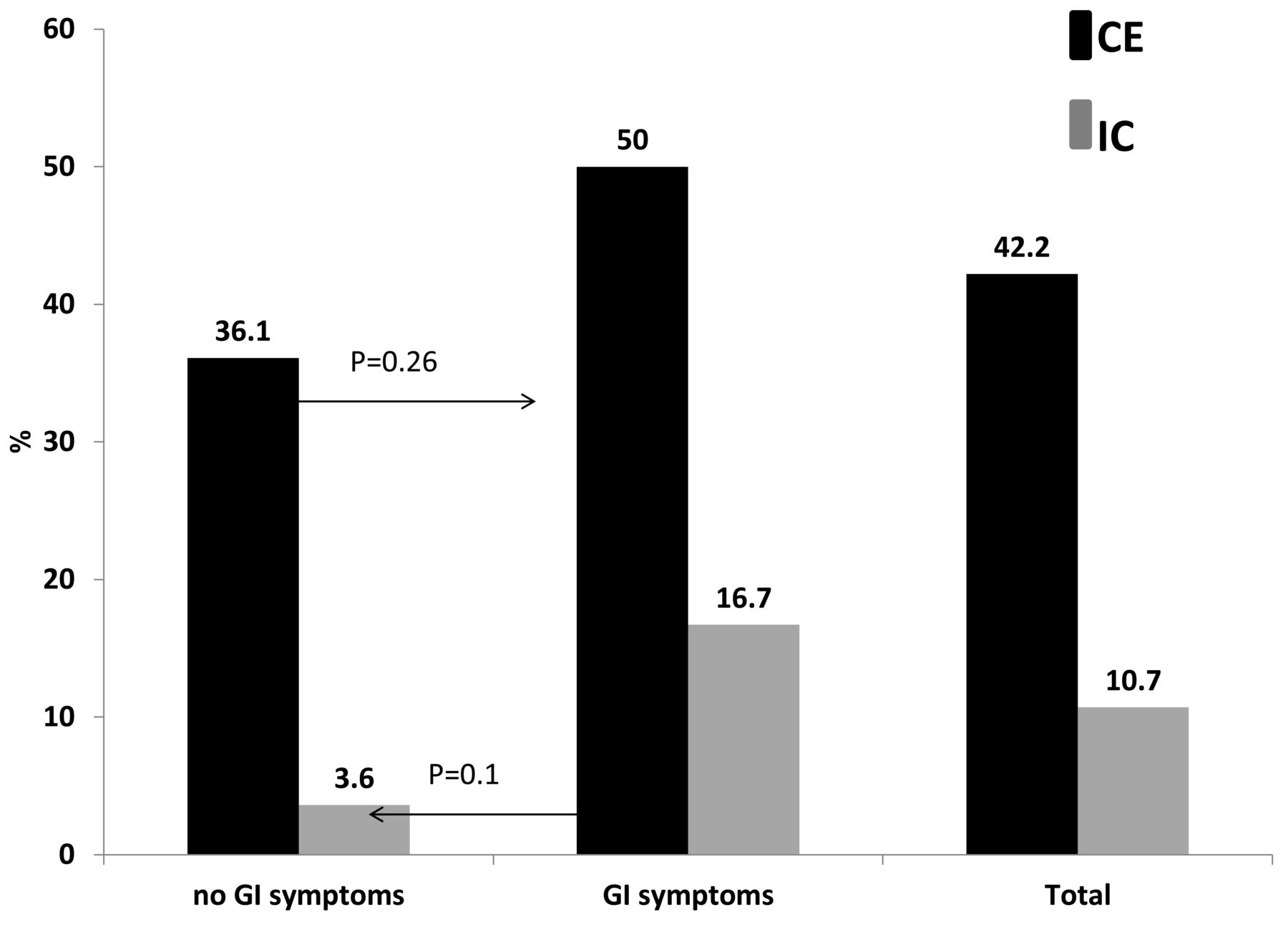
CE revealed SBI consistent with CD (LS ≥ 300) in 27 (42.2%) patients (Figure 1). In 8 of these (29.6%), CD only involved the proximal small bowel (first and/or second tertiles), sparing the distal ileum. The mean ± SD LS in CE-positive patients was 855 ± 361. In 12 (18.7%), moderate to severe inflammation (LS ≥ 790) was observed. In 15 (23.4%), mild to moderate inflammation (135 < LS < 790) was detected. CE in 11 (17.1%) revealed only mild, nonspecific inflammation (135 ≤ LS < 300) and the CE was deemed negative. CE was normal (LS < 135) in the rest. All CE examinations were complete with the capsule reaching the cecum. There were no cases of capsule retention.

Capsule endoscopy and ileocolonoscopy findings in spondyloarthropathy patients with and without gastrointestinal complaints. GI: gastrointestinal; IC: ileocolonoscopy; CE: video capsule endoscopy.
A complete colonoscopy, defined as successful intubation and biopsy of the TI, was achieved in 59 subjects (92.2%). The colonoscopy reached the cecum in the 5 others. Colonoscopy findings consistent with IBD were revealed in 7/64 (10.9%; p = 0.035 vs CE). In 4 patients, terminal ileal ulcers were seen, with histological confirmation of CD in 3. In another 4, mucosal inflammation was detected by colonoscopy in the TI and colon. In all 4, CD was confirmed by histology as well as detected by CE. In 6 patients, mild colonic inflammation without TI involvement was seen by colonoscopy (2 with proctitis; 4 with multifocal colonic ulcers); 2 of the patients (none with proctitis) had SBI on CE. Overall, small bowel CD was diagnosed by CE in 6/7 patients (85.7%) diagnosed with CD on colonoscopy. CD was detected by colonoscopy in 6/30 (20%) of CD detected by CE. On followup, 1 additional patient was found to have CD restricted to perianal area (recurrent ischiorectal abscesses). CE and IC and biopsies were negative in this patient with perianal CD.
GI complaints
GI symptoms were reported in 56.3% of the patients (HBI index > 4). None of the individual GI symptoms or their combination was associated with CD.
Classification of SpA and assessment of clinical disease activity
In the axSpA group, CD was found in 20/50 cases, versus 7/14 with pSpA. This 10% difference did not reach statistical significance (p = 0.46), possibly because of the relatively small number of cases in the peripheral group.
Data were collected to determine BASDAI and BASFI. For each, a numerical rating scale was used (range 0–10), expressed as mean ± SD. In the group found to have CD in association with AS, BASDAI (5.9 ± 1.4) was significantly higher than in those with SpA only (3.2 ± 1.5; p < 0.05). Unfortunately, too few questionnaires were available to analyze BASFI.
Inflammatory biomarkers
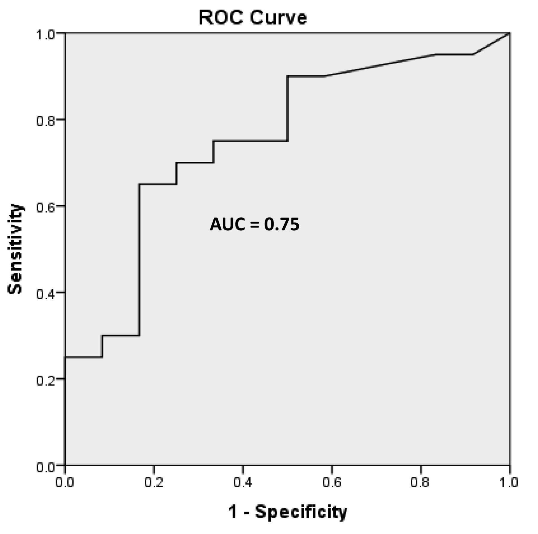
Complete blood counts, hemoglobin, and C-reactive protein (CRP) levels were not significantly associated with CD. An elevated FCP (> 100 µg/g) was significantly associated with small bowel CD (OR 4.5, 95% CI 1.01–19.9, p = 0.042; Figure 2). The area under the curve of FCP for prediction of small bowel CD by CE was 0.75 (Figure 3), with the optimal cutoff value of 132 µg/g (sensitivity = 66.7%, specificity = 76.9%).

Ability of biomarkers to predict small bowel inflammation in spondyloarthitis. SGI panel comprises these markers: serology — IgA and IgG ASCA, IgA OmpC, IBD-specific pANCA, DNA-sensitive pANCA, and antiflagellin antibodies (IgA CBir1, IgG anti-A4-Fla2, anti-FlaX); genetic — ATG16L1 SNP (rs2241880), EMC1 SNP (rs3737240), NKX2-3 SNP (rs10883365), and STAT3 SNP (rs744166); inflammation — ICAM-1, VCAM-1, VEGF, CRP, and SAA. SGI: serology, genetics, and inflammation; ASCA: anti-Saccharomyces cerevisiae antibodies; CRP: C-reactive protein; IBD: inflammatory bowel disease; ICAM-1: intercellular adhesion molecule; OmpC: anti-outer membrane porin C of E. coli; pANCA: perinuclear antineutrophil cytoplasmic antibody; SAA: serum amyloid A; SNP: single-nucleotide polymorphism; VCAM-1: vascular cell adhesion molecule; VEGF: vascular endothelial growth factor; NS: not significant.

ROC analysis of fecal calprotectin for prediction of significant small bowel inflammation on video capsule endoscopy. AUC: area under the curve; ROC: receiver-operated curve.
SGI panel results
SGI results were predictive of CD in 11/40 (27.5%; 4 CD, 4 UC, and 3 indeterminate colitis). There was no correlation between SGI results and either CE or colonoscopy results (Figure 2). For correlation with CE, the sensitivity of SGI was 15% and specificity 95% (r = 0.17, p = 0.3). If all abnormal SGI results consistent with IBD were grouped as positive, the sensitivity was 40% and specificity 85% (r = 0.28, p = 0.08). For colonoscopy, the correlation was nonsignificant (r = 0.025, p = 0.6). In 4 patients predicted as having UC by SGI, only 1 had mild, nonspecific colonic lesions not confirmed as IBD histologically. No individual serological, genetic, or inflammatory SGI panel variables, analyzed independently, was associated with improved accuracy (Figure 2). The prevalence of mutations in the single-nucleotide polymorphism (SNP) included in the SGI panel in our cohort was high [ATG16L1 SNP (rs2241880) 80.8%; EMC1 SNP (rs3737240) 47.5%; NKX2-3 SNP (rs10883365) 80.8%; and STAT3 SNP (rs744166) 61.5%]. However, the prevalence of these polymorphisms was similar in patients with and without SBI.
NOD2 mutations were detected in 7/46 (15.2%) of the patients. In 5, R702W polymorphism was detected; and G908R and 1007fs were detected in 1 patient each. The prevalence of NOD2 mutations was somewhat higher in patients with CD; however, the difference did not reach significance (26.3% vs 11.1%, p = 0.18).
Changes in management in patients with SpA and IBD
Among the 27 patients diagnosed with CD, 23 were seen at 3 months after undergoing CE. A change in management (excluding cessation of NSAID alone) was made in 15/23 (65.2%; Table 2). All treatment decisions were made by the referring rheumatologist, in consultation with the gastroenterologist. The most common change was initiating anti-TNF therapy. In those patients receiving ETN, it was switched to another anti-TNF established to treat SpA and CD.
Effect of the diagnosis of Crohn disease on management of patients with SpA.
DISCUSSION
Our results confirm a high prevalence of SBI as a result of CD in patients with SpA. However, relatively few cases were uncovered by colonoscopy, the customary standard investigation. The incremental yield of CE was significantly greater compared to colonoscopy for detection of CD, consistent with our hypothesis. GI complaints were comparably common in patients with a normal small bowel and colon, suggesting the symptoms were often a result of functional bowel disorder [irritable bowel syndrome (IBS)].
Several studies have suggested that a significant number of patients with SpA have asymptomatic intestinal inflammation. The prevalence of abnormal findings was as high as 49% for macroscopic and 69% for microscopic abnormalities5. De Vos, et al25 prospectively examined the longterm evolution of gut inflammation in patients with SpA. Repeat colonoscopy was carried out after ≥ 2 years in 49 patients. Macroscopic ulcerations or histological inflammation persisted in the majority. Overt clinical IBD developed in 7.3%. Persistent gut inflammation correlated with articular inflammation and diarrhea. Articular remission was associated with endoscopic and histological remission. However, the majority were taking NSAID at initial and followup colonoscopy. The prevalence of lesions would have been significantly lower had NSAID been withdrawn, as in our study. The same limitation applies to most other colonoscopy studies5. NSAID treatment, even if short term, may result in small bowel lesions indistinguishable from CD26,27.
Some studies reported a similar prevalence of colonic and small bowel findings in patients with SpA28,29, while others described mostly ileal distribution16. The most noteworthy difference between earlier reports and our study is the use of CE, a superior modality for diagnosing small bowel pathologies compared to colonoscopy and cross-sectional imaging17,18,19,20,30. CE enables the detection of lesions outside the reach of endoscopic techniques. To date, 1 pilot study using CE in patients with SpA20 reported a similar distribution of findings to our present study, with evidence of SBI in 9/20 (45%) and colonic inflammation in 5%.
Biomarkers could be useful to predict the coexistence of IBD in SpA. Antibodies targeting bacterial antigens have been extensively evaluated for diagnosis, classification, and prognostication in IBD31. The prevalence of positive IBD-related serological biomarkers [anti-Saccharomyces cerevisiae antibodies (ASCA) IgA and IgG, anti-outer membrane porin C of E. coli (OmpC), and anti-CBir-1] is higher in SpA than controls32,33. A serological panel combining 7 of these antibodies [ASCA (IgA and IgG), CBir-1 (IgA), OmpC -1 (IgA), and perinuclear antineutrophil cytoplasmic antibody (autoantibody ELISA, IFA perinuclear pattern, anti–DNAse sensitivity)] analyzed collectively using a computerized diagnostic algorithm was introduced to screen for IBD (Prometheus Labs IBD Serology 7)34. This panel was expanded to include 2 additional antiflagellin antibodies (anti-A4-Fla2 IgG, anti-FlaX IgG ELISA), an array of genetic (SNP rs2241880 in the ATG16L1 gene, SNP rsl0883365 in the NKX2–3 gene, SNP rs3737240 in the ECM1 gene, and SNP rs744166 in the STAT3 gene) and inflammation markers [intercellular adhesion molecule (ICAM)-1, vascular cell adhesion molecule (VCAM)-1, vascular endothelial growth factor (VEGF), CRP, and serum amyloid A]. In a validation study, the sensitivity and specificity of this SGI panel (Prometheus Labs) were 73.6% and 89.6% for detection of IBD, 88.9% and 81% for detection of CD, and 97.7% and 83.5% for detection of UC, respectively23. In our study, the SGI panel had a low sensitivity for predicting IBD. However, our SpA cohort was very different from the one included in the aforementioned studies. Further study in larger cohorts is needed.
There are studies that reported polymorphisms common to AS and CD. Although the most significant genetic association for SpA is with the genes related to the MHC (including HLA-B27), several polymorphisms outside the MHC were identified, including IL-23R, STAT3, PSMG1, IL12B, CDKAL1, LRRK2/MUC19, and ERAP1/2 genes13,35,36. Certain mutations in the NOD2 gene were reported to be equally prevalent in SpA patients with subclinical bowel inflammation and CD36. As part of the SGI panel, the prevalence of prespecified genetic polymorphisms described above was determined in our cohort. Mutations were detected in ≥ 50% of the patients for each of the evaluated SNP. Two of these polymorphisms (ECM1 and NkX2-3) were not previously described in SpA. However, these polymorphisms did not correlate with bowel inflammation. Our study was underpowered to address the significance of these findings.
The management of patients with SpA and CD poses challenges. Clinicians must consider the potential systemic characteristics of both disorders, musculoskeletal and GI manifestations, and the risk/benefit of available therapies3,37. Therapies for SpA and IBD overlap, but effectiveness in the gut and joints can differ10. Anti-TNF agents, including adalimumab, ETN, and infliximab (IFX), are all effective and cost-efficient in treating SpA and AS3,37,38. However, ETN is not effective for IBD39. Moreover, new-onset IBD was reported in patients taking ETN for SpA40,41. Braun, et al reported that IFX, not ETN, prevented IBD activity in SpA42. In our study, CD was discovered in 2/7 patients taking ETN for SpA. Adalimumab and IFX are both effective in treating CD and SpA. Our study supports use of molecules with proven therapeutic success. A change in management was made in 65.2% of patients found to have CD and SpA (Table 2). The most common change was the initiation of an anti-TNF, in 10/23 cases (43.5%). In a single-center retrospective study of the effect of CE in IBD43, a change in management was prescribed in 61.6% of CD cases 3 months after CE, similar to our study. Although guidelines for CD do not generally recommend anti-TNF therapy for asymptomatic patients, exceptions exist. Examples include patients who have other extraintestinal disorders, such as recurrent uveitis and severe psoriasis. Another consideration is the presence of extensive, unresectable small bowel CD, a known risk factor for aggressive disease.
In our study, FCP was a significant predictor of bowel inflammation (Figure 2). A recent longitudinal study also reported that an elevated FCP was the strongest predictor of the development of CD in AS44. As in our study, all the IBD cases uncovered were CD rather than UC. There is a strong correlation between the FCP levels and inflammatory findings in the gut; the association appears to be somewhat stronger in cases with colonic involvement45. In a recent metaanalysis, the pooled sensitivity and specificity of FCP for discrimination between IBD and IBS was 0.9346.
Our study has potential limitations. The longterm clinical significance of mild SBI in patients with SpA is unclear. LS of 135 was initially proposed as a cutoff value in patients with clinical suspicion of IBD47. However, mild small bowel lesions can be detected frequently in healthy controls or in NSAID users26. In patients with SpA, continuation of NSAID has been associated with elevated FCP levels44. Although patients were instructed to discontinue NSAID for at least 4 weeks, some concealed use cannot be excluded. We predetermined a higher cutoff for definition of significant SBI using LS ≥ 300 to avoid overdiagnosis of patients with very mild lesions of unclear significance. In addition, the results of the SGI diagnostic panel were available in 40/64 patients, because the test was available only as of 2013. However, our results suggest a low correlation of the SGI results with CE findings. It is uncertain that the correlation would have improved with a larger number of patients.
An unanticipated feature of our study cohort was the relatively high proportion of women. Although initially considered a diagnosis almost exclusive to men, the proportion of women with AS has increased since the discovery of HLA–B27 and the development of improved diagnostic and classification criteria. AS in women is still considered underdiagnosed and associated with a longer delay in diagnosis. Other recent studies have not uniformly confirmed male predominance in SpA. To clarify sex differences, a cohort of 708 patients with early inflammatory back pain suggestive of axSpA was recruited for a prospective multicenter French study48. Among the 475 patients diagnosed with SpA, 50.3% were men and 49.7% women. Other factors that may have affected the sex ratio of patients we recruited include disease duration and the restriction to anti-TNF–naive cases. In another study, Reveille, et al suggested that the high prevalence of female patients with SpA was due in part to the early onset inflammatory back pain and the reliance on clinical rather than imaging criteria for the diagnosis49.
Our study confirmed a high prevalence of SBI consistent with CD in SpA. CE was significantly more sensitive in detecting lesions compared to colonoscopy. Small bowel CD was poorly correlated with GI complaints and biomarker panel results. Two factors predictive of CD in association with SpA were higher disease activity, as reflected by the BASDAI, and increased FCP when not taking NSAID. Our findings are thus in keeping with other studies44,50. Anti-TNF therapy has been shown to induce and maintain remission of CD, while at the same time treating severe active SpA51, suggesting that it should be the preferred drug for the treatment of active and severe SpA associated with active or quiescent CD. These results are likely to have an important effect on the selection of anti-TNF therapy in patients with SpA.
Footnotes
Full Release Article. For details see Reprints and Permissions at jrheum.org
Financial support for our research study was provided by Abbvie Canada as an unrestricted grant. In-kind research support (EGS) was provided by Given Imaging/Medtronic Inc., and Prometheus Laboratories. Salary support (EGS) funded by the Canada Research Chair in Immune-mediated Gastrointestinal Disorders and the Bruce Kaufman Endowed Chair in Inflammatory Bowel Diseases (IBD) at McGill University. Fellowship awards funded by the McGill IBD Research Group (Dr. Kopylov) and the Fondation Litta of Geneva, Switzerland (MG).
- Accepted for publication October 11, 2017.
Free online via JRheum Full Release option


