
Main menu

  • Home
  • Content
    • First Release
    • Current
    • Archives
    • Collections
    • Audiovisual Rheum
    • 50th Volume Reprints
  • Resources
    • Guide for Authors
    • Submit Manuscript
    • Payment
    • Reviewers
    • Advertisers
    • Classified Ads
    • Reprints and Translations
    • Permissions
    • Meetings
    • FAQ
    • Policies
  • Subscribers
    • Subscription Information
    • Purchase Subscription
    • Your Account
    • Terms and Conditions
  • About Us
    • About Us
    • Editorial Board
    • Letter from the Editor
    • Duncan A. Gordon Award
    • Privacy/GDPR Policy
    • Accessibility
  • Contact Us
  • JRheum Supplements
  • Services

User menu

  • My Cart
  • Log In

Search

  • Advanced search
The Journal of Rheumatology
  • JRheum Supplements
  • Services
  • My Cart
  • Log In
The Journal of Rheumatology

Advanced Search

  • Home
  • Content
    • First Release
    • Current
    • Archives
    • Collections
    • Audiovisual Rheum
    • 50th Volume Reprints
  • Resources
    • Guide for Authors
    • Submit Manuscript
    • Payment
    • Reviewers
    • Advertisers
    • Classified Ads
    • Reprints and Translations
    • Permissions
    • Meetings
    • FAQ
    • Policies
  • Subscribers
    • Subscription Information
    • Purchase Subscription
    • Your Account
    • Terms and Conditions
  • About Us
    • About Us
    • Editorial Board
    • Letter from the Editor
    • Duncan A. Gordon Award
    • Privacy/GDPR Policy
    • Accessibility
  • Contact Us
  • Follow Jrheum on BlueSky
  • Follow jrheum on Twitter
  • Visit jrheum on Facebook
  • Follow jrheum on LinkedIn
  • Follow jrheum on YouTube
  • Follow jrheum on Instagram
  • Follow jrheum on RSS
Research ArticlePsoriatic Arthritis

Utility of Power Doppler Ultrasound–detected Synovitis for the Prediction of Short-term Flare in Psoriatic Patients with Arthritis in Clinical Remission

Santiago Ruta, Josefina Marin, Maria Laura Acosta Felquer, Leandro Ferreyra-Garrot, Javier Rosa, Ricardo García-Monaco and Enrique R. Soriano
The Journal of Rheumatology July 2017, 44 (7) 1018-1023; DOI: https://doi.org/10.3899/jrheum.161347
Santiago Ruta
From the Rheumatology Unit, Internal Medicine Service, and Radiology Department, Hospital Italiano de Buenos Aires; University Institute from Hospital Italiano de Buenos Aires; Fundación Dr. Pedro M. Catoggio para el Progreso de la Reumatología, Buenos Aires, Argentina.
  • Find this author on Google Scholar
  • Find this author on PubMed
  • Search for this author on this site
  • For correspondence: santiago.ruta@hospitalitaliano.org.ar
Josefina Marin
  • Find this author on Google Scholar
  • Find this author on PubMed
  • Search for this author on this site
Maria Laura Acosta Felquer
  • Find this author on Google Scholar
  • Find this author on PubMed
  • Search for this author on this site
Leandro Ferreyra-Garrot
  • Find this author on Google Scholar
  • Find this author on PubMed
  • Search for this author on this site
Javier Rosa
  • Find this author on Google Scholar
  • Find this author on PubMed
  • Search for this author on this site
Ricardo García-Monaco
  • Find this author on Google Scholar
  • Find this author on PubMed
  • Search for this author on this site
Enrique R. Soriano
  • Find this author on Google Scholar
  • Find this author on PubMed
  • Search for this author on this site
  • Article
  • Figures & Data
  • Info & Metrics
  • References
  • PDF
PreviousNext
Loading

Abstract

Objective. Power Doppler ultrasound (PDUS) has been shown to detect subclinical synovitis in psoriatic arthritis (PsA), but its value is not yet fully understood. The aim of this study was to evaluate PDUS features at joint level in patients with PsA in clinical remission and to investigate its value for predicting short-term flares.

Methods. Consecutive patients with PsA in clinical remission according to the attending rheumatologist and who fulfill minimal disease activity criteria and/or 28-joint Disease Activity Score in remission criteria underwent PDUS examination of 18 joints. All patients were followed up for 6 months. Disease flare was defined as any increase of disease activity generating the need of any of the following changes in therapy with disease-modifying antirheumatic drugs (DMARD) by the attending rheumatologist: dose increase, switch or addition of a different DMARD, and/or switch or addition of biological therapies.

Results. Among 54 patients with PsA in clinical remission, 15 (27.8%) experienced a flare within the next 6 months. Twenty patients had at least 1 joint with PDUS synovitis at baseline, and 13 (65%) of these had a disease flare during the followup period compared with only 2 of the 34 patients (5.9%) without baseline PDUS synovitis (relative risk = 11, 95% CI 2.8–44, p < 0.001). On logistic regression analysis, the only variables associated with short-term flares were baseline PDUS synovitis and the use of nonbiologic DMARD.

Conclusion. Among patients with PsA in clinical remission, PDUS-detected synovitis was a strong predictor of short-term flare of the disease.

Key Indexing Terms:
  • ULTRASONOGRAPHY
  • PSORIATIC ARTHRITIS
  • REMISSION
  • SYNOVITIS

Psoriatic arthritis (PsA) is a chronic inflammatory disease associated with cutaneous psoriasis with wide variation in the incidence and prevalence among different regions of the world1,2. PsA frequently has a chronic and progressive course. However, an early diagnosis, tight control of the disease, and the treat-to-target methodology would be effective measures to achieve a better prognosis in these patients3. The European League Against Rheumatism (EULAR) working group and the Group for Research and Assessment of Psoriasis and Psoriatic Arthritis set out recommendations based on evidence for the treatment of PsA4,5. Both recommendations support the concept that treatment should aim for remission, or alternatively, minimal/low disease activity. The concept of remission and of sustained remission as the goal of treatment is shared by most experts around the world6,7,8,9,10,11,12. Flares imply a loss of remission status with return to active inflammatory stages, and ideally should be avoided. Remission criteria are based mainly on clinical evaluation. However, clinical assessment has limitations because of its low sensitivity to detect true synovitis, tenosynovitis, dactylitis, and enthesitis, potentially underestimating the inflammatory process13,14,15,16,17. Imaging techniques, such as ultrasound (US) and magnetic resonance imaging, have the ability to visualize the inflammatory process. US has become increasingly accessible for rheumatologists and is widely used in the evaluation of patients with inflammatory joint diseases. There is evidence of presence of subclinical synovitis (detected by US) in patients with rheumatoid arthritis (RA) in clinical remission and its association with worse clinical and radiological outcomes18,19,20. Similarly, some studies have demonstrated the ability of US to detect subclinical inflammation in patients with PsA, leading both to a disparity between clinical composite scores and US findings17, and also to reclassification of patients with clinical oligoarthritis (< 5 swollen joints) to US polyarthritis (≥ 5 swollen joints)21. The significance of these subclinical findings in patients with PsA in clinical remission is not clear because there is scarce information on the value of US features as predictors of clinical and functional outcomes on followup.

The main aim of our present study was to evaluate power Doppler US (PDUS) features at joint level in patients with PsA in clinical remission and to investigate its value for predicting short-term flares.

MATERIALS AND METHODS

Consecutive patients with a diagnosis of PsA according to the ClASsification for Psoriatic ARthritis criteria (CASPAR)22 and in clinical remission were included. Clinical remission was defined as the absence of disease activity determined by the attending rheumatologist and by fulfilling the minimal disease activity (MDA) criteria23 and/or the Disease Activity Score in 28 joints (DAS28) remission criteria (< 2.6)24, regardless of the treatment received.

The study was conducted according to the Declaration of Helsinki and local regulations. Ethical approval for the study was obtained from the hospital’s local ethics committee (Comite de Etica de Protocolos de Investigacion, approval number: 1955), and informed consent was obtained from all patients.

Baseline clinical assessment

All necessary information to complete MDA and DAS28-erythrocyte sedimentation rate was collected. Demographic characteristics, including age and sex, were assessed. Disease duration and duration of remission was also documented. Type of disease-modifying antirheumatic drugs (DMARD) received at the time of evaluation was recorded. Severity of psoriasis was scored according to the Psoriasis Area and Severity Index. Nails were assessed on each patient and psoriatic nail involvement was graded using the modified Nail Psoriasis Severity Index. Musculoskeletal assessment was performed according to standard clinical procedures and included tender joint count in 68 joints, swollen joint count in 66 joints, visual analog scale with respect to patient’s global perception of disease activity, Leeds Enthesitis Index, and Leeds Dactylitis Index. Function was assessed by the Argentinean version of the Health Assessment Questionnaire-Disability Index25.

Baseline US examination

All US examinations were performed by the same rheumatologist experienced with this imaging technique, blinded to clinical data, on the same day as the clinical assessment. Patients were asked not to talk with the operator during the US examination of their clinical condition. A MyLab 70 machine (Esaote) with 2 different multifrequency linear transducers (4–13 and 6–18 MHz) was used. The following joints were assessed bilaterally: wrist, second and third metacarpophalangeal, second and third interphalangeal, knee, ankle, and second and fifth metatarsophalangeal, giving a total of 18 assessed joints per patient. The standardized scanning method recommended by EULAR was used and joint cavity widening, due to the presence of synovial fluid and/or synovial hypertrophy (greyscale synovitis) according to the Outcome Measures in Rheumatology preliminary definitions, was evaluated at each joint26,27. All joints were evaluated with PD technique to assess the presence of increased abnormal synovial vascularization. Intraarticular PD signal was scored on a semiquantitative scale from 0 to 3 (grade 0 = no intraarticular PD signal; grade 1 = presence of a single PD signal; grade 2 = 2 or more confluent foci of PD signal, but occupying < 50% of intraarticular area; grade 3 = PD signal in > 50% of the intraarticular area). To maximize PD sensitivity and try to avoid the presence of artifacts, the settings of PD were adjusted as follows: low pulse frequency repetition (between 500 Hz and 1000 Hz), dynamic range 20–40 dB, low wall filters (2–3), and PD gain below the level at which color noise appeared in the underlying bone28,29,30,31.

An US score (range 0–54) was constructed by summing PD values of each of the individual joints assessed.

Followup

All patients were followed for 6 months after baseline clinical and US examinations to assess disease flares. A disease flare was defined as any increase of disease activity generating the need for any of the following changes in therapy with DMARD by the attending rheumatologist: dose increase, switch or addition of a different DMARD, and/or switch or addition of biological therapies19,32.

Statistical analysis

Descriptive statistics were used to summarize patients’ characteristics. Continuous variables were expressed as medians and interquartile range (IQR) or as means and SD, and categorical variables were expressed as percentages with their corresponding 95% CI. Comparisons were performed using parametric and nonparametric tests for continuous variables and the chi-square test for categorical variables. Relative risk (RR) and 95% CI for the occurrence of disease flare was calculated between patients with and without baseline PD signal. A multivariate logistic regression analysis using the presence of disease flares as the outcome variable, and the presence of PD signal, demographic characteristics, disease activity, and treatment as independent variables, was also conducted. Sensitivity, specificity, and positive and negative likelihood ratios (LR) of PD signal for detecting short-term flare in patients with PsA in remission were also calculated.

RESULTS

Baseline demographic and clinical features

A total of 54 patients with PsA were included (Table 1). All patients were in clinical remission according to their attending rheumatologist, and all met at least 1 of the remission criteria (MDA and/or DAS28). Forty-seven (87%) were in remission according to MDA criteria, 36 (67%) according to DAS28, and 29 (54%) by both criteria (MDA and DAS28). The median time in remission at baseline was 5.5 months (IQR 2–12). Most patients (n = 40, 74%) had no swollen joints. Of the 14 (26%) remaining patients, 10 had only 1 swollen joint, 3 had 2 swollen joints, and 1 had 3 swollen joints. Thirty-five patients (65%) were treated with DMARD, with most patients receiving methotrexate (31 out of these 35 patients) and 12 (22%) receiving biologic therapy with tumor necrosis factor-α inhibitors (TNFi). All patients receiving DMARD or TNFi continued treatment during the 6-month followup period or until they had a flare. Only 7 patients were in clinical remission and without treatment.

View this table:
  • View inline
  • View popup
Table 1.

Baseline demographic, clinical, and US features of patients with PsA (n = 54). Values are mean (SD) unless otherwise specified.

Baseline US features

Joint cavity widening, due to the presence of synovial fluid and/or synovial hypertrophy (greyscale synovitis), in at least 1 joint was detected in 30 patients (55.5%). Twenty patients (37%) had synovitis with PD grade ≥ 1 in at least 1 of the examined joints (Table 1). Eight of these 20 patients had only 1 joint with PD signal, while the remaining 12 had 2 or more joints with positive PD. From a total of 972 joints evaluated, 38 (4%) showed increased abnormal vascularization. PD grade 1 was detected in 19/38 joints (50%), PD grade 2 in 17/38 joints (44.7%), and PD grade 3 was seen in only 2/38 joints (5.3%). The most frequently involved joint was the wrist (34.2%, 13/38; Figure 1). The mean (SD) of the US score was 1 (1.9; Table 1).

Figure 1.
  • Download figure
  • Open in new tab
  • Download powerpoint
Figure 1.

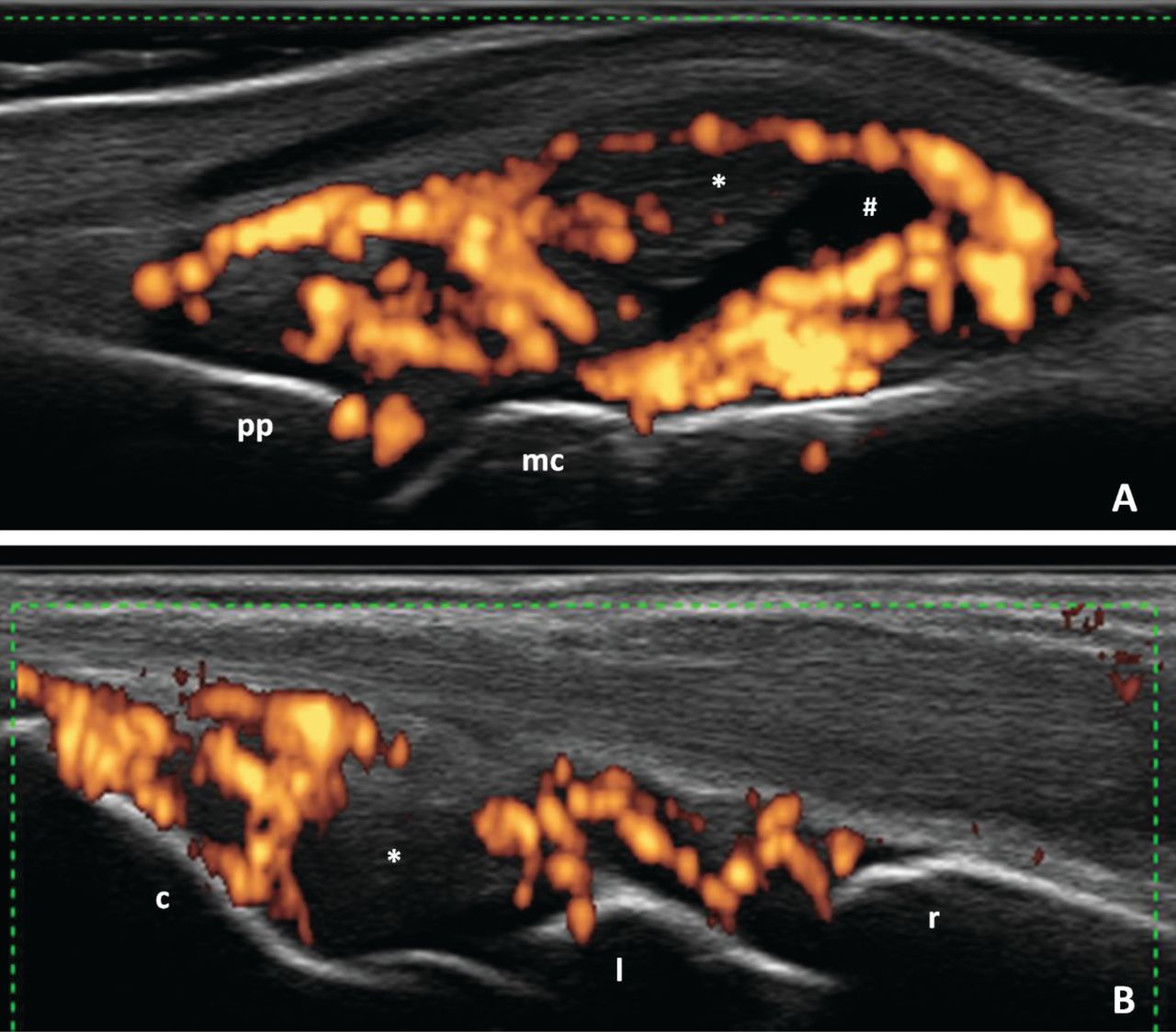
Representative ultrasound images obtained during the study. (A) Metacarpophalangeal joint. Longitudinal dorsal scan. # Joint cavity widening with synovial fluid. * Synovial hypertrophy with increased intraarticular abnormal vascularization by power Doppler. pp: proximal phalanx; mc: metacarpal head. (B) Wrist. Longitudinal dorsal scan. * Joint cavity widening with predominant synovial hypertrophy and increased intraarticular abnormal vascularization by power Doppler. r: radius; l: lunate; c: capitate.

Flares and associated factors

Fifteen of the 54 patients (27.8%) had a disease flare during the followup period of 6 months (Table 2). Eleven out of 15 (73.3%) disease flares were characterized by inflammatory articular involvement, 3/15 (20%) by both inflammatory articular and enthesis involvement, and 1/15 (6.6%) by the presence of dactylitis. Patients who had a flare were significantly older and were more frequently treated with DMARD than those who did not have a flare (p = 0.011 and p = 0.037, respectively; Table 3). Twenty patients (37%) had at least 1 joint with PD grade ≥ 1 at baseline; 13 (65%) of these patients had a disease flare during followup period. Only 2 of the 34 patients (5.9%) without baseline US synovitis with PD grade ≥ 1 had a flare of the disease during followup (RR = 11, 95% CI 2.8–44, p < 0.001). Eleven patients had at least 1 joint with PD grade ≥ 2 at baseline; 8 (72.7%) of these had a disease flare during followup period. Among 43 patients without US synovitis with PD grade ≥ 2, 7 (13.9%) had a disease flare during followup (RR = 4.5, 95% CI 2.0–9.6, p < 0.001; Table 4).

View this table:
  • View inline
  • View popup
Table 2.

Percentage of disease flares according to the different applied remission criteria. Values are n (%; 95% CI) unless otherwise specified.

View this table:
  • View inline
  • View popup
Table 3.

Comparative analysis of patients’ baseline features according to the presence of disease flares during the 6-month followup period. Values are mean (SD) unless otherwise specified.

View this table:
  • View inline
  • View popup
Table 4.

Univariate analysis of baseline PDUS features and their relationship with disease flares during the followup period.

In univariate analysis, no other variable (not even greyscale synovitis) was associated with an increased risk of flare (Table 3).

In multivariate analysis, the only variables that were associated with the occurrence of flares were US synovitis with PD grade ≥ 1 in at least 1 joint (OR 35.5, 95% CI 4.5–283) and the concomitant use of DMARD (OR 12.7, 95% CI 1.03–158).

Diagnostic test properties of the PD signal for detecting flares

Sensitivity and specificity of the presence of intraarticular PD signal at baseline for the detection of flare was 87% (95% CI 59.5–98) and 82% (95% CI 62.5–92.5), respectively. The positive LR was 4.8 (2.4–9.7), while the negative LR was 0.1 (0.4–0.6).

US score and flares

The mean (SD) US score in patients who had a flare was 3 (2.5) and in those without flare was 0.3 (0.9; p < 0.001). The area under the receiver-operating characteristic curve of the US score for the prediction of flare was 0.88 (95% CI 0.77–0.95). An US score ≥ 1 showed the best cutoff point, with a sensitivity of 86.7% and a specificity of 87.2% for predicting flare (positive LR: 6.7, negative LR: 0.1).

DISCUSSION

One of the most important goals in the treatment of patients with PsA is to reach a state of sustained clinical remission. A major challenge is to recognize that patients who are in remission are at an increased risk of experiencing a disease flare. US has been shown to detect subclinical inflammation in patients in clinical remission, but the prognostic value of this feature was not proved until now. Therefore, we evaluated the predictive value of PDUS for short-term flare in patients with PsA in clinical remission.

The percentage of patients with PsA in clinical remission with synovitis detected by PDUS in our cohort (37%) was lower than the one reported by Nguyen, et al from a metaanalysis of patients with RA (44%) in clinical remission20. It was higher than the one reported by Araujo, et al in patients with PsA in clinical remission33. In our cohort of patients with PsA in clinical remission, around a quarter (27.8%) experienced a flare during the 6-month followup period. This was low compared with the high incidence of disease recurrence reported by Araujo, et al (76.9%)33. This difference occurred possibly because in the study by Araujo, et al, the incidence of disease recurrence was evaluated after discontinuation of DMARD treatment, while the majority of patients in our study (47/54) remained with their corresponding DMARD or TNFi therapy during the followup period.

The main result of our study was that US-detected synovitis with PD grade ≥ 1 at baseline in patients with PsA in clinical remission was a strong predictor of flare during short-term followup (RR: 11, 95% CI 2.8–44, p < 0.0001). The presence of US synovitis with positive PD signal despite clinical remission suggests that flares may be related to an incomplete suppression of inflammation, undetectable by the clinical indices used in daily rheumatology practice. Therefore, once the patients have achieved clinical remission, an US examination with PD technique could predict the probability of remaining in remission. A small percentage of patients (5.9%) without PDUS synovitis at baseline showed a flare, so a negative result on PDUS would provide an adequate degree of confidence of sustained remission during short-term followup.

We also found that the use of DMARD was associated with increased risk of disease flares compared with patients not treated with DMARD and with patients receiving biologic therapy. Although there is not a clear explanation for this, our interpretation is that patients not receiving DMARD are patients with mild disease less prone to flare, and patients with biologics are treated more intensively and less likely to flare.

Our study has some limitations. First, the choice of the joints to be evaluated by US was arbitrary and based on only the frequency of joint involvement in PsA and previous studies in patients with RA and PsA. Second, there is no universally accepted definition of flare in PsA. The definition we used was extrapolated from RA and has not been validated in PsA. Third, we did not evaluate by US the presence of enthesitis and/or dactylitis as main features of PsA. This could underestimate the inflammatory involvement detected by PDUS at baseline. Finally, we did not assess the interobserver and intraobserver reliability regarding US findings that could increase the internal validity of our study.

Among patients with PsA in clinical remission, PDUS-detected synovitis was a strong predictor of short-term flare of the disease.

Footnotes

  • Supported by a PANLAR (Pan-American League of Associations of Rheumatology) Prize and by an investigator-originated study grant from Abbvie.

  • Accepted for publication March 22, 2017.

REFERENCES

  1. 1.↵
    1. Alamanos Y,
    2. Voulgari PV,
    3. Drosos AA
    . Incidence and prevalence of psoriatic arthritis: a systematic review. J Rheumatol 2008;35:1354–8.
    OpenUrlAbstract/FREE Full Text
  2. 2.↵
    1. Soriano ER,
    2. Rosa J,
    3. Velozo E,
    4. Schpilberg M,
    5. Imamura PM,
    6. Diaz J,
    7. et al.
    Incidence and prevalence of psoriatic arthritis in Buenos Aires, Argentina: a 6-year health management organization-based study. Rheumatology 2011;50:729–34.
    OpenUrlAbstract/FREE Full Text
  3. 3.↵
    1. Coates LC,
    2. Navarro-Coy N,
    3. Brown SR,
    4. Brown S,
    5. McParland L,
    6. Collier H,
    7. et al.
    The TICOPA protocol (TIght COntrol of Psoriatic Arthritis): a randomised controlled trial to compare intensive management versus standard care in early psoriatic arthritis. BMC Musculoskelet Disord 2013;14:101.
    OpenUrlCrossRefPubMed
  4. 4.↵
    1. Gossec L,
    2. Smolen JS,
    3. Gaujoux-Viala C,
    4. Ash Z,
    5. Marzo-Ortega H,
    6. van der Heijde D,
    7. et al;
    8. European League Against Rheumatism
    . European League Against Rheumatism recommendations for the management of psoriatic arthritis with pharmacological therapies. Ann Rheum Dis 2012;71:4–12.
    OpenUrlAbstract/FREE Full Text
  5. 5.↵
    1. Coates LC,
    2. Kavanaugh A,
    3. Mease PJ,
    4. Soriano ER,
    5. Laura Acosta-Felquer M,
    6. Armstrong AW,
    7. et al.
    Group for Research and Assessment of Psoriasis and Psoriatic Arthritis 2015 Treatment Recommendations for Psoriatic Arthritis. Arthritis Rheumatol 2016;68:1060–71.
    OpenUrl
  6. 6.↵
    1. American Academy of Dermatology Work Group,
    2. Menter A,
    3. Korman NJ,
    4. Elmets CA,
    5. Feldman SR,
    6. Gelfand JM,
    7. et al.
    Guidelines of care for the management of psoriasis and psoriatic arthritis: section 6. Guidelines of care for the treatment of psoriasis and psoriatic arthritis: case-based presentations and evidence-based conclusions. J Am Acad Dermatol 2011;65:137–74.
    OpenUrlCrossRefPubMed
  7. 7.↵
    1. Menter A,
    2. Korman NJ,
    3. Elmets CA,
    4. Feldman SR,
    5. Gelfand JM,
    6. Gordon KB,
    7. et al.
    Guidelines of care for the management of psoriasis and psoriatic arthritis: section 4. Guidelines of care for the management and treatment of psoriasis with traditional systemic agents. J Am Acad Dermatol 2009;61:451–85.
    OpenUrlCrossRefPubMed
  8. 8.↵
    1. Gottlieb A,
    2. Korman NJ,
    3. Gordon KB,
    4. Feldman SR,
    5. Lebwohl M,
    6. Koo JY,
    7. et al.
    Guidelines of care for the management of psoriasis and psoriatic arthritis: Section 2. Psoriatic arthritis: overview and guidelines of care for treatment with an emphasis on the biologics. J Am Acad Dermatol 2008;58:851–64.
    OpenUrlCrossRefPubMed
  9. 9.↵
    1. Gladman DD,
    2. Mease PJ
    . Towards international guidelines for the management of psoriatic arthritis. J Rheumatol 2006;33:1228–30.
    OpenUrlFREE Full Text
  10. 10.↵
    1. Smolen JS,
    2. Braun J,
    3. Dougados M,
    4. Emery P,
    5. Fitzgerald O,
    6. Helliwell P,
    7. et al.
    Treating spondyloarthritis, including ankylosing spondylitis and psoriatic arthritis, to target: recommendations of an international task force. Ann Rheum Dis 2014;73:6–16.
    OpenUrlAbstract/FREE Full Text
  11. 11.↵
    1. Soriano ER
    . Defining remission in psoriatic arthritis: are we getting closer? J Rheumatol 2015;42:907–8.
    OpenUrlFREE Full Text
  12. 12.↵
    1. Acosta Felquer ML,
    2. Ferreyra Garrott L,
    3. Marin J,
    4. Catay E,
    5. Scolnik M,
    6. Scaglioni V,
    7. et al.
    Remission criteria and activity indices in psoriatic arthritis. Clin Rheumatol 2014;33:1323–30.
    OpenUrlCrossRefPubMed
  13. 13.↵
    1. Balint PV,
    2. Kane D,
    3. Wilson H,
    4. McInnes IB,
    5. Sturrock RD
    . Ultrasonography of entheseal insertions in the lower limb in spondyloarthropathy. Ann Rheum Dis 2002;61:905–10.
    OpenUrlAbstract/FREE Full Text
  14. 14.↵
    1. D’Agostino MA,
    2. Said-Nahal R,
    3. Hacquard-Bouder C,
    4. Brasseur JL,
    5. Dougados M,
    6. Breban M
    . Assessment of peripheral enthesitis in the spondylarthropathies by ultrasonography combined with power Doppler: a cross-sectional study. Arthritis Rheum 2003;48:523–33.
    OpenUrlCrossRefPubMed
  15. 15.↵
    1. Ruta S,
    2. Gutierrez M,
    3. Pena C,
    4. Garcia M,
    5. Arturi A,
    6. Filippucci E,
    7. et al.
    Prevalence of subclinical enthesopathy in patients with spondyloarthropathy: an ultrasound study. J Clin Rheumatol 2011;17:18–22.
    OpenUrlPubMed
  16. 16.↵
    1. Naredo E,
    2. Möller I,
    3. de Miguel E,
    4. Batlle-Gualda E,
    5. Acebes C,
    6. Brito E,
    7. et al;
    8. Ultrasound School of the Spanish Society of Rheumatology and Spanish ECO-APs Group
    . High prevalence of ultrasonographic synovitis and enthesopathy in patients with psoriasis without psoriatic arthritis: a prospective case-control study. Rheumatology 2011;50:1838–48.
    OpenUrlAbstract/FREE Full Text
  17. 17.↵
    1. Husic R,
    2. Gretler J,
    3. Felber A,
    4. Graninger WB,
    5. Duftner C,
    6. Hermann J,
    7. et al.
    Disparity between ultrasound and clinical findings in psoriatic arthritis. Ann Rheum Dis 2014;73:1529–36.
    OpenUrlAbstract/FREE Full Text
  18. 18.↵
    1. Brown AK,
    2. Conaghan PG,
    3. Karim Z,
    4. Quinn MA,
    5. Ikeda K,
    6. Peterfy CG,
    7. et al.
    An explanation for the apparent dissociation between clinical remission and continued structural deterioration in rheumatoid arthritis. Arthritis Rheum 2008;58:2958–67.
    OpenUrlCrossRefPubMed
  19. 19.↵
    1. Saleem B,
    2. Brown AK,
    3. Quinn M,
    4. Karim Z,
    5. Hensor EM,
    6. Conaghan P,
    7. et al.
    Can flare be predicted in DMARD treated RA patients in remission, and is it important? A cohort study. Ann Rheum Dis 2012;71:1316–21.
    OpenUrlAbstract/FREE Full Text
  20. 20.↵
    1. Nguyen H,
    2. Ruyssen-Witrand A,
    3. Gandjbakhch F,
    4. Constantin A,
    5. Foltz V,
    6. Cantagrel A
    . Prevalence of ultrasound-detected residual synovitis and risk of relapse and structural progression in rheumatoid arthritis patients in clinical remission: a systematic review and meta-analysis. Rheumatology 2014;53:2110–8.
    OpenUrlAbstract/FREE Full Text
  21. 21.↵
    1. Freeston JE,
    2. Coates LC,
    3. Nam JL,
    4. Moverley AR,
    5. Hensor EM,
    6. Wakefield RJ,
    7. et al.
    Is there subclinical synovitis in early psoriatic arthritis? A clinical comparison with gray-scale and power Doppler ultrasound. Arthritis Care Res 2014;66:432–9.
    OpenUrlCrossRef
  22. 22.↵
    1. Taylor W,
    2. Gladman D,
    3. Helliwell P,
    4. Marchesoni A,
    5. Mease P,
    6. Mielants H;
    7. CASPAR Study Group
    . Classification criteria for psoriatic arthritis: development of new criteria from a large international study. Arthritis Rheum 2006;54:2665–73.
    OpenUrlCrossRefPubMed
  23. 23.↵
    1. Coates LC,
    2. Helliwell PS
    . Validation of minimal disease activity criteria for psoriatic arthritis using interventional trial data. Arthritis Care Res 2010;62:965–9.
    OpenUrlCrossRef
  24. 24.↵
    1. Prevoo ML,
    2. van ‘t Hof MA,
    3. Kuper HH,
    4. van Leeuwen MA,
    5. van de Putte LB,
    6. van Riel PL
    . Modified disease activity scores that include twenty-eight-joint counts. Development and validation in a prospective longitudinal study of patients with rheumatoid arthritis. Arthritis Rheum 1995;38:44–8.
    OpenUrlCrossRefPubMed
  25. 25.↵
    1. Citera G,
    2. Arriola MS,
    3. Maldonado-Cocco JA,
    4. Rosemffet MG,
    5. Sanchez MM,
    6. Goñi MA,
    7. et al.
    Validation and crosscultural adaptation of an Argentine Spanish version of the health assessment questionnaire disability index. J Clin Rheumatol 2004;10:110–5.
    OpenUrlCrossRefPubMed
  26. 26.↵
    1. Backhaus M,
    2. Burmester GR,
    3. Gerber T,
    4. Grassi W,
    5. Machold KP,
    6. Swen WA,
    7. et al;
    8. Working Group for Musculoskeletal Ultrasound in the EULAR Standing Committee on International Clinical Studies including Therapeutic Trials
    . Guidelines for musculoskeletal ultrasound in rheumatology. Ann Rheum Dis 2001;60:641–9.
    OpenUrlFREE Full Text
  27. 27.↵
    1. Wakefield RJ,
    2. Balint PV,
    3. Szkudlarek M,
    4. Filippucci E,
    5. Backhaus M,
    6. D’Agostino MA,
    7. et al.
    Musculoskeletal ultrasound including definitions for ultrasonographic pathology. J Rheumatol 2005;32:2485–7.
    OpenUrlAbstract/FREE Full Text
  28. 28.↵
    1. Gutierrez M,
    2. Filippucci E,
    3. Salaffi F,
    4. Di Geso L,
    5. Grassi W
    . Differential diagnosis between rheumatoid arthritis and psoriatic arthritis: the value of ultrasound findings at metacarpophalangeal joints level. Ann Rheum Dis 2011;70:1111–4.
    OpenUrlAbstract/FREE Full Text
  29. 29.↵
    1. Naredo E,
    2. Bonilla G,
    3. Gamero F,
    4. Uson J,
    5. Carmona L,
    6. Laffon A
    . Assessment of inflammatory activity in rheumatoid arthritis: a comparative study of clinical evaluation with grey scale and power Doppler ultrasonography. Ann Rheum Dis 2005;64:375–81.
    OpenUrlAbstract/FREE Full Text
  30. 30.↵
    1. Naredo E,
    2. Gamero F,
    3. Bonilla G,
    4. Uson J,
    5. Carmona L,
    6. Laffon A
    . Ultrasonographic assessment of inflammatory activity in rheumatoid arthritis: comparison of extended versus reduced joint evaluation. Clin Exp Rheumatol 2005;23:881–4.
    OpenUrlPubMed
  31. 31.↵
    1. Naredo E,
    2. Möller I,
    3. Cruz A,
    4. Carmona L,
    5. Garrido J
    . Power Doppler ultrasonographic monitoring of response to anti-tumor necrosis factor therapy in patients with rheumatoid arthritis. Arthritis Rheum 2008;58:2248–56.
    OpenUrlCrossRefPubMed
  32. 32.↵
    1. Bingham CO 3rd,
    2. Pohl C,
    3. Woodworth TG,
    4. Hewlett SE,
    5. May JE,
    6. Rahman MU,
    7. et al.
    Developing a standardized definition for disease “flare” in rheumatoid arthritis (OMERACT 9 Special Interest Group). J Rheumatol 2009;36:2335–41.
    OpenUrlAbstract/FREE Full Text
  33. 33.↵
    1. Araujo EG,
    2. Finzel S,
    3. Englbrecht M,
    4. Schreiber DA,
    5. Faustini F,
    6. Hueber A,
    7. et al.
    High incidence of disease recurrence after discontinuation of disease-modifying antirheumatic drug treatment in patients with psoriatic arthritis in remission. Ann Rheum Dis 2015;74:655–60.
    OpenUrlAbstract/FREE Full Text
View Abstract
PreviousNext
Back to top

In this issue

The Journal of Rheumatology
Vol. 44, Issue 7
1 Jul 2017
  • Table of Contents
  • Table of Contents (PDF)
  • Index by Author
  • Editorial Board (PDF)
Print
Download PDF
Article Alerts
Sign In to Email Alerts with your Email Address
Email Article

Thank you for your interest in spreading the word about The Journal of Rheumatology.

NOTE: We only request your email address so that the person you are recommending the page to knows that you wanted them to see it, and that it is not junk mail. We do not capture any email address.

Enter multiple addresses on separate lines or separate them with commas.
Utility of Power Doppler Ultrasound–detected Synovitis for the Prediction of Short-term Flare in Psoriatic Patients with Arthritis in Clinical Remission
(Your Name) has forwarded a page to you from The Journal of Rheumatology
(Your Name) thought you would like to see this page from the The Journal of Rheumatology web site.
CAPTCHA
This question is for testing whether or not you are a human visitor and to prevent automated spam submissions.
Citation Tools
Utility of Power Doppler Ultrasound–detected Synovitis for the Prediction of Short-term Flare in Psoriatic Patients with Arthritis in Clinical Remission
Santiago Ruta, Josefina Marin, Maria Laura Acosta Felquer, Leandro Ferreyra-Garrot, Javier Rosa, Ricardo García-Monaco, Enrique R. Soriano
The Journal of Rheumatology Jul 2017, 44 (7) 1018-1023; DOI: 10.3899/jrheum.161347

Citation Manager Formats

  • BibTeX
  • Bookends
  • EasyBib
  • EndNote (tagged)
  • EndNote 8 (xml)
  • Medlars
  • Mendeley
  • Papers
  • RefWorks Tagged
  • Ref Manager
  • RIS
  • Zotero

 Request Permissions

Share
Utility of Power Doppler Ultrasound–detected Synovitis for the Prediction of Short-term Flare in Psoriatic Patients with Arthritis in Clinical Remission
Santiago Ruta, Josefina Marin, Maria Laura Acosta Felquer, Leandro Ferreyra-Garrot, Javier Rosa, Ricardo García-Monaco, Enrique R. Soriano
The Journal of Rheumatology Jul 2017, 44 (7) 1018-1023; DOI: 10.3899/jrheum.161347
del.icio.us logo Twitter logo Facebook logo  logo Mendeley logo
  • Tweet Widget
  •  logo
Bookmark this article

Jump to section

  • Article
    • Abstract
    • MATERIALS AND METHODS
    • RESULTS
    • DISCUSSION
    • Footnotes
    • REFERENCES
  • Figures & Data
  • Info & Metrics
  • References
  • PDF

Keywords

ULTRASONOGRAPHY
PSORIATIC ARTHRITIS
REMISSION
SYNOVITIS

Related Articles

Cited By...

More in this TOC Section

  • Degenerative Disc Disease in Young Adults With Psoriatic Arthritis
  • Risk of Mortality of People With Psoriasis and Psoriatic Arthritis in Taiwan: A Nationwide Cohort Study
  • Tight Control and Radiological Progression: The Radiographic Outcomes of the TICOPA Study
Show more Psoriatic Arthritis

Similar Articles

Keywords

  • ULTRASONOGRAPHY
  • psoriatic arthritis
  • remission
  • SYNOVITIS

Content

  • First Release
  • Current
  • Archives
  • Collections
  • Audiovisual Rheum
  • COVID-19 and Rheumatology

Resources

  • Guide for Authors
  • Submit Manuscript
  • Author Payment
  • Reviewers
  • Advertisers
  • Classified Ads
  • Reprints and Translations
  • Permissions
  • Meetings
  • FAQ
  • Policies

Subscribers

  • Subscription Information
  • Purchase Subscription
  • Your Account
  • Terms and Conditions

More

  • About Us
  • Contact Us
  • My Alerts
  • My Folders
  • Privacy/GDPR Policy
  • RSS Feeds
The Journal of Rheumatology
The content of this site is intended for health care professionals.
Copyright © 2025 by The Journal of Rheumatology Publishing Co. Ltd.
Print ISSN: 0315-162X; Online ISSN: 1499-2752
Powered by HighWire