
Main menu

  • Home
  • Content
    • First Release
    • Current
    • Archives
    • Collections
    • Audiovisual Rheum
    • COVID-19 and Rheumatology
    • 50th Volume Reprints
  • Resources
    • Guide for Authors
    • Submit Manuscript
    • Payment
    • Reviewers
    • Advertisers
    • Classified Ads
    • Reprints and Translations
    • Permissions
    • Meetings
    • FAQ
    • Policies
  • Subscribers
    • Subscription Information
    • Purchase Subscription
    • Your Account
    • Terms and Conditions
  • About Us
    • About Us
    • Editorial Board
    • Letter from the Editor
    • Duncan A. Gordon Award
    • Privacy/GDPR Policy
    • Accessibility
  • Contact Us
  • JRheum Supplements
  • Services

User menu

  • My Cart
  • Log In

Search

  • Advanced search
The Journal of Rheumatology
  • JRheum Supplements
  • Services
  • My Cart
  • Log In
The Journal of Rheumatology

Advanced Search

  • Home
  • Content
    • First Release
    • Current
    • Archives
    • Collections
    • Audiovisual Rheum
    • COVID-19 and Rheumatology
    • 50th Volume Reprints
  • Resources
    • Guide for Authors
    • Submit Manuscript
    • Payment
    • Reviewers
    • Advertisers
    • Classified Ads
    • Reprints and Translations
    • Permissions
    • Meetings
    • FAQ
    • Policies
  • Subscribers
    • Subscription Information
    • Purchase Subscription
    • Your Account
    • Terms and Conditions
  • About Us
    • About Us
    • Editorial Board
    • Letter from the Editor
    • Duncan A. Gordon Award
    • Privacy/GDPR Policy
    • Accessibility
  • Contact Us
  • Follow jrheum on Twitter
  • Visit jrheum on Facebook
  • Follow jrheum on LinkedIn
  • Follow jrheum on YouTube
  • Follow jrheum on Instagram
  • Follow jrheum on RSS
Research ArticleSpondyloarthritis

Are Ultrasound Findings Similar in Patients with Axial Spondyloarthritis and in Athlete Entheses?

Marie-Alix Lanfranchi, Olivier Leluc, Alice Tavano, Cécile Wormser, Sophie Morange, Christophe Chagnaud, Vincent Pradel, Pierre Lafforgue and Thao Pham
The Journal of Rheumatology May 2017, 44 (5) 609-612; DOI: https://doi.org/10.3899/jrheum.160715
Marie-Alix Lanfranchi
From the Assistance Publique—Hôpitaux de Marseille (APHM), CHU Sainte Marguerite, Rheumatology Department, Aix-Marseille University; CHU Conception, Radiology Department, Clinical Investigation Center; CHU Sainte Marguerite, Public Health Department; Aix-Marseille University, Marseille, France.
  • Find this author on Google Scholar
  • Find this author on PubMed
  • Search for this author on this site
Olivier Leluc
  • Find this author on Google Scholar
  • Find this author on PubMed
  • Search for this author on this site
Alice Tavano
  • Find this author on Google Scholar
  • Find this author on PubMed
  • Search for this author on this site
Cécile Wormser
  • Find this author on Google Scholar
  • Find this author on PubMed
  • Search for this author on this site
Sophie Morange
  • Find this author on Google Scholar
  • Find this author on PubMed
  • Search for this author on this site
Christophe Chagnaud
  • Find this author on Google Scholar
  • Find this author on PubMed
  • Search for this author on this site
Vincent Pradel
  • Find this author on Google Scholar
  • Find this author on PubMed
  • Search for this author on this site
Pierre Lafforgue
  • Find this author on Google Scholar
  • Find this author on PubMed
  • Search for this author on this site
Thao Pham
  • Find this author on Google Scholar
  • Find this author on PubMed
  • Search for this author on this site
  • For correspondence: thao.pham@ap-hm.fr
  • Article
  • Figures & Data
  • Info & Metrics
  • References
  • PDF
  • eLetters
PreviousNext
Loading

Abstract

Objective. Enthesitis is the spondyloarthritis (SpA) landmark, but can also be seen after entheses overuse, such as during intensive sport.

Methods. We aimed to compare entheses ultrasound (US) findings in a prospective cross-sectional study of 30 axial SpA cases, 30 athletes, and 29 controls.

Results. Mean (SD) MAdrid Sonographic Enthesis Index (MASEI) score was 26.3 (13), 12.2 (7), and 10.4 (6) in patients with SpA, athletes, and non-athlete control groups, respectively (p < 0.0001).

Conclusion. The MASEI score was significantly higher in patients with SpA compared with healthy controls, athletes, and non-athletes, and can be of value to distinguish SpA from healthy subjects, whatever their physical activity.

Key Indexing Terms:
  • SPONDYLOARTHROPATHY
  • ULTRASONOGRAPHY
  • ENTHESIS
  • ATHLETE

Spondyloarthritis (SpA) is characterized by inflammatory and structural changes in the entheses.

In a cross-sectional study, D’Agostino, et al1 compared ultrasound (US) examination of the major entheses in 164 patients with SpA and 64 control subjects [34 mechanical low back pain and 30 rheumatoid arthritis (RA)]. At least 1 enthesitis was observed in 161 of the 164 patients with SpA (98%) and in 33 of the 64 control patients (52%)1. US enthesitis was most commonly distributed in the distal portion of the lower limbs, irrespective of clinical symptoms.

Similarly, Ruta, et al confirmed the high prevalence of asymptomatic lower-limb enthesitis (almost 60% of subclinical US enthesitis) in 60 patients with SpA compared with patients with RA and healthy subjects2.

However, enthesitis is not only observed in SpA, but can also be seen after entheses overuse, such as during intensive sport.

Several studies have demonstrated that sport can lead to structural changes of the entheses3,4,5,6,7. Enthesis vascularization detected with power Doppler US (PDUS) can also be modified by sport. Boesen, et al observed a significant increase in entheses vascularization as indicated by color Doppler activity in both the Achilles and patella tendons after loading during high-level badminton matches6. Similarly, 634 asymptomatic runners were tested for Achilles tendon thickness, vascularization, and structural abnormalities using a high-resolution PDUS device8. Their mean training volume was 4.0 ± 2.5 h per week. Neovascularization was observed on PDUS in 38%.

The MAdrid Sonographic Enthesis Index (MASEI) is an US score developed by de Miguel, et al for SpA diagnosis and assessment9. A score ≥ 18 performed well in discriminating patients with SpA from healthy controls. The sensitivity, specificity, positive, and negative likelihood ratios (LR+, LR–) were 83.3%, 82.8%, 4.8%, and 0.2%, respectively. This cutoff point was established with an a priori sedentary control group.

Our aim was to compare US findings of entheses among 3 groups: patients with axial SpA (axSpA), athletes, and non-athlete healthy controls, and to assess the discriminant ability of the MASEI score.

MATERIALS AND METHODS

Patients

We conducted a cross-sectional study including 30 patients with axSpA, 30 athletes, and 29 non-athlete healthy controls.

All patients with axSpA fulfilled the 2009 Assessment of Spondyloarthritis international Society (ASAS) criteria10. Athletes were defined as subjects playing a sport resulting in a strain on the lower limbs, such as running or soccer, ≥ 6 h per week. Subjects were recruited among affiliates of a local athletics club. Non-athlete healthy controls had to play a sport < 1 h per week. They were recruited from university students and hospital personnel.

To participate in the study, all healthy subjects, athletes, and non-athletes had to have no musculoskeletal disorder, particularly heel or back pain. A history of surgery and recent corticosteroid infiltration of the heel or the ankle were also noninclusion criteria.

The study was conducted according to the Declaration of Helsinki and French legislation. Before study inclusion, each subject gave informed consent for research use and publication of the results. The local ethics committee CPP Sud Méditerranée I (13-44) approved the study (NCT02038426).

Clinical and biochemical evaluation

Clinical, biological, and imaging evaluations were performed on the same day for patients with SpA and healthy subjects, athletes, and non-athletes.

We collected the following demographic data: age, sex, weight, medical and surgical history, and ongoing treatments (including nonsteroidal anti-inflammatory drugs and oral corticosteroids). As part of the consultation, the clinician (MAL) completed a standardized questionnaire covering the presence or absence of clinical features included in the ASAS classification criteria for SpA, and performed a standardized examination of the heels. Enthesis was defined if the patient reported heel pain during 1 of the following 3 tests: passive stretching, contraction against a resistance exerted by the examiner, and/or Achilles tendon palpation.

Scores on the Bath Ankylosing Spondylitis Disease Activity Index (BASDAI) and the Ankylosing Spondylitis Disease Activity Score (ASDAS) were collected for the patients with SpA.

Blood samples were analyzed for HLA-B27 and C-reactive protein (CRP). Levels of CRP > 5 mg/l were considered elevated. A pregnancy urinary test was performed for all women of childbearing age.

Radiographs, an anteroposterior view of the pelvis and 1 oblique view of each sacroiliac joint, were graded using the New York classification scale. We also assessed profile calcaneus radiographs, searching for heel spur.

US evaluation

The physicians who performed the clinical examination and the radiologists performing US scanning were blinded to one another’s results. Patients with SpA and healthy subjects had at least a 15-min rest in the room before US evaluation. US scanning was performed by 3 trained radiologists (OL, AT, CW) using a Toshiba Aplio 500 equipped with linear transducer, frequency of 6–18 MHz. The following enthesis sites included in the MASEI scoring9 were scanned bilaterally, using both greyscale and PD: proximal plantar fascia, distal Achilles tendon, distal and proximal patellar tendon insertion, distal quadriceps tendon, and distal brachial triceps tendon. Each tendon was scanned in both longitudinal and transverse planes, and the scan images were stored. At each site, the following elementary lesions were assessed: bursitis, calcification, erosion, PD, thickening of tendon, and structural change.

Interobserver reliability was assessed by reading 20 images of the study subjects, randomly selected, based on a reading consensus for the MASEI positivity scoring ≥ 18/136.

Analysis

Continuous data are described by mean (SD) and categorical variables and expressed as frequencies and percentages. To compare groups, we used the chi-square test (or Fisher’s exact test when chi-square was not possible) and 1-way ANOVA (depending on categorical/continuous variables) with Bonferroni correction for posthoc tests. Interreader reliability was assessed with Cohen’s κ. SPSS 17.0 version was used for management and statistical analysis.

RESULTS

Patient and control demographic and clinical characteristics are shown in Table 1.

View this table:
  • View inline
  • View popup
Table 1.

Patients and control characteristics. Values are n (%) unless otherwise specified.

All of the patients with SpA fulfilled the axial 2009 ASAS criteria10. Their mean disease duration was 9.6 years. BASDAI and ASDAS mean (SD) scores were 3.14 ± 1.9 and 1.78 ± 1.01, respectively.

No athletes and 3 non-athlete controls (10%) reported chronic low back pain history. Heel pain history was reported in 56.3%, 20.0%, and 0.0% in the SpA, athlete, and non-athlete groups, respectively. Achilles tendon palpation was painful in 23.3%, 6.9%, and 0.0% of the SpA, athlete, and non-athlete subjects, respectively.

Difference in US enthesis scores among the groups

The mean MASEI and individual subitem scores were significantly higher in the patients with SpA than in both healthy control groups (Figure 1). There was no significant difference between the athlete and non-athlete healthy groups. The percentage of patients with a MASEI score ≥ 18 was 70.0%, 16.7%, and 13.7% in the patients with SpA, athletes, and non-athlete control groups, respectively. Each elementary lesion assessed with the MASEI score was significantly more frequent in the patients with SpA than in the controls, except for bursitis (Table 2). The interreader reliability κ value was 0.68.

Figure 1.
  • Download figure
  • Open in new tab
  • Download powerpoint
Figure 1.

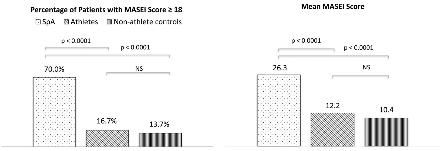
Percentage of patients with a MASEI score ≥ 18 and mean MASEI score in patients with SpA, athletes, and non-athlete controls. MASEI: MAdrid Sonographic Enthesis Index; SpA: spondyloarthritis; NS: not significant.

View this table:
  • View inline
  • View popup
Table 2.

MASEI elementary lesions in patients with SpA, athletes, and non-athlete controls. Values are mean (SD).

Correlation between heel clinical and US findings

The prevalence of Achilles enthesitis was 23%, 3%, and 0% (p < 0.006) in the SpA, athlete, and non-athlete groups, respectively. No correlation was found in any of the 3 groups between heel enthesis and the MASEI total score, or with PD+ at the calcaneal entheses. In the SpA group, no significant correlation between the BASDAI or ASDAS and MASEI score was found.

DISCUSSION

To our knowledge, no study has ever compared the prevalence of US enthesitis in athletes playing a sport with strains in the lower limbs with those observed in patients with SpA.

In our study, we aimed to analyze the usefulness of the MASEI score in differentiating patients with axSpA from healthy controls, whatever their daily physical/sports activity. We chose the MASEI score because it is the only validated scoring system, including a PD assessment, with a proposed cutoff point9,11. In their articles, de Miguel, et al did not describe the physical activity of the controls.

In our study, the MASEI score was significantly higher in patients with SpA compared with the healthy controls, athletes, and non-athletes. Even if the MASEI score was somewhat higher in the athlete control group than in the non-athlete group, the difference was not significant. These results confirm the diagnostic accuracy of the MASEI score, whatever the physical activity of the subjects.

One of our concerns was the prevalence of Achilles enthesitis in the athlete group. We demonstrated a clear difference between the patients with SpA and the athletes (23% vs 3%, p = 0.052). There was, therefore, a significant proportion of PD+ Achilles tendon enthesitis in patients with SpA, but we were surprised by the low prevalence in the athlete group despite a state of regular overuse. Indeed, we previously showed that a simple walking exercise significantly increases the MASEI score12. These findings suggest that the increase could be reversible and probably related to mechanical stress13. They also emphasize the need to propose systematic rest before any US assessment of enthesis.

The MASEI score was significantly higher in patients with SpA compared with healthy controls, athletes, and non-athletes. The MASEI appears to be of value to distinguish SpA from healthy subjects, whatever their physical activity.

Acknowledgment

The authors are grateful to Peter Tucker for his careful reading of the manuscript.

Footnotes

  • Supported by a grant from APHM, France.

  • Accepted for publication January 4, 2017.

REFERENCES

  1. 1.↵
    1. D’Agostino MA,
    2. Said-Nahal R,
    3. Hacquard-Bouder C,
    4. Brasseur JL,
    5. Dougados M,
    6. Breban M
    . Assessment of peripheral enthesitis in the spondylarthropathies by ultrasonography combined with power Doppler: a cross-sectional study. Arthritis Rheum 2003;48:523–33.
    OpenUrlCrossRefPubMed
  2. 2.↵
    1. Ruta S,
    2. Gutierrez M,
    3. Pena C,
    4. Garcia M,
    5. Arturi A,
    6. Filippucci E,
    7. et al.
    Prevalence of subclinical enthesopathy in patients with spondyloarthropathy: an ultrasound study. J Clin Rheumatol 2011;17:18–22.
    OpenUrlPubMed
  3. 3.↵
    1. Comin J,
    2. Cook JL,
    3. Malliaras P,
    4. McCormack M,
    5. Calleja M,
    6. Clarke A,
    7. et al.
    The prevalence and clinical significance of sonographic tendon abnormalities in asymptomatic ballet dancers: a 24-month longitudinal study. Br J Sports Med 2013;47:89–92.
    OpenUrlAbstract/FREE Full Text
  4. 4.↵
    1. van Drongelen S,
    2. Boninger ML,
    3. Impink BG,
    4. Khalaf T
    . Ultrasound imaging of acute biceps tendon changes after wheelchair sports. Arch Phys Med Rehabil 2007;88:381–5.
    OpenUrlCrossRefPubMed
  5. 5.↵
    1. Major NM,
    2. Helms CA
    . MR imaging of the knee: findings in asymptomatic collegiate basketball players. AJR Am J Roentgenol 2002;179:641–4.
    OpenUrlCrossRefPubMed
  6. 6.↵
    1. Boesen AP,
    2. Boesen MI,
    3. Koenig MJ,
    4. Bliddal H,
    5. Torp-Pedersen S,
    6. Langberg H
    . Evidence of accumulated stress in Achilles and anterior knee tendons in elite badminton players. Knee Surg Sports Traumatol Arthrosc 2011;19:30–7.
    OpenUrlCrossRefPubMed
  7. 7.↵
    1. Emerson C,
    2. Morrissey D,
    3. Perry M,
    4. Jalan R
    . Ultrasonographically detected changes in Achilles tendons and self reported symptoms in elite gymnasts compared with controls—an observational study. Man Ther 2010;15:37–42.
    OpenUrlCrossRefPubMed
  8. 8.↵
    1. Hirschmüller A,
    2. Frey V,
    3. Konstantinidis L,
    4. Baur H,
    5. Dickhuth HH,
    6. Südkamp NP,
    7. et al.
    Prognostic value of Achilles tendon Doppler sonography in asymptomatic runners. Med Sci Sports Exerc 2012;44:199–205.
    OpenUrlCrossRefPubMed
  9. 9.↵
    1. de Miguel E,
    2. Cobo T,
    3. Muñoz-Fernández S,
    4. Naredo E,
    5. Usón J,
    6. Acebes JC,
    7. et al.
    Validity of enthesis ultrasound assessment in spondyloarthropathy. Ann Rheum Dis 2009;68:169–74.
    OpenUrlAbstract/FREE Full Text
  10. 10.↵
    1. Rudwaleit M,
    2. van der Heijde D,
    3. Landewé R,
    4. Listing J,
    5. Akkoc N,
    6. Brandt J,
    7. et al.
    The development of Assessment of SpondyloArthritis international Society classification criteria for axial spondyloarthritis (part II): validation and final selection. Ann Rheum Dis 2009;68:777–83.
    OpenUrlAbstract/FREE Full Text
  11. 11.↵
    1. de Miguel E,
    2. Muñoz-Fernández S,
    3. Castillo C,
    4. Cobo-Ibáñez T,
    5. Martín-Mola E
    . Diagnostic accuracy of enthesis ultrasound in the diagnosis of early spondyloarthritis. Ann Rheum Dis 2011;7:434–9.
    OpenUrl
  12. 12.↵
    1. Méric JC,
    2. Grandgeorge Y,
    3. Lotito G,
    4. Pham T
    . Walking before an ultrasound assessment increases the enthesis score significantly. J Rheumatol 2011;38:961.
    OpenUrlFREE Full Text
  13. 13.↵
    1. Malliaras P,
    2. Chan O,
    3. Simran G,
    4. Martinez de Albornoz P,
    5. Morrissey D,
    6. Maffulli N
    . Doppler ultrasound signal in Achilles tendinopathy reduces immediately after activity. Int J Sports Med 2012;33:480–4.
    OpenUrlPubMed
PreviousNext
Back to top

In this issue

The Journal of Rheumatology
Vol. 44, Issue 5
1 May 2017
  • Table of Contents
  • Table of Contents (PDF)
  • Index by Author
  • Editorial Board (PDF)
Print
Download PDF
Article Alerts
Sign In to Email Alerts with your Email Address
Email Article

Thank you for your interest in spreading the word about The Journal of Rheumatology.

NOTE: We only request your email address so that the person you are recommending the page to knows that you wanted them to see it, and that it is not junk mail. We do not capture any email address.

Enter multiple addresses on separate lines or separate them with commas.
Are Ultrasound Findings Similar in Patients with Axial Spondyloarthritis and in Athlete Entheses?
(Your Name) has forwarded a page to you from The Journal of Rheumatology
(Your Name) thought you would like to see this page from the The Journal of Rheumatology web site.
CAPTCHA
This question is for testing whether or not you are a human visitor and to prevent automated spam submissions.
Citation Tools
Are Ultrasound Findings Similar in Patients with Axial Spondyloarthritis and in Athlete Entheses?
Marie-Alix Lanfranchi, Olivier Leluc, Alice Tavano, Cécile Wormser, Sophie Morange, Christophe Chagnaud, Vincent Pradel, Pierre Lafforgue, Thao Pham
The Journal of Rheumatology May 2017, 44 (5) 609-612; DOI: 10.3899/jrheum.160715

Citation Manager Formats

  • BibTeX
  • Bookends
  • EasyBib
  • EndNote (tagged)
  • EndNote 8 (xml)
  • Medlars
  • Mendeley
  • Papers
  • RefWorks Tagged
  • Ref Manager
  • RIS
  • Zotero

 Request Permissions

Share
Are Ultrasound Findings Similar in Patients with Axial Spondyloarthritis and in Athlete Entheses?
Marie-Alix Lanfranchi, Olivier Leluc, Alice Tavano, Cécile Wormser, Sophie Morange, Christophe Chagnaud, Vincent Pradel, Pierre Lafforgue, Thao Pham
The Journal of Rheumatology May 2017, 44 (5) 609-612; DOI: 10.3899/jrheum.160715
Reddit logo Twitter logo Facebook logo Mendeley logo
  • Tweet Widget
  • Facebook Like
  • Google Plus One
Bookmark this article

Jump to section

  • Article
    • Abstract
    • MATERIALS AND METHODS
    • RESULTS
    • DISCUSSION
    • Acknowledgment
    • Footnotes
    • REFERENCES
  • Figures & Data
  • Info & Metrics
  • References
  • PDF
  • eLetters

Keywords

SPONDYLOARTHROPATHY
ULTRASONOGRAPHY
ENTHESIS
ATHLETE

Related Articles

Cited By...

More in this TOC Section

  • Prevalence and Associated Factors of Facet Joint Ankylosis in Patients With Axial Spondyloarthritis
  • Clinimetric Validation of the Assessment of Spondyloarthritis International Society Health Index in Patients With Radiographic Axial Spondyloarthritis in Ixekizumab Trials
  • Real-World Retention and Clinical Effectiveness of Secukinumab for Axial Spondyloarthritis: Results From the Canadian Spondyloarthritis Research Network
Show more Spondyloarthritis

Similar Articles

Keywords

  • spondyloarthropathy
  • ultrasonography
  • enthesis
  • ATHLETE

Content

  • First Release
  • Current
  • Archives
  • Collections
  • Audiovisual Rheum
  • COVID-19 and Rheumatology

Resources

  • Guide for Authors
  • Submit Manuscript
  • Author Payment
  • Reviewers
  • Advertisers
  • Classified Ads
  • Reprints and Translations
  • Permissions
  • Meetings
  • FAQ
  • Policies

Subscribers

  • Subscription Information
  • Purchase Subscription
  • Your Account
  • Terms and Conditions

More

  • About Us
  • Contact Us
  • My Alerts
  • My Folders
  • Privacy/GDPR Policy
  • RSS Feeds
The Journal of Rheumatology
The content of this site is intended for health care professionals.
Copyright © 2022 by The Journal of Rheumatology Publishing Co. Ltd.
Print ISSN: 0315-162X; Online ISSN: 1499-2752
Powered by HighWire