
Main menu

  • Home
  • Content
    • First Release
    • Current
    • Archives
    • Collections
    • Audiovisual Rheum
    • COVID-19 and Rheumatology
  • Resources
    • Guide for Authors
    • Submit Manuscript
    • Payment
    • Reviewers
    • Advertisers
    • Classified Ads
    • Reprints and Translations
    • Permissions
    • Meetings
    • FAQ
    • Policies
  • Subscribers
    • Subscription Information
    • Purchase Subscription
    • Your Account
    • Terms and Conditions
  • About Us
    • About Us
    • Editorial Board
    • Letter from the Editor
    • Duncan A. Gordon Award
    • Privacy/GDPR Policy
    • Accessibility
  • Contact Us
  • JRheum Supplements
  • Services

User menu

  • My Cart
  • Log In

Search

  • Advanced search
The Journal of Rheumatology
  • JRheum Supplements
  • Services
  • My Cart
  • Log In
The Journal of Rheumatology

Advanced Search

  • Home
  • Content
    • First Release
    • Current
    • Archives
    • Collections
    • Audiovisual Rheum
    • COVID-19 and Rheumatology
  • Resources
    • Guide for Authors
    • Submit Manuscript
    • Payment
    • Reviewers
    • Advertisers
    • Classified Ads
    • Reprints and Translations
    • Permissions
    • Meetings
    • FAQ
    • Policies
  • Subscribers
    • Subscription Information
    • Purchase Subscription
    • Your Account
    • Terms and Conditions
  • About Us
    • About Us
    • Editorial Board
    • Letter from the Editor
    • Duncan A. Gordon Award
    • Privacy/GDPR Policy
    • Accessibility
  • Contact Us
  • Follow jrheum on Twitter
  • Visit jrheum on Facebook
  • Follow jrheum on LinkedIn
  • Follow jrheum on YouTube
  • Follow jrheum on Instagram
  • Follow jrheum on RSS
Case ReportImages in Rheumatology

Two Cases of Gouty Sacroiliitis Evaluated by Dual-energy Computed Tomography

TOMOKA AO, HIROFUMI SHODA and KAZUHIKO YAMAMOTO
The Journal of Rheumatology June 2016, 43 (6) 1146-1147; DOI: https://doi.org/10.3899/jrheum.151454
TOMOKA AO
Department of Allergy and Rheumatology, Graduate School of Medicine, University of Tokyo;
  • Find this author on Google Scholar
  • Find this author on PubMed
  • Search for this author on this site
HIROFUMI SHODA
Department of Allergy and Rheumatology, Graduate School of Medicine, University of Tokyo;
  • Find this author on Google Scholar
  • Find this author on PubMed
  • Search for this author on this site
  • For correspondence: shoda-tky@umin.ac.jp
KAZUHIKO YAMAMOTO
Department of Allergy and Rheumatology, Graduate School of Medicine, University of Tokyo, Tokyo, Japan.
  • Find this author on Google Scholar
  • Find this author on PubMed
  • Search for this author on this site
  • Article
  • Figures & Data
  • References
  • Info & Metrics
  • PDF
  • eLetters
PreviousNext
Loading

Dual-energy computed tomography (DECT) is a method to detect urate crystals in gouty joints. DECT uses 2 separate radiography tubes producing differing energy levels, which allows an identification of urate crystals in articular and periarticular locations1. The usefulness of DECT has been reported for a diagnosis of gouty spondyloarthritis2,3. We report 2 cases of gouty sacroiliitis evaluated by DECT.

Case 1

A 44-year-old man was admitted because of multiple peripheral joint swelling, tophus, and tenderness in his sacroiliac joints (SIJ). He had a 14-year history of gout, and an 8-year history of inflammatory back pain. Laboratory tests showed elevated C-reactive protein (CRP; 14.26 mg/dl) and hyperuricemia (9.9 mg/dl). His HLA-B27 was positive. A joint fluid aspiration from ankles contained urate crystals. Magnetic resonance imaging (MRI) showed bone erosion, subchondral osteomyelitis, and synovitis in bilateral SIJ. DECT demonstrated urate crystals attached to SIJ (Figure 1A and 1B).

Figure 1.
  • Download figure
  • Open in new tab
  • Download powerpoint
Figure 1.

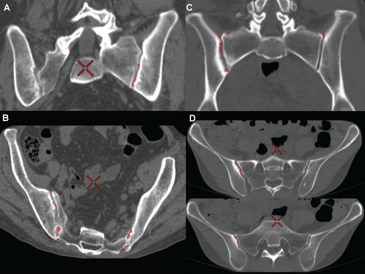
DECT of sacroiliac joints in gouty sacroiliitis. DECT was performed by GE Discovery CT750HD in accordance with manufacturer’s protocol. Red spots indicate monosodium urate crystal deposits. (A and B) Case 1. (C and D) Case 2. DECT: dual-energy computed tomography.

Case 2

A 40-year-old woman with a 10-year history of untreated hyperuricemia was admitted because of peripheral arthritis and tophus. She also claimed low back pain. Laboratory tests showed elevated CRP (39.9 mg/dl) and hyperuricemia (8.2 mg/dl). HLA-B27 was negative. A joint fluid aspiration from elbows contained urate crystals and neutrophils. Sacroiliitis was suspected by sclerosis in computed tomography and synovitis in MRI. DECT showed urate crystals attached to the right SIJ (Figure 1C and 1D). Both cases were treated by steroid and febuxostat; however, they showed recurrences of gouty attacks.

In this way, DECT provides important evidence for a diagnosis of gouty spondyloarthritis with minimal invasion.

Acknowledgment

We thank Dr. Yusuke Sugimori, Dr. Oh Sasaki, Dr. Hiroaki Harada, and Dr. Keishi Fujio from the Department of Allergy and Rheumatology for their diagnosis and assistance with this report.

REFERENCES

  1. 1.↵
    1. Nicolaou S,
    2. Yong-Hing CJ,
    3. Galea-Soler S,
    4. Hou DJ,
    5. Louis L,
    6. Munk P
    . Dual-energy CT as a potential new diagnostic tool in the management of gout in the acute setting. AJR Am J Roentgenol 2010;194:1072–8.
    OpenUrlCrossRefPubMed
  2. 2.↵
    1. Dhaese S,
    2. Stryckers M,
    3. Van Der Meersch H,
    4. Terryn W,
    5. Van Laecke S
    . Gouty arthritis of the spine in a renal transplant patient: a clinical case report: an unusual presentation of a common disorder. Medicine 2015;94:e676.
    OpenUrlCrossRefPubMed
  3. 3.↵
    1. Parikh P,
    2. Butendieck R,
    3. Kransdorf M,
    4. Calamia K
    . Detection of lumbar facet joint gouty arthritis using dual-energy computed tomography. J Rheumatol 2010;37:2190–1.
    OpenUrlFREE Full Text
View Abstract
PreviousNext
Back to top

In this issue

The Journal of Rheumatology
Vol. 43, Issue 6
1 Jun 2016
  • Table of Contents
  • Table of Contents (PDF)
  • Index by Author
  • Editorial Board (PDF)
Print
Download PDF
Article Alerts
Sign In to Email Alerts with your Email Address
Email Article

Thank you for your interest in spreading the word about The Journal of Rheumatology.

NOTE: We only request your email address so that the person you are recommending the page to knows that you wanted them to see it, and that it is not junk mail. We do not capture any email address.

Enter multiple addresses on separate lines or separate them with commas.
Two Cases of Gouty Sacroiliitis Evaluated by Dual-energy Computed Tomography
(Your Name) has forwarded a page to you from The Journal of Rheumatology
(Your Name) thought you would like to see this page from the The Journal of Rheumatology web site.
CAPTCHA
This question is for testing whether or not you are a human visitor and to prevent automated spam submissions.
Citation Tools
Two Cases of Gouty Sacroiliitis Evaluated by Dual-energy Computed Tomography
TOMOKA AO, HIROFUMI SHODA, KAZUHIKO YAMAMOTO
The Journal of Rheumatology Jun 2016, 43 (6) 1146-1147; DOI: 10.3899/jrheum.151454

Citation Manager Formats

  • BibTeX
  • Bookends
  • EasyBib
  • EndNote (tagged)
  • EndNote 8 (xml)
  • Medlars
  • Mendeley
  • Papers
  • RefWorks Tagged
  • Ref Manager
  • RIS
  • Zotero

 Request Permissions

Share
Two Cases of Gouty Sacroiliitis Evaluated by Dual-energy Computed Tomography
TOMOKA AO, HIROFUMI SHODA, KAZUHIKO YAMAMOTO
The Journal of Rheumatology Jun 2016, 43 (6) 1146-1147; DOI: 10.3899/jrheum.151454
del.icio.us logo Digg logo Reddit logo Twitter logo CiteULike logo Facebook logo Google logo Mendeley logo
  • Tweet Widget
  • Facebook Like
  • Google Plus One
Bookmark this article

Jump to section

  • Article
    • Case 1
    • Case 2
    • Acknowledgment
    • REFERENCES
  • Figures & Data
  • References
  • Info & Metrics
  • PDF
  • eLetters

Related Articles

Cited By...

More in this TOC Section

  • IgG4-related Disease With Destructive Nasal Bone Involvement Leading to Saddle Nose Deformity
  • A Case of Rowell Syndrome Manifested on Hands: Targetoid-like Lesions and Nailfold Capillaries
  • Hide-bound Bowel Sign in Systemic Sclerosis
Show more Images in Rheumatology

Similar Articles

Content

  • First Release
  • Current
  • Archives
  • Collections
  • Audiovisual Rheum
  • COVID-19 and Rheumatology

Resources

  • Guide for Authors
  • Submit Manuscript
  • Author Payment
  • Reviewers
  • Advertisers
  • Classified Ads
  • Reprints and Translations
  • Permissions
  • Meetings
  • FAQ
  • Policies

Subscribers

  • Subscription Information
  • Purchase Subscription
  • Your Account
  • Terms and Conditions

More

  • About Us
  • Contact Us
  • My Alerts
  • My Folders
  • Privacy/GDPR Policy
  • RSS Feeds
The Journal of Rheumatology
The content of this site is intended for health care professionals.
Copyright © 2022 by The Journal of Rheumatology Publishing Co. Ltd.
Print ISSN: 0315-162X; Online ISSN: 1499-2752
Powered by HighWire