

Acute calcium deposits cause significant pain and swelling of abrupt onset in an otherwise asymptomatic patient, usually in the shoulder joint. Diagnosis can be difficult; the condition is sometimes mistaken for infection, malignancy, or other crystal arthropathy.
A 77-year-old, right hand-dominant woman presented with a 2-day history of acute right wrist pain and swelling. She was constitutionally well with no history of local trauma. She was unable to move her right wrist and had increased pain with finger flexion. She had normal limits of white blood cell count, serum uric acid, calcium, inorganic phosphorus, and creatinine. Radiograph showed amorphous calcification (Figure 1). Ultrasound of right wrist revealed multiple confluent-appearing lobulated echogenic lesions throughout radiocarpal and midcarpal joints, most prominently over volar aspect with trace fluid within wrist joint. There was no evidence of tenosynovitis of the flexors or extensors. She was given a volar wrist splint. She was discharged home while receiving prednisone 25 mg, to be tapered by 5 mg every 3 days. She had slow improvement in her wrist pain and range of motion.
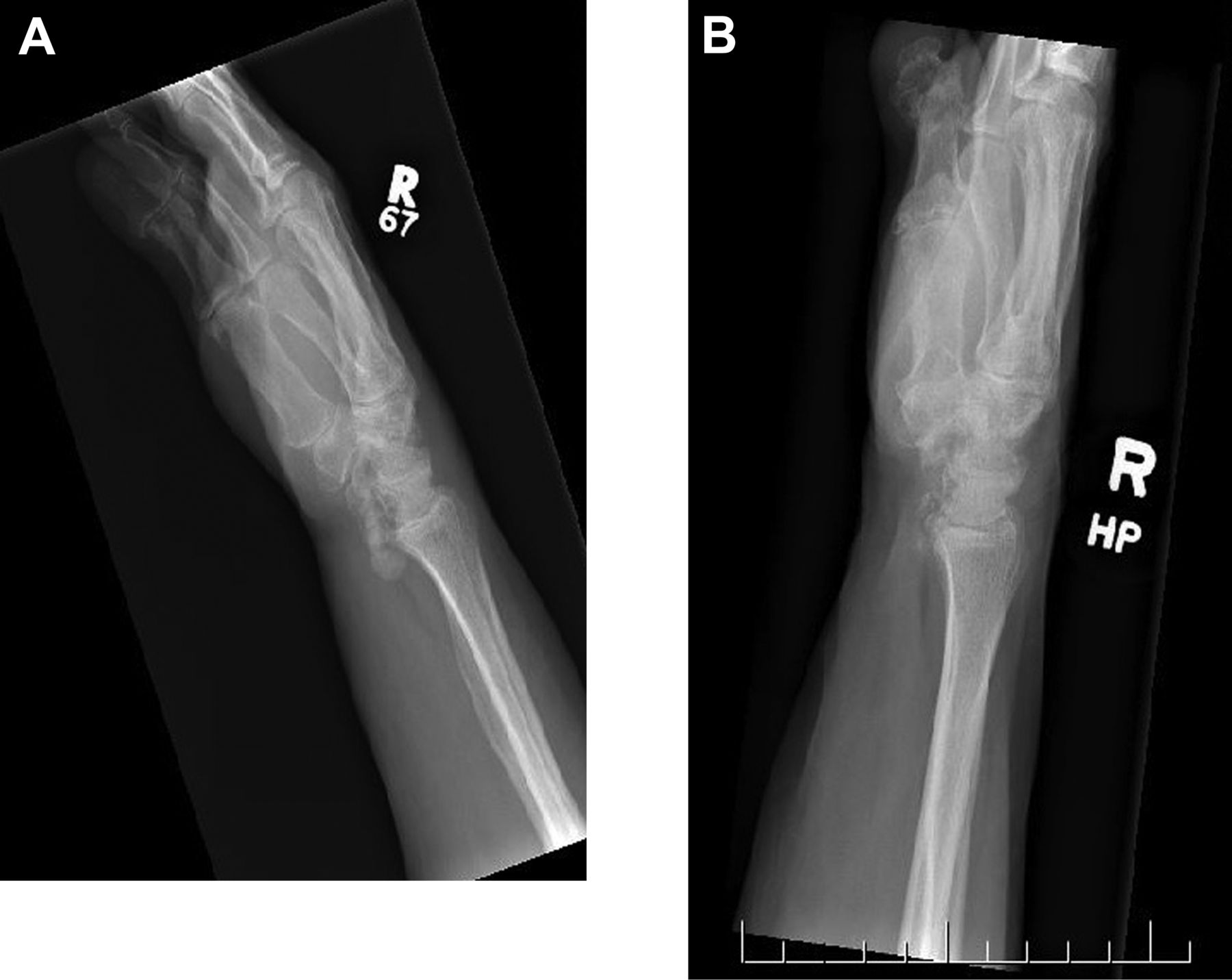
A. Radiograph of the right wrist at initial presentation revealed amorphous calcification volar to the wrist joint. B. Repeat radiographs of the patient’s hands 2 weeks later revealed near resolution of the calcification.
Acute calcium deposits, consisting of calcium hydroxyapatite crystals within soft tissue, present with significant pain and swelling in an otherwise asymptomatic patient1,2,3,4,5. This can often be mistaken for acute bacterial infection, malignancy, or other crystal arthropathies (gout and pseudogout).
The shoulder joint is the most commonly affected structure, followed by hip, knee, elbow, wrist, and hand2. Crystal deposition may occur within a joint as well as periarticular structures, including tendons and ligaments3. Affected patients are treated with splint immobilization, steroid injection, or antiinflammatories and rarely require surgery1,3,5. Radiographic changes resolve over 2–3 weeks with resorption of deposition by macrophages over time4,5.


