

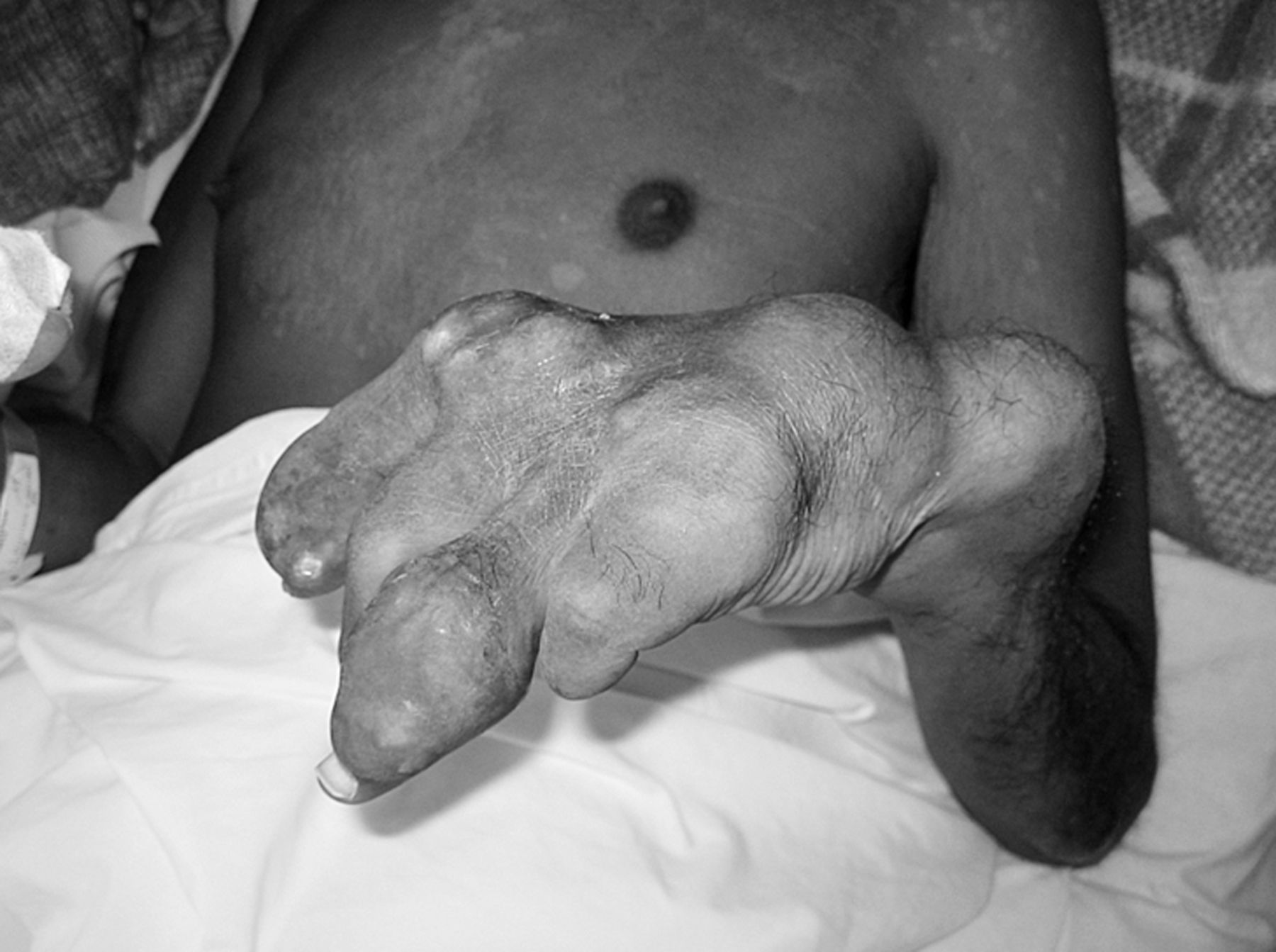
A 44-year-old man presented to our hospital with severe polyarticular familial gout. He experienced recurrent attacks on a monthly basis, and multiple tophaceous deformations appeared over the years as serum urate levels remained above 10 mg/dl in spite of a daily treatment by allopurinol 600 mg. This led to dramatic infirmity as demonstrated by the deformities because of tophi development (Figure 1), leaving him entirely dependent on his family for everyday care. The presented radiograph examination of the left foot demonstrated massive calcified soft tissue urate deposits and intrabone tophi (Figure 2). Parathormone, calcium, and phosphate levels were normal.

Tophaceous deformations of the left hand.

Plain radiography of diffuse calcified tophi of the left foot.
Prevalence of gout in Polynesia is high because of the combination of the adoption of occidental food diets and genetic predisposing factors, especially the mutation of renal urate transporter genes leading to decreased urate excretion1,2. Tophaceous gout affects 15% to 20% of patients with gout3. Tophi are classically radio-transparent4, but such case illustrates the possibility of associated calcic crystals, especially in cases of renal impairment such as the one affecting our patient (estimated glomerular filtration rate 47 ml/min). These calcifications could be the result of a persistent inflammation taking place within tophi as proven by histological studies5. Severe cases of tophaceous gout are responsible for joint destruction and severe disability. Prolonged treatment by urate-lowering therapies can maintain serum uric acid levels below urate crystallization levels and eventually deplete urate deposits, which ultimately leads to tophi regression.


