

A rare skin manifestation of familial Mediterranean fever (FMF) usually appears in the lower extremities and can be brought on by physical exertion.
A 19-year-old white male was presented to the emergency room with a chief complaint of a painful red discoloration of the ankles of 3 days’ duration with accompanying fever.
The patient had been diagnosed with FMF at the age of 6 years, based on recurrent episodes of fever, abdominal pain, and foot pain beginning at the age of 3 years. Each episode lasted for a few days. The family history was significant; 2 of 11 siblings also carried a diagnosis of FMF. Neither the patient nor his affected siblings had been tested for MEFV gene mutations. Treatment with colchicine was instituted at a dose of 0.5 mg per day, and later increased to 0.75 mg per day. The patient had recently joined the army and begun basic training 4 days before presentation. On the following day, he began to experience fever and pain over his left ankle, followed by similar symptoms over the right ankle.
The sedimentation rate was 29 and the C-reactive protein was 8.25 mg/dl. Complete blood count was within normal limits. A diagnosis of erysipelas-like erythema (ELE) was made, and the patient was treated with bed rest and nonsteroidal antiinflammatory agents. The colchicine dose was increased to 1.0 mg per day.
ELE is the only pathognomonic skin manifestation of FMF. It was neither included in the widely used 1997 Tel Hashomer criteria for FMF1 because it did not contribute to sensitivity, nor was it included in more recent criteria2. It is said to be more common in early onset FMF and in the Turkish population3. It has been described predominantly in the dermatology literature4,5. Lesions are characterized by tender erythematous plaques, usually located over the distal lower extremities. Histologically, it is characterized by edema of the papillary dermis, a lymphocytic infiltration of the superficial and deep dermis, perivascular infiltrates, and an absence of changes in the epidermis4. Fever and leukocytosis may accompany this condition. ELE may be triggered by physical exertion and subside spontaneously within 48 h to 72 h with bed rest.
REFERENCES
Figures

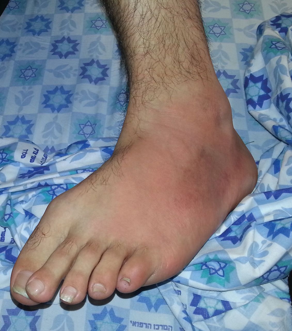
After 4 days of basic training in the army, the patient began to experience fever and pain in his right foot.

Left ankle, with redness and swelling.


