
Main menu

  • Home
  • Content
    • First Release
    • Current
    • Archives
    • Collections
    • Audiovisual Rheum
    • COVID-19 and Rheumatology
  • Resources
    • Guide for Authors
    • Submit Manuscript
    • Payment
    • Reviewers
    • Advertisers
    • Classified Ads
    • Reprints and Translations
    • Permissions
    • Meetings
    • FAQ
    • Policies
  • Subscribers
    • Subscription Information
    • Purchase Subscription
    • Your Account
    • Terms and Conditions
  • About Us
    • About Us
    • Editorial Board
    • Letter from the Editor
    • Duncan A. Gordon Award
    • Privacy/GDPR Policy
    • Accessibility
  • Contact Us
  • JRheum Supplements
  • Services

User menu

  • My Cart
  • Log In

Search

  • Advanced search
The Journal of Rheumatology
  • JRheum Supplements
  • Services
  • My Cart
  • Log In
The Journal of Rheumatology

Advanced Search

  • Home
  • Content
    • First Release
    • Current
    • Archives
    • Collections
    • Audiovisual Rheum
    • COVID-19 and Rheumatology
  • Resources
    • Guide for Authors
    • Submit Manuscript
    • Payment
    • Reviewers
    • Advertisers
    • Classified Ads
    • Reprints and Translations
    • Permissions
    • Meetings
    • FAQ
    • Policies
  • Subscribers
    • Subscription Information
    • Purchase Subscription
    • Your Account
    • Terms and Conditions
  • About Us
    • About Us
    • Editorial Board
    • Letter from the Editor
    • Duncan A. Gordon Award
    • Privacy/GDPR Policy
    • Accessibility
  • Contact Us
  • Follow jrheum on Twitter
  • Visit jrheum on Facebook
  • Follow jrheum on LinkedIn
  • Follow jrheum on YouTube
  • Follow jrheum on Instagram
  • Follow jrheum on RSS
Research ArticlePediatric Rheumatology

Risk Factors for Temporomandibular Joint Arthritis in Children with Juvenile Idiopathic Arthritis

MATTHEW L. STOLL, TYLER SHARPE, TIMOTHY BEUKELMAN, JENNIFER GOOD, DANIEL YOUNG and RANDY Q. CRON
The Journal of Rheumatology September 2012, 39 (9) 1880-1887; DOI: https://doi.org/10.3899/jrheum.111441
MATTHEW L. STOLL
From the Department of Pediatrics and the Department of Radiology, University of Alabama at Birmingham, Birmingham, Alabama; and the Carolinas Oral and Facial Surgery Center, Wilmington, North Carolina, USA.
  • Find this author on Google Scholar
  • Find this author on PubMed
  • Search for this author on this site
  • For correspondence: mstoll@uab.edu
TYLER SHARPE
  • Find this author on Google Scholar
  • Find this author on PubMed
  • Search for this author on this site
TIMOTHY BEUKELMAN
  • Find this author on Google Scholar
  • Find this author on PubMed
  • Search for this author on this site
JENNIFER GOOD
  • Find this author on Google Scholar
  • Find this author on PubMed
  • Search for this author on this site
DANIEL YOUNG
  • Find this author on Google Scholar
  • Find this author on PubMed
  • Search for this author on this site
RANDY Q. CRON
  • Find this author on Google Scholar
  • Find this author on PubMed
  • Search for this author on this site
  • Article
  • Figures & Data
  • Info & Metrics
  • References
  • PDF
  • eLetters
PreviousNext
Loading

Article Figures & Data

Figures

  • Tables
  • Figure 1.
    • Download figure
    • Open in new tab
    • Download powerpoint
    Figure 1.

    Normal magnetic resonance imaging of the temporomandibular joint (TMJ). Coronal (A) and sagittal (B) postcontrast T1-weighted fat-saturated images of the TMJ. The condylar heads (labeled C) are well situated in their respective fossae.

  • Figure 2.
    • Download figure
    • Open in new tab
    • Download powerpoint
    Figure 2.

    Active temporomandibular joint synovitis. Sagittal T2-weighted fat-saturated (FS) precontrast (A) and T1-weighted postcontrast FS (B) images show synovial fluid (A, arrows) and enhancement (B, large arrows). Erosive changes at the condylar head are also evident on B (small arrows).
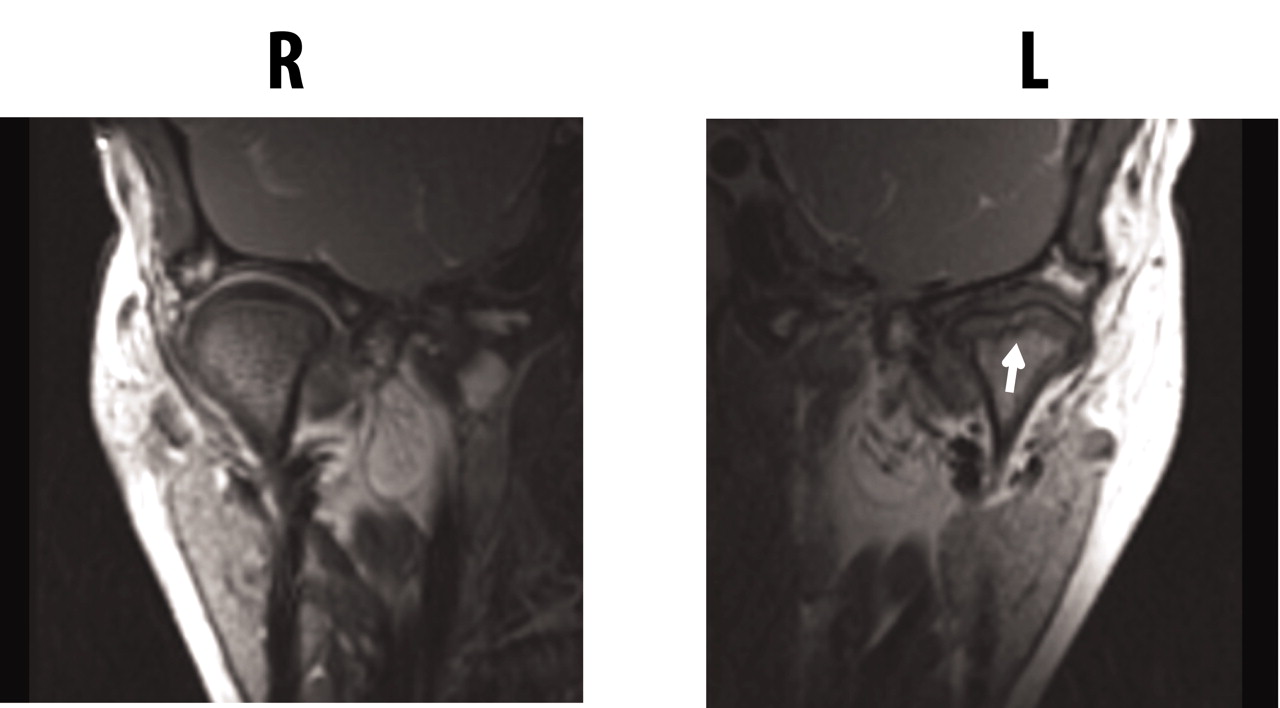
  • Figure 3.
    • Download figure
    • Open in new tab
    • Download powerpoint
    Figure 3.

    Destructive changes of the temporomandibular joint (TMJ). Coronal T1-weighted image of the TMJ shows flattening and erosive changes of the left condylar head (arrow); the right TMJ is normal.

  • Figure 4.
    • Download figure
    • Open in new tab
    • Download powerpoint
    Figure 4.

    Percentage of patients with abnormal MRI of the TMJ, by JIA subtype. Current results are compared with those published by Cannizzaro, et al16. Oligo: oligoarticular JIA; poly: polyarticular JIA.
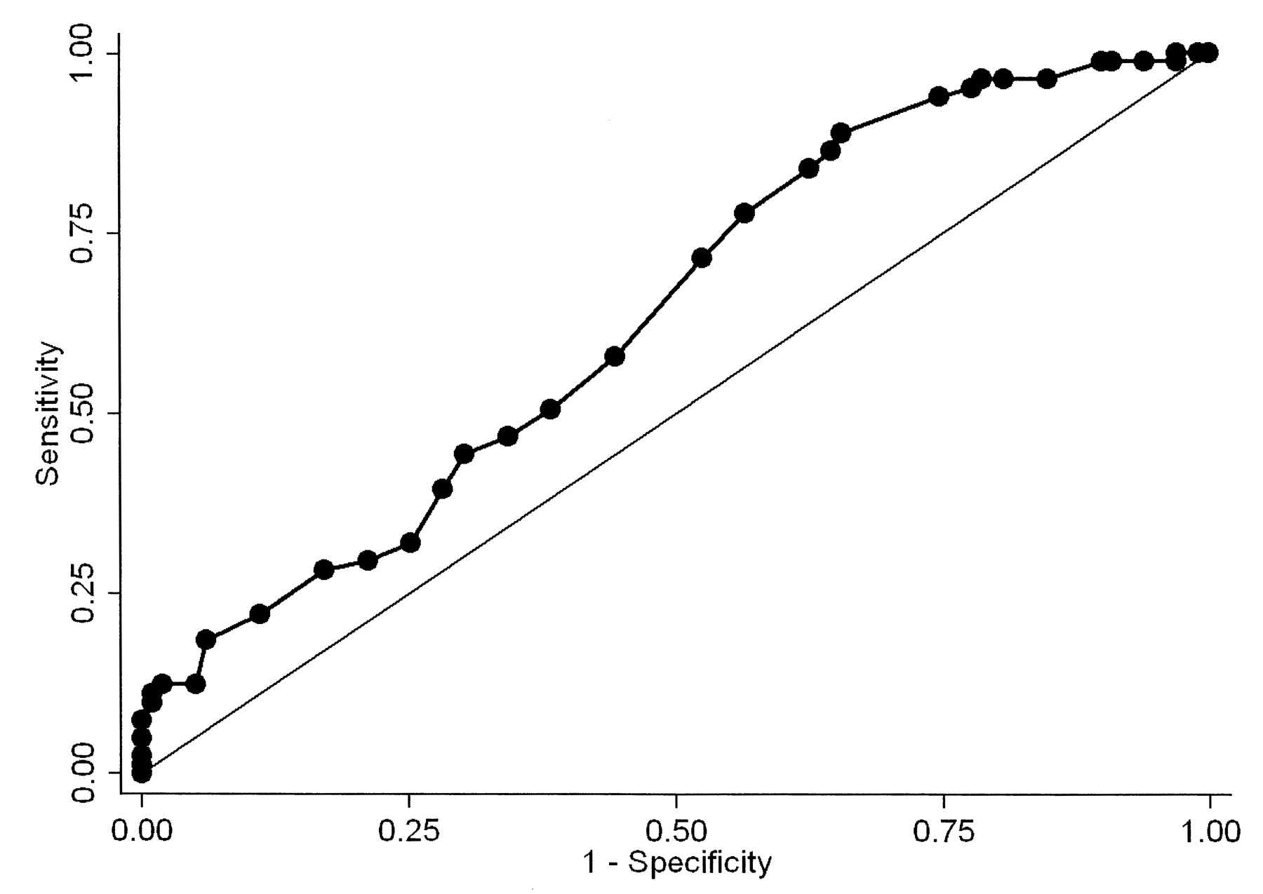
  • Figure 5.
    • Download figure
    • Open in new tab
    • Download powerpoint
    Figure 5.

    Receiver-operator curve analysis for the ability of maximal incisal opening to predict TMJ arthritis.

Tables

  • Figures
    • View popup
    Table 1.

    Patient population. Statistically significant differences (p < 0.05) are depicted in bold type.

    FeatureEntire Group, n = 187TMJ Arthritis-negative, n = 106TMJ Arthritis-positive, n = 81p
    Demographics
      Age at diagnosis, yrs; mean, SEM, range6.7, 0.31, 1–156.0, 0.40, 1–157.6, 0.46, 1–150.006
      Disease duration*, yrs; mean, SEM, range2.5, 0.3, 0–173.1, 0.38, 0–141.6, 0.31, 0–170.005
      Newly diagnosed (< 3 mo)56, 3028, 2628, 350.228
      Age at study, yrs; mean, SEM, range9.1, 0.34, 1.7–219.1, 0.50, 1.7–2.19.2, 0.45, 2–180.794
      Female, n, %116, 6263, 5953, 650.402
      Race, n (%)0.981
        White162, 8792, 8770, 86
        African-American23, 1213, 1210, 12
        Other2, 1.01, 0.91, 1.2
    JIA subtype, n, %0.251
      Oligoarticular JIA56, 3031, 2925, 31
      RF– polyarticular JIA35, 1918, 1717, 21
      RF+ polyarticular JIA3, 1.61, 0.92, 2.5
      Psoriatic JIA37, 2023, 2214, 17
      Enthesitis-related arthritis40, 2125, 2415, 18
      Systemic-onset JIA12, 6.48, 7.54, 4.9
      Undifferentiated4, 2.104, 4.9
    Laboratory studies
      Positive ANA39/164, 2423/92, 2516/72, 220.678
      Positive RF6/118, 5.13/63, 4.83/55, 5.51.000
      ACPA4/50, 8.01/23, 4.33/27, 110.617
      Positive HLA-B2717/104, 1611/54, 206/50, 120.249
    Medications, n, %
      NSAID125, 6777, 7348, 590.054
      MTX110, 5968, 6442, 520.090
      Sulfasalazine2, 1.102, 2.50.186
      Leflunomide2, 1.12, 1.900.506
      Any anti-TNF84, 4550, 4734, 420.479
      Anakinra9, 4.85, 4.74, 4.91.000
      Combination DMARD80, 4349, 4631, 380.276
        MTX and anti-TNF70, 3742, 4028, 350.479
        MTX and anakinra8, 4.35, 4.73, 3.71.000
      Any DMARD126, 6776, 7250, 620.150
      Oral corticosteroids32, 1719, 1813, 16
    Physical examination, n, %
      Mouth opening deviation53, 2813, 1240, 49< 0.001
      MIO, mm; mean, SEM, range
        Age 0–7.941.3, 0.74, 23–5443.1, 0.86, 34–5438.9, 1.2, 23–490.004
        Age 8+45.8, 0.77, 30–6547.8, 1.1, 31–6543.6, 0.97, 30–620.005
      Low MIO for age**48, 2721/99, 2127/81, 330.067
      Mouth opening deviation or low MIO81, 4330, 2851, 63< 0.001
      Overall joint count†; mean, SEM, range2.2, 0.32, 0–342.0, 0.31, 0–162.6, 0.61, 0–340.358
      Joint count 0†73, 3937, 3536, 440.185
      Joint count 1 or lower†115, 6263, 5952, 640.507
      Joint count 4 or lower†165, 8894, 8971, 880.829
    • ↵* Elapsed time between diagnosis of JIA and the initial MRI.

    • ↵** Defined as < 2 SD below norm for age20.

    • ↵† Total joint count is exclusive of the TMJ itself. TMJ: temporomandibular joint; ANA: antinuclear antibody; ACPA: anticitrullinated protein antibodies; DMARD: disease-modifying antirheumatic drug (includes methotrexate, sulfasalazine, leflunomide, and biologic); JIA: juvenile idiopathic arthritis; MIO: maximal incisal opening; MTX: methotrexate; NSAID: nonsteroidal antiinflammatory drugs; RF: rheumatoid factor; TNF: tumor necrosis factor.

    • View popup
    Table 2.

    MRI findings of TMJ arthritis. Data are limited to patients with contrast-enhanced MRI (n = 185).

    FindingnPercentage of TotalPercentage of Abnormal
    By individual TMJ370NANA
      Active or chronic arthritis14238NA
        Active arthritis, any1353695
          Synovial fluid alone195.114
          Enhancement alone852363
          Synovial fluid and enhancement318.423
        Chronic arthritis, any267.018
          Erosions alone123.246
          Condylar flattening alone123.246
          Combination of findings of chronic arthritis20.57.6
        Acute arthritis without chronic changes1153181
        Chronic arthritis without acute changes61.64.2
        Acute and chronic changes205.414
    By individual patient185NANA
      Any abnormality8143NA
      Active arthritis without chronic changes593273
      Chronic arthritis without acute changes21.12.4
      Active and chronic changes201124
    • MRI: magnetic resonance imaging; TMJ: temporomandibular joint. NA: not applicable.

    • View popup
    Table 3.

    Multivariable analysis of predictors of TMJ synovitis. Data are OR (95% CI).

    FactorUnivariate ORMultivariable OR
    Mouth opening deviation6.98 (3.38–14.4)6.21 (2.87–13.4)
    Maximal incisal opening0.93 (0.89–0.97)0.94 (0.90–0.99)
    Age at diagnosis1.10 (1.03–1.19)NS
    Disease duration0.87 (0.79–0.96)0.87 (0.78–0.97)
    • TMJ: temporomandibular joint; NS: not significant.

PreviousNext
Back to top

In this issue

The Journal of Rheumatology
Vol. 39, Issue 9
1 Sep 2012
  • Table of Contents
  • Table of Contents (PDF)
  • Index by Author
  • Editorial Board (PDF)
Print
Download PDF
Article Alerts
Sign In to Email Alerts with your Email Address
Email Article

Thank you for your interest in spreading the word about The Journal of Rheumatology.

NOTE: We only request your email address so that the person you are recommending the page to knows that you wanted them to see it, and that it is not junk mail. We do not capture any email address.

Enter multiple addresses on separate lines or separate them with commas.
Risk Factors for Temporomandibular Joint Arthritis in Children with Juvenile Idiopathic Arthritis
(Your Name) has forwarded a page to you from The Journal of Rheumatology
(Your Name) thought you would like to see this page from the The Journal of Rheumatology web site.
CAPTCHA
This question is for testing whether or not you are a human visitor and to prevent automated spam submissions.
Citation Tools
Risk Factors for Temporomandibular Joint Arthritis in Children with Juvenile Idiopathic Arthritis
MATTHEW L. STOLL, TYLER SHARPE, TIMOTHY BEUKELMAN, JENNIFER GOOD, DANIEL YOUNG, RANDY Q. CRON
The Journal of Rheumatology Sep 2012, 39 (9) 1880-1887; DOI: 10.3899/jrheum.111441

Citation Manager Formats

  • BibTeX
  • Bookends
  • EasyBib
  • EndNote (tagged)
  • EndNote 8 (xml)
  • Medlars
  • Mendeley
  • Papers
  • RefWorks Tagged
  • Ref Manager
  • RIS
  • Zotero

 Request Permissions

Share
Risk Factors for Temporomandibular Joint Arthritis in Children with Juvenile Idiopathic Arthritis
MATTHEW L. STOLL, TYLER SHARPE, TIMOTHY BEUKELMAN, JENNIFER GOOD, DANIEL YOUNG, RANDY Q. CRON
The Journal of Rheumatology Sep 2012, 39 (9) 1880-1887; DOI: 10.3899/jrheum.111441
Reddit logo Twitter logo Facebook logo Mendeley logo
  • Tweet Widget
  • Facebook Like
  • Google Plus One
Bookmark this article

Jump to section

  • Article
    • Abstract
    • MATERIALS AND METHODS
    • RESULTS
    • DISCUSSION
    • Acknowledgment
    • Footnotes
    • REFERENCES
  • Figures & Data
  • Info & Metrics
  • References
  • PDF
  • eLetters

Related Articles

Cited By...

More in this TOC Section

  • Modified Juvenile Spondyloarthritis Disease Activity Index in the Childhood Arthritis and Rheumatology Research Alliance (CARRA) Registry
  • Treatment Response to Tumor Necrosis Factor Inhibitors and Methotrexate Monotherapy in Adults With Juvenile Idiopathic Arthritis: Data From NOR-DMARD
  • Canadian Rheumatology Association Recommendations for the Screening, Monitoring, and Treatment of Juvenile Idiopathic Arthritis-Associated Uveitis
Show more Pediatric Rheumatology

Similar Articles

Content

  • First Release
  • Current
  • Archives
  • Collections
  • Audiovisual Rheum
  • COVID-19 and Rheumatology

Resources

  • Guide for Authors
  • Submit Manuscript
  • Author Payment
  • Reviewers
  • Advertisers
  • Classified Ads
  • Reprints and Translations
  • Permissions
  • Meetings
  • FAQ
  • Policies

Subscribers

  • Subscription Information
  • Purchase Subscription
  • Your Account
  • Terms and Conditions

More

  • About Us
  • Contact Us
  • My Alerts
  • My Folders
  • Privacy/GDPR Policy
  • RSS Feeds
The Journal of Rheumatology
The content of this site is intended for health care professionals.
Copyright © 2022 by The Journal of Rheumatology Publishing Co. Ltd.
Print ISSN: 0315-162X; Online ISSN: 1499-2752
Powered by HighWire