

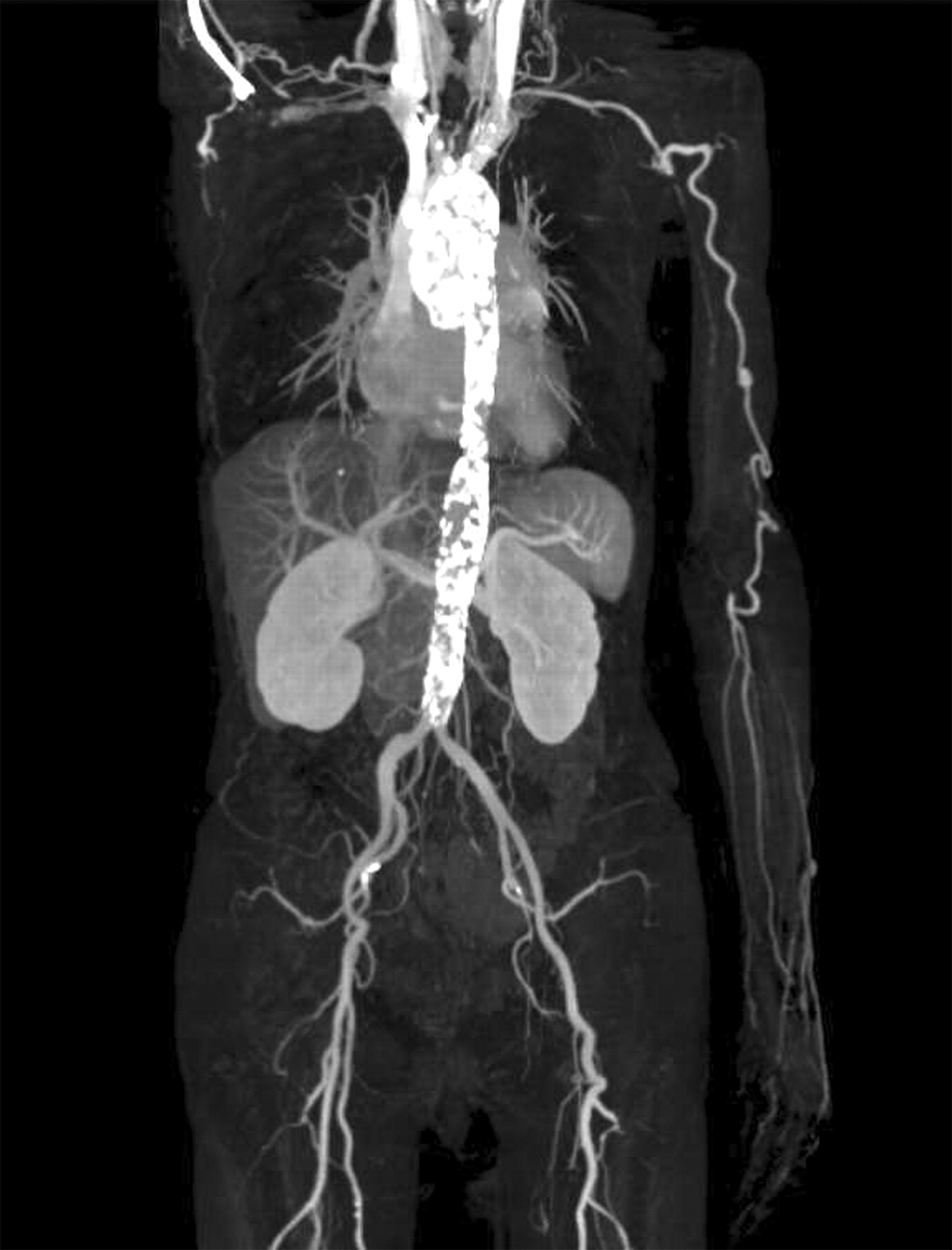
Persistent inflammation is a serious risk factor for arteriosclerosis and arteriosclerotic disease in Takayasu arteritis (TA)1,2. A female patient was diagnosed with TA at 32 years of age. She underwent treatment with glucocorticoids for about 1 year, and then the medication was discontinued. At 57 years of age, she presented to our hospital complaining of left back pain on exertion, lateral abdominal pain, and melena and was admitted for evaluation. She was negative for arteriosclerosis risk factors such as hypertension, diabetes mellitus, hyperlipidemia, and smoking. Examination revealed dissociation of blood pressures of both arms (right 87/44 mm Hg, left 129/76 mm Hg) and bruits on the abdominal aorta. Blood tests revealed no sign of fever or inflammation, with C-reactive protein (CRP) 0.2 mg/l and erythrocyte sedimentation rate (ESR) 11 mm/h. Chest-abdominal contrast computed tomography revealed calcification of the whole aorta (Figure 1). Stenotic lesions were noted in the right subclavian artery, at the root of the right coronary artery, the left subclavian artery, and the root of the inferior mesenteric artery. 18FFluorodeoxyglucose positron emission tomography suggested no inflammatory lesions in these vessels. These findings prompted a diagnosis of ischemic heart disease and ischemic colitis.

Chest-abdominal contrast computed tomography reveals calcification of the whole aorta. Stenotic lesions are visible in the right subclavian artery, at the root of the right coronary artery, the left subclavian artery, and the root of the inferior mesenteric artery.
TA is a chronic, idiopathic, and inflammatory disease that principally affects the aorta and its primary branches. Although the patient had experienced no symptoms or inflammatory signs for 25 years, arteriosclerosis was considered to have developed in the whole aorta. Arterial wall inflammation can persist even with normal ESR and CRP in TA3. Thus it is important that patients with TA receive routine followup for many years even after the major symptoms have disappeared.


