
Main menu

  • Home
  • Content
    • First Release
    • Current
    • Archives
    • Collections
    • Audiovisual Rheum
    • COVID-19 and Rheumatology
  • Resources
    • Guide for Authors
    • Submit Manuscript
    • Payment
    • Reviewers
    • Advertisers
    • Classified Ads
    • Reprints and Translations
    • Permissions
    • Meetings
    • FAQ
    • Policies
  • Subscribers
    • Subscription Information
    • Purchase Subscription
    • Your Account
    • Terms and Conditions
  • About Us
    • About Us
    • Editorial Board
    • Letter from the Editor
    • Duncan A. Gordon Award
    • Privacy/GDPR Policy
    • Accessibility
  • Contact Us
  • JRheum Supplements
  • Services

User menu

  • My Cart
  • Log In

Search

  • Advanced search
The Journal of Rheumatology
  • JRheum Supplements
  • Services
  • My Cart
  • Log In
The Journal of Rheumatology

Advanced Search

  • Home
  • Content
    • First Release
    • Current
    • Archives
    • Collections
    • Audiovisual Rheum
    • COVID-19 and Rheumatology
  • Resources
    • Guide for Authors
    • Submit Manuscript
    • Payment
    • Reviewers
    • Advertisers
    • Classified Ads
    • Reprints and Translations
    • Permissions
    • Meetings
    • FAQ
    • Policies
  • Subscribers
    • Subscription Information
    • Purchase Subscription
    • Your Account
    • Terms and Conditions
  • About Us
    • About Us
    • Editorial Board
    • Letter from the Editor
    • Duncan A. Gordon Award
    • Privacy/GDPR Policy
    • Accessibility
  • Contact Us
  • Follow jrheum on Twitter
  • Visit jrheum on Facebook
  • Follow jrheum on LinkedIn
  • Follow jrheum on YouTube
  • Follow jrheum on Instagram
  • Follow jrheum on RSS
Research ArticleOMERACT 10: 10th International Consensus Conference on Outcome Measures in Rheumatology, Kota Kinabalu, Borneo - May 4–8, 2010

Patterns of Magnetic Resonance Imaging Bone Erosion in Rheumatoid Arthritis — Which Bones Are Most Frequently Involved and Show the Most Change?

MIKKEL ØSTERGAARD, UFFE MØLLER DØHN, ANNE DUER-JENSEN, MERETE LUND HETLAND, KIM HØRSLEV-PETERSEN, KRISTIAN STENGAARD-PEDERSEN, PETER JUNKER, JAN PØDENPHANT and BO EJBJERG
The Journal of Rheumatology September 2011, 38 (9) 2014-2017; DOI: https://doi.org/10.3899/jrheum.110416
MIKKEL ØSTERGAARD
  • Find this author on Google Scholar
  • Find this author on PubMed
  • Search for this author on this site
  • For correspondence: mo@dadlnet.dk
UFFE MØLLER DØHN
  • Find this author on Google Scholar
  • Find this author on PubMed
  • Search for this author on this site
ANNE DUER-JENSEN
  • Find this author on Google Scholar
  • Find this author on PubMed
  • Search for this author on this site
MERETE LUND HETLAND
  • Find this author on Google Scholar
  • Find this author on PubMed
  • Search for this author on this site
KIM HØRSLEV-PETERSEN
  • Find this author on Google Scholar
  • Find this author on PubMed
  • Search for this author on this site
KRISTIAN STENGAARD-PEDERSEN
  • Find this author on Google Scholar
  • Find this author on PubMed
  • Search for this author on this site
PETER JUNKER
  • Find this author on Google Scholar
  • Find this author on PubMed
  • Search for this author on this site
JAN PØDENPHANT
  • Find this author on Google Scholar
  • Find this author on PubMed
  • Search for this author on this site
BO EJBJERG
  • Find this author on Google Scholar
  • Find this author on PubMed
  • Search for this author on this site
  • Article
  • Figures & Data
  • Info & Metrics
  • References
  • PDF
  • eLetters
PreviousNext
Loading

Abstract

Objective. To investigate by magnetic resonance imaging (MRI) which bones in wrists and metacarpophalangeal (MCP) joints most frequently show bone erosions, and which most frequently demonstrate erosive progression, in early and established rheumatoid arthritis (RA).

Methods. MRI datasets from 258 RA patients [126 with early RA (disease duration < 6 months)] were analyzed, of whom 223, including 126 with early RA, had 1-year followup MRI. All patients had MRI of one wrist, whereas 86 patients had additional images of 2nd–5th MCP joints, and 46 patients additional images of the contralateral wrist. MRI were evaluated blinded by one reader, according to the OMERACT RA MRI scoring system (RAMRIS) for erosions, and presence/absence of erosions was noted in each bone, as was presence/absence of erosive progression.

Results. The capitate, ulna, lunate, triquetrum, and scaphoid were the 5 bones with both most frequent baseline erosions and most frequently demonstrated erosive progression. No bones were without erosions. Patterns of erosions and progression were similar in early and established RA. No major difference between dominant and nondominant wrists was detected. In the fingers, the 2nd–3rd MCP joint most frequently displayed erosions and erosive progression.

Conclusion. The distribution and frequency of bone erosion and erosive progression as detected by MRI in RA wrists and MCP joints were identified. No pattern differences between early versus established disease and dominant versus nondominant sides were detected. No bones showed erosive progression. Thus, no self-evident simplification of the RAMRIS erosion score was identified. Bone involvement patterns may be considered, when joints are selected for MRI protocols for clinical trials and practice.

Key Indexing Terms:
  • RHEUMATOID ARTHRITIS
  • MAGNETIC RESONANCE IMAGING
  • BONE EROSION
  • WRIST
  • METACARPOPHALANGEAL JOINT

Magnetic resonance imaging (MRI) has been shown to be more sensitive for identifying early bone damage than conventional radiography1,2,3,4, and is increasingly used as an outcome measure of structural joint damage in rheumatoid arthritis (RA) clinical trials5,6,7,8. MRI of unilateral wrist and 2nd–5th metacarpophalangeal (MCP) joints is more sensitive to change for erosive progression than radiography of both hands, wrists and forefeet9. However, data on the exact distribution of bone erosions, i.e., the pattern of bone involvement, are minimal.

The aim of this study was to investigate which bones in wrists and MCP joints most frequently show bone erosions and which most frequently demonstrate erosive progression in early and established RA.

MATERIALS AND METHODS

MRI datasets from 258 RA patients [126 with early RA (disease duration < 6 months)] were analyzed. One-year followup MRI were available in 223 patients (126 with early RA). All patients had MRI of one wrist, whereas 86 had additional MCP joint MR images and 46 additional images of the contralateral wrist. The MRI data originated from 5 investigator-initiated clinical studies9,10,11,12,13,14, in which details on population characteristics and MRI techniques are described.

MRI (including T1-weighted images in coronal and axial planes) were evaluated for bone erosions according to the OMERACT RA MRI scoring system (RAMRIS), i.e., an erosion was defined as a sharply marginated bone lesion, with correct juxtaarticular localization and typical signal characteristics, visible in 2 planes, with a cortical break seen in at least one plane5.

Presence/absence of erosions at baseline was noted in each bone, as was presence/absence of erosive progression. All images were read blinded by the same reader (BE), who has extensive experience in reading MRI of RA patients and has demonstrated high levels of intra- and interobserver reproducibility6,9.

RESULTS

The distribution of baseline erosions per wrist bone is shown in Figure 1A, whereas the distribution of bones with progression at Year 1 is seen in Figure 1B. The capitate, ulna, lunate, triquetrum, and scaphoid were the 5 bones most frequently demonstrating both baseline erosions and erosive progression. Patterns were similar in early and in established RA. No bones were without erosions. In patients with available bilateral wrist MRI, no major differences between dominant and nondominant hands were found (269 vs 277 bones with baseline erosions; 73 vs 85 bones with erosive progression).

Figure 1.
  • Download figure
  • Open in new tab
  • Download powerpoint
Figure 1.

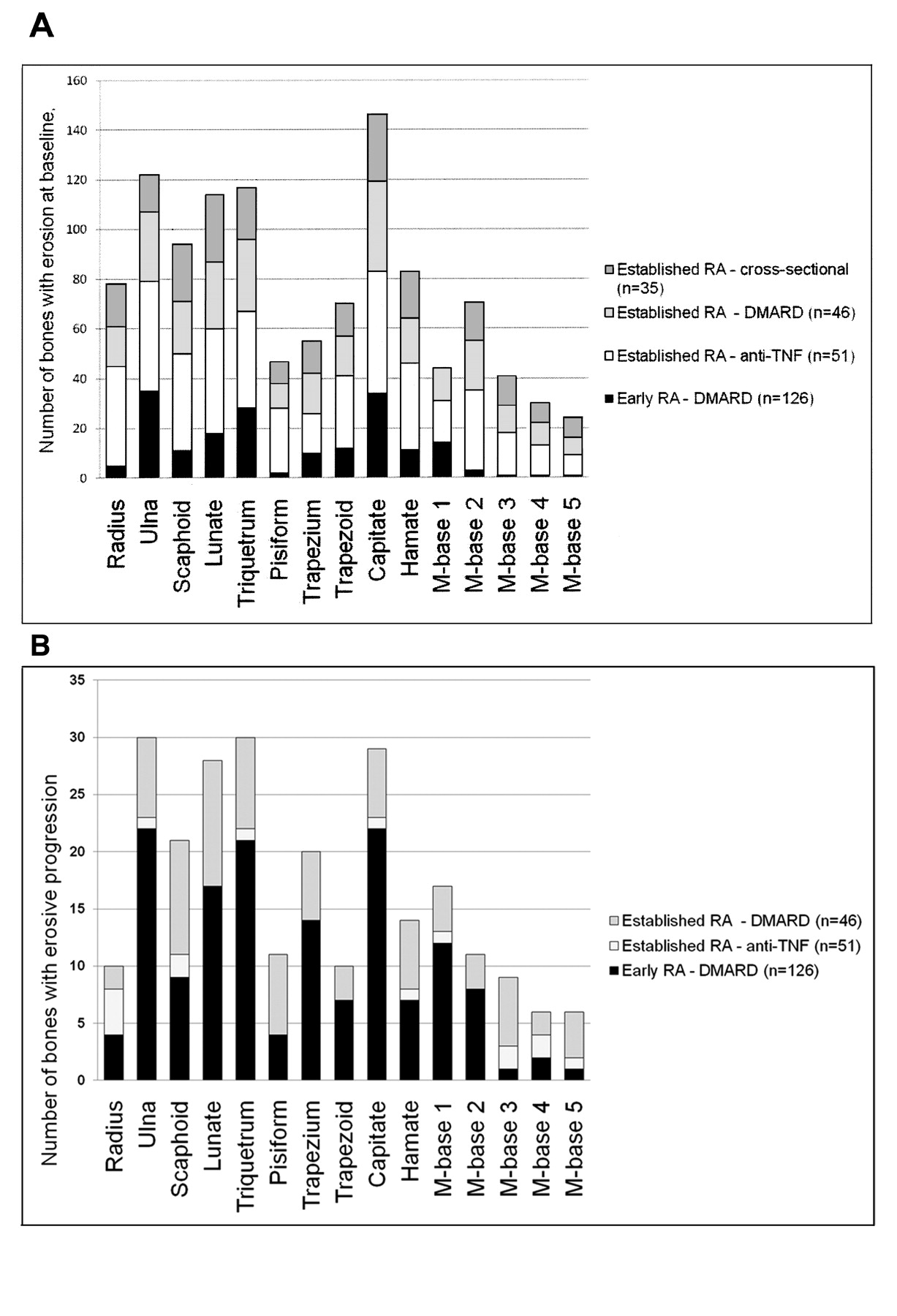
A. Number of bones with bone erosion, per bone (n = 258). B. Number of bones with erosive progression, per bone (n = 233).

In the patients in whom both wrist and MCP joint images were available, baseline erosions were still most frequently found in the 5 previously mentioned wrist bones, whereas the head of the 2nd metacarpal bone was the most frequently involved MCP joint bone. Second and 3rd MCP joints most frequently showed erosive progression.

DISCUSSION

Our study documents the frequency and distribution of the presence and progression of MRI bone erosions in a large number of RA patients from different populations. Based on data from 258 RA patients, patterns of MRI bone erosion in wrists and MCP joints could be identified. The capitate, ulna, lunate, triquetrum, and scaphoid were most frequently involved and showed, together with the 2nd and 3rd MCP joints, the most change over time. No obvious differences in erosive patterns between early and established RA, or between dominant and nondominant wrists, were identified.

The images were obtained and scored in agreement with the OMERACT RAMRIS. This method has been extensively validated and is now the standard MRI method in RA clinical trials5,6,7,8. Comparisons with computed tomography as a “gold standard” reference for bony destruction have demonstrated a very high level of agreement and, consequently, confirmed that MRI bone erosions are true erosions15,16,17. MRI has been documented to be more sensitive to change in erosive joint damage than radiography of both hands, wrists, and forefeet, as evaluated by the Sharp-van der Heijde method8,9. The present study did not include assessment of interobserver reliability, but all images were read by the same experienced reader (BE).

This MRI study of 258 RA patients demonstrated the distribution of erosions and erosive progression in wrist and MCP joints. No pattern differences between early versus established disease and dominant versus nondominant sides were detected. No bones ever showed erosive progression. Thus, no evident reductions in the number of bones to be assessed for erosions could be identified. Future studies should test whether the RAMRIS erosion score can be simplfied to include only the most frequently involved areas (capitate, ulna, lunate, triquetrum, and scaphoid and the 2nd–3rd MCP joint) without losing sensitivity to change. Bone involvement patterns may be considered when joints are selected for MRI protocols for clinical trials and practice.

Acknowledgment

Physicians, study nurses, and patients who took part in the RA cohorts are acknowledged for their contributions.

REFERENCES

  1. 1.↵
    1. McQueen FM,
    2. Stewart N,
    3. Crabbe J,
    4. Robinson E,
    5. Yeoman S,
    6. Tan PLJ,
    7. et al.
    Magnetic resonance imaging of the wrist in early rheumatoid arthritis reveals progression of erosions despite clinical improvement. Ann Rheum Dis 1999;58:156–63.
    OpenUrlAbstract/FREE Full Text
  2. 2.↵
    1. Klarlund M,
    2. Østergaard M,
    3. Jensen KE,
    4. Madsen JL,
    5. Skjødt H;
    6. the TIRA group
    . Magnetic resonance imaging, radiography, and scintigraphy of the finger joints: one year follow up of patients with early arthritis. Ann Rheum Dis 2000;59:521–8.
    OpenUrlAbstract/FREE Full Text
  3. 3.↵
    1. Quinn MA,
    2. Conaghan PG,
    3. O’Connor PJ,
    4. Karim Z,
    5. Greenstein A,
    6. Brown A,
    7. et al.
    Very early treatment with infliximab in addition to methotrexate in early, poor-prognosis rheumatoid arthritis reduces magnetic resonance imaging evidence of synovitis and damage, with sustained benefit after infliximab withdrawal: results from a twelve-month randomized, double-blind, placebo-controlled trial. Arthritis Rheum 2005;52:27–35.
    OpenUrlCrossRefPubMed
  4. 4.↵
    1. Ejbjerg BJ,
    2. Vestergaard A,
    3. Jacobsen S,
    4. Thomsen H,
    5. Østergaard M
    . Conventional radiography requires a MRI-estimated bone volume loss of 20% to 30% to allow certain detection of bone erosions in rheumatoid arthritis metacarpophalangeal joints. Arthritis Res Ther 2006;8:R59.
    OpenUrlCrossRefPubMed
  5. 5.↵
    1. Østergaard M,
    2. Peterfy C,
    3. Conaghan P,
    4. McQueen F,
    5. Bird P,
    6. Ejbjerg B,
    7. et al.
    OMERACT Rheumatoid Arthritis Magnetic Resonance Imaging Studies. Core set of MRI acquisitions, joint pathology definitions, and the OMERACT RA-MRI scoring system. J Rheumatol 2003;30:1385–6.
    OpenUrlPubMed
  6. 6.↵
    1. Haavardsholm EA,
    2. Østergaard M,
    3. Ejbjerg BJ,
    4. Kvan NP,
    5. Uhlig TA,
    6. Lilleas FG,
    7. et al.
    Reliability and sensitivity to change of the OMERACT rheumatoid arthritis magnetic resonance imaging score in a multireader, longitudinal setting. Arthritis Rheum 2005;52:3860–7.
    OpenUrlCrossRefPubMed
  7. 7.↵
    1. Cohen SB,
    2. Dore RK,
    3. Lane NE,
    4. Ory PA,
    5. Peterfy CG,
    6. Sharp JT,
    7. et al.
    Denosumab treatment effects on structural damage, bone mineral density, and bone turnover in rheumatoid arthritis: a twelve-month, multicenter, randomized, double-blind, placebo-controlled, phase II clinical trial. Arthritis Rheum 2008;58:1299–309.
    OpenUrlCrossRefPubMed
  8. 8.↵
    1. Østergaard M,
    2. Emery P,
    3. Conaghan PG,
    4. Fleischmann R,
    5. Xu W,
    6. Hsia EC,
    7. et al.
    Golimumab and methotrexate combination therapy significantly improves synovitis, osteitis and bone erosion compared to methotrexate alone — a magnetic resonance imaging study of 318 methotrexate-naïve rheumatoid arthritis patients [abstract]. Arthritis Rheum 2011;62 Suppl:S952.
    OpenUrl
  9. 9.↵
    1. Ejbjerg BJ,
    2. Vestergaard A,
    3. Jacobsen S,
    4. Thomsen HS,
    5. Østergaard M
    . The smallest detectable difference and sensitivity to change of magnetic resonance imaging and radiographic scoring of structural joint damage in rheumatoid arthritis finger, wrist, and toe joints: a comparison of the OMERACT rheumatoid arthritis magnetic resonance imaging score applied to different joint combinations and the Sharp/van der Heijde radiographic score. Arthritis Rheum 2005;52:2300–6.
    OpenUrlCrossRefPubMed
  10. 10.↵
    1. Hetland ML,
    2. Stengaard-Pedersen K,
    3. Junker P,
    4. Lottenburger T,
    5. Ellingsen T,
    6. Andersen LS,
    7. et al.
    Combination treatment with methotrexate, cyclosporine, and intraarticular betamethasone compared with methotrexate and intraarticular betamethasone in early active rheumatoid arthritis: an investigator-initiated, multicenter, randomized, double-blind, parallel-group, placebo-controlled study. Arthritis Rheum 2006;54:1401–9.
    OpenUrlCrossRefPubMed
  11. 11.↵
    1. Hetland ML,
    2. Stengaard-Pedersen K,
    3. Junker P,
    4. Ostergaard M,
    5. Ejbjerg BJ,
    6. Jacobsen S,
    7. et al.
    Radiographic progression and remission rates in early rheumatoid arthritis — MRI bone oedema and anti-CCP predicted radiographic progression in the 5-year extension of the double-blind randomised CIMESTRA trial. Ann Rheum Dis 2010;69:1789–95.
    OpenUrlAbstract/FREE Full Text
  12. 12.↵
    1. Duer-Jensen A,
    2. Vestergaard A,
    3. Døhn UM,
    4. Ejbjerg B,
    5. Hetland ML,
    6. Albrecht-Beste E,
    7. et al.
    Detection of rheumatoid arthritis bone erosions by two different dedicated extremity MRI units and conventional radiography. Ann Rheum Dis 2008;67:998–1003.
    OpenUrlAbstract/FREE Full Text
  13. 13.↵
    1. Duer-Jensen A,
    2. Ejbjerg B,
    3. Albrecht-Beste E,
    4. Vestergaard A,
    5. Dohn UM,
    6. Hetland ML,
    7. et al.
    Does low-field dedicated extremity MRI (E-MRI) reliably detect bone erosions in rheumatoid arthritis? A comparison of two different E-MRI units and conventional radiography with high-resolution CT scanning. Ann Rheum Dis 2009;68:1296–302.
    OpenUrlAbstract/FREE Full Text
  14. 14.↵
    1. Dohn UM,
    2. Ejbjerg B,
    3. Boonen A,
    4. Hetland ML,
    5. Hansen MS,
    6. Knudsen LS,
    7. et al.
    No overall progression and occasional repair of erosions despite persistent inflammation in adalimumab-treated rheumatoid arthritis patients: results from a longitudinal comparative MRI, ultrasonography, CT and radiography study. Ann Rheum Dis 2011;70:252–8.
    OpenUrlAbstract/FREE Full Text
  15. 15.↵
    1. Døhn UM,
    2. Ejbjerg BJ,
    3. Court-Payen M,
    4. Hasselquist M,
    5. Narvestad E,
    6. Szkudlarek M,
    7. et al.
    Are bone erosions detected by magnetic resonance imaging and ultrasonography true erosions? A comparison with computed tomography in rheumatoid arthritis metacarpophalangeal joints. Arthritis Res Ther 2006;8:R110.
    OpenUrlCrossRefPubMed
  16. 16.↵
    1. Døhn UM,
    2. Ejbjerg BJ,
    3. Hasselquist M,
    4. Narvestad E,
    5. Court-Payen,
    6. Szkudlarek M,
    7. et al.
    Rheumatoid arthritis bone erosion volumes on CT and MRI: reliability and correlations with erosion scores on CT, MRI and radiography. Ann Rheum Dis 2007;66:1388–92.
    OpenUrlAbstract/FREE Full Text
  17. 17.↵
    1. Døhn UM,
    2. Ejbjerg BJ,
    3. Hasselquist M,
    4. Narvestad E,
    5. Møller J,
    6. Thomsen HS,
    7. et al.
    Detection of bone erosions in rheumatoid arthritis wrist joints with magnetic resonance imaging, computed tomography and radiography. Arthritis Res Ther 2008;10:R25.
    OpenUrlCrossRefPubMed
PreviousNext
Back to top

In this issue

The Journal of Rheumatology
Vol. 38, Issue 9
1 Sep 2011
  • Table of Contents
  • Table of Contents (PDF)
  • Index by Author
  • Editorial Board (PDF)
Print
Download PDF
Article Alerts
Sign In to Email Alerts with your Email Address
Email Article

Thank you for your interest in spreading the word about The Journal of Rheumatology.

NOTE: We only request your email address so that the person you are recommending the page to knows that you wanted them to see it, and that it is not junk mail. We do not capture any email address.

Enter multiple addresses on separate lines or separate them with commas.
Patterns of Magnetic Resonance Imaging Bone Erosion in Rheumatoid Arthritis — Which Bones Are Most Frequently Involved and Show the Most Change?
(Your Name) has forwarded a page to you from The Journal of Rheumatology
(Your Name) thought you would like to see this page from the The Journal of Rheumatology web site.
CAPTCHA
This question is for testing whether or not you are a human visitor and to prevent automated spam submissions.
Citation Tools
Patterns of Magnetic Resonance Imaging Bone Erosion in Rheumatoid Arthritis — Which Bones Are Most Frequently Involved and Show the Most Change?
MIKKEL ØSTERGAARD, UFFE MØLLER DØHN, ANNE DUER-JENSEN, MERETE LUND HETLAND, KIM HØRSLEV-PETERSEN, KRISTIAN STENGAARD-PEDERSEN, PETER JUNKER, JAN PØDENPHANT, BO EJBJERG
The Journal of Rheumatology Sep 2011, 38 (9) 2014-2017; DOI: 10.3899/jrheum.110416

Citation Manager Formats

  • BibTeX
  • Bookends
  • EasyBib
  • EndNote (tagged)
  • EndNote 8 (xml)
  • Medlars
  • Mendeley
  • Papers
  • RefWorks Tagged
  • Ref Manager
  • RIS
  • Zotero

 Request Permissions

Share
Patterns of Magnetic Resonance Imaging Bone Erosion in Rheumatoid Arthritis — Which Bones Are Most Frequently Involved and Show the Most Change?
MIKKEL ØSTERGAARD, UFFE MØLLER DØHN, ANNE DUER-JENSEN, MERETE LUND HETLAND, KIM HØRSLEV-PETERSEN, KRISTIAN STENGAARD-PEDERSEN, PETER JUNKER, JAN PØDENPHANT, BO EJBJERG
The Journal of Rheumatology Sep 2011, 38 (9) 2014-2017; DOI: 10.3899/jrheum.110416
del.icio.us logo Digg logo Reddit logo Twitter logo CiteULike logo Facebook logo Google logo Mendeley logo
  • Tweet Widget
  • Facebook Like
  • Google Plus One
Bookmark this article

Jump to section

  • Article
    • Abstract
    • MATERIALS AND METHODS
    • RESULTS
    • DISCUSSION
    • Acknowledgment
    • REFERENCES
  • Figures & Data
  • Info & Metrics
  • References
  • PDF
  • eLetters

Related Articles

Cited By...

More in this TOC Section

OMERACT 10: 10th International Consensus Conference on Outcome Measures in Rheumatology, Kota Kinabalu, Borneo - May 4–8, 2010

  • OMERACT 10 Sharp Symposium: Important Findings in Examination of Imaging Methods for Measurement of Joint Damage in Rheumatoid Arthritis
  • Magnetic Resonance Imaging in Rheumatoid Arthritis Clinical Trials: Emerging Patterns Based on Recent Experience
  • Damage and Progression on Radiographs in Individual Joints: Data from Pivotal Randomized Controlled Trials
Show more OMERACT 10: 10th International Consensus Conference on Outcome Measures in Rheumatology, Kota Kinabalu, Borneo - May 4–8, 2010

Biomarker and Imaging Outcomes

  • OMERACT Magnetic Resonance Imaging Initiative on Structural and Inflammatory Lesions in Ankylosing Spondylitis — Report of a Special Interest Group at OMERACT 10 on Sacroiliac Joint and Spine Lesions
  • Synovitis and Osteitis Are Very Frequent in Rheumatoid Arthritis Clinical Remission: Results from an MRI Study of 294 Patients in Clinical Remission or Low Disease Activity State
  • The OMERACT Psoriatic Arthritis Magnetic Resonance Imaging Score (PsAMRIS) Is Reliable and Sensitive to Change: Results from an OMERACT Workshop
Show more Biomarker and Imaging Outcomes

Similar Articles

Content

  • First Release
  • Current
  • Archives
  • Collections
  • Audiovisual Rheum
  • COVID-19 and Rheumatology

Resources

  • Guide for Authors
  • Submit Manuscript
  • Author Payment
  • Reviewers
  • Advertisers
  • Classified Ads
  • Reprints and Translations
  • Permissions
  • Meetings
  • FAQ
  • Policies

Subscribers

  • Subscription Information
  • Purchase Subscription
  • Your Account
  • Terms and Conditions

More

  • About Us
  • Contact Us
  • My Alerts
  • My Folders
  • Privacy/GDPR Policy
  • RSS Feeds
The Journal of Rheumatology
The content of this site is intended for health care professionals.
Copyright © 2022 by The Journal of Rheumatology Publishing Co. Ltd.
Print ISSN: 0315-162X; Online ISSN: 1499-2752
Powered by HighWire