
Main menu

  • Home
  • Content
    • First Release
    • Current
    • Archives
    • Collections
    • Audiovisual Rheum
    • COVID-19 and Rheumatology
  • Resources
    • Guide for Authors
    • Submit Manuscript
    • Payment
    • Reviewers
    • Advertisers
    • Classified Ads
    • Reprints and Translations
    • Permissions
    • Meetings
    • FAQ
    • Policies
  • Subscribers
    • Subscription Information
    • Purchase Subscription
    • Your Account
    • Terms and Conditions
  • About Us
    • About Us
    • Editorial Board
    • Letter from the Editor
    • Duncan A. Gordon Award
    • Privacy/GDPR Policy
    • Accessibility
  • Contact Us
  • JRheum Supplements
  • Services

User menu

  • My Cart
  • Log In
  • Log Out

Search

  • Advanced search
The Journal of Rheumatology
  • JRheum Supplements
  • Services
  • My Cart
  • Log In
  • Log Out
The Journal of Rheumatology

Advanced Search

  • Home
  • Content
    • First Release
    • Current
    • Archives
    • Collections
    • Audiovisual Rheum
    • COVID-19 and Rheumatology
  • Resources
    • Guide for Authors
    • Submit Manuscript
    • Payment
    • Reviewers
    • Advertisers
    • Classified Ads
    • Reprints and Translations
    • Permissions
    • Meetings
    • FAQ
    • Policies
  • Subscribers
    • Subscription Information
    • Purchase Subscription
    • Your Account
    • Terms and Conditions
  • About Us
    • About Us
    • Editorial Board
    • Letter from the Editor
    • Duncan A. Gordon Award
    • Privacy/GDPR Policy
    • Accessibility
  • Contact Us
  • Follow jrheum on Twitter
  • Visit jrheum on Facebook
  • Follow jrheum on LinkedIn
  • Follow jrheum on YouTube
  • Follow jrheum on Instagram
  • Follow jrheum on RSS
Research ArticleArticle

Repair of Radiographic Joint Damage Following Treatment with Etanercept in Psoriatic Arthritis Is Demonstrable by 3 Radiographic Methods

LIHI EDER, VINOD CHANDRAN and DAFNA D. GLADMAN
The Journal of Rheumatology June 2011, 38 (6) 1066-1070; DOI: https://doi.org/10.3899/jrheum.100919
LIHI EDER
  • Find this author on Google Scholar
  • Find this author on PubMed
  • Search for this author on this site
VINOD CHANDRAN
  • Find this author on Google Scholar
  • Find this author on PubMed
  • Search for this author on this site
DAFNA D. GLADMAN
  • Find this author on Google Scholar
  • Find this author on PubMed
  • Search for this author on this site
  • For correspondence: dafna.gladman@utoronto.ca
  • Article
  • Figures & Data
  • References
  • Info & Metrics
  • PDF
  • eLetters
PreviousNext
Loading

Abstract

Objective. Psoriatic arthritis (PsA) is characterized by varied radiographic features. We describe a patient with PsA with severe radiographic damage that improved significantly following treatment with etanercept. The improvement was documented by several methods of radiographic assessment.

Methods. Etanercept was introduced in September 2005. Radiographs of the hands and feet were read using 3 methods: the modified Steinbrocker method, the van der Heijde (vdH) modification of the Sharp method, and the Ratingen scoring system.

Results. In July 2009, radiographs of the hands and feet showed improvement in erosion score and joint space narrowing, while bony proliferation remained the same [43 by modified Steinbrocker, 26 by the vdH Sharp score (12 for erosions and 14 for joint space narrowing), and 56 by the Ratingen (18 for erosion and 38 for proliferation].

Conclusion. The 3 radiographic methods were useful in demonstrating improvement in joint scores. The modified Steinbrocker method, which is the simplest, was able to reveal improvement in our patient.

Key Indexing Terms:
  • PSORIATIC ARTHRITIS
  • REPAIR
  • RADIOGRAPHY
  • ANTI-TUMOR NECROSIS FACTOR AGENTS

Psoriatic arthritis (PsA) is characterized by varied radiographic features including bone resorption in the form of large eccentric erosions and pencil-in-cup changes along with features of bone formation such as periostitis and ankylosis1. Until recently these changes were considered irreversible. We describe a patient with PsA with severe radiographic damage that has improved significantly following treatment with etanercept. We demonstrate that the improvement could be documented by several methods of radiographic assessment.

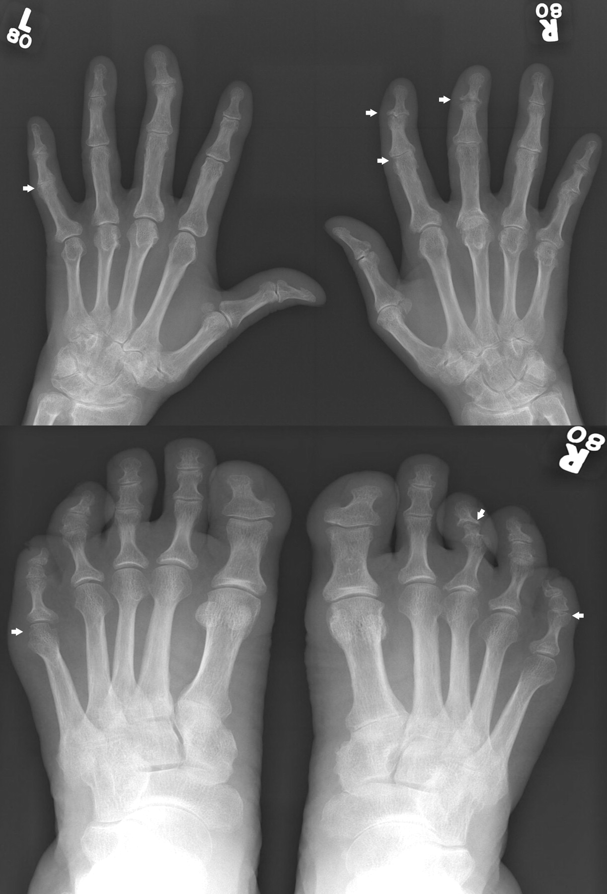
A 49-year-old woman with psoriasis since the age of 25 years developed pain and swelling in her fingers, wrists, and ankles and was diagnosed with PsA at age 42 years. Prior to her first visit to the PsA Clinic in 1996, she had been treated with and failed several nonsteroidal antiinflammatory medications as well as intramuscular gold and oral methotrexate (MTX) 15 mg per week. At her first visit to our PsA Clinic she was noted to have asymmetric polyarthritis with dactylitis and 2 clinically damaged joints. Radiographs demonstrated erosions in 7 joints, fluffy periostitis, and bilateral grade 1 sacroiliitis. The diagnosis of PsA was confirmed and the dose of MTX was increased, first orally and later by subcutaneous injections, to a maximal dose of 25 mg per week. Despite this she continued to have active polyarthritis and accumulated joint damage. Followup was irregular as she lived at a distance from the Clinic. In May 2005 she was found to have 21 actively inflamed joints, 10 of which were swollen; she also had 11 clinically damaged joints with 7 flail joints. She had accumulated extensive radiographic joint damage, with fluffy periostitis and numerous erosive changes, mainly in the fingers and toes, including some joints with a total destruction in the form of pencil-in-cup change (Figure 1). Erythrocyte sedimentation rate (ESR) at that time was high at 72 mm/h.

Figure 1.
  • Download figure
  • Open in new tab
  • Download powerpoint
Figure 1.

2005: Severe radiographic joint damage prior to starting anti-TNF therapy; large erosions, pencil-in-cup deformity, and periostitis.

MATERIALS AND METHODS

Etanercept was introduced in September 2005 by subcutaneous injections at a dose of 25 mg twice a week. Within several weeks of initiation of etanercept therapy, there was marked improvement in the peripheral joints symptomatically, and on physical examination there were no signs of active arthritis. ESR decreased to normal levels. She was able to stop treatment with MTX.

Radiographic assessments

Radiographs of the hands and feet were read by 3 observers by consensus using 3 methods: the modified Steinbrocker method, which is used in our clinic2; the van der Heijde (vdH) modification of the Sharp methods3; and the Ratingen scoring system4.

RESULTS

Radiographic damage scores are presented in Table 1. Plain radiographs of the hands and feet from May 2005 (Figure 1) scored 61 on the modified Steinbrocker, 59 by the vdH Sharp score (36 for erosions and 23 for joint space narrowing), and 86 by the Ratingen system (45 for erosion and 41 for proliferation). Radiographs from 2007 (Figure 2) showed marked improvement in erosion and proliferation scoring, with scores of 58 by the modified Steinbrocker, 38 by vdH Sharp score (16 for erosions and 22 for joint space narrowing), and 63 by the Ratingen (26 for erosion and 37 for proliferation). The patient remained in remission without any significant change in physical examination findings at her next visit to the clinic in July 2009. At that visit, radiographs of the hands and feet (Figure 3) showed additional improvement in erosion score and joint space narrowing, while bony proliferation remained the same [43 by modified Steinbrocker, 26 by the vdH Sharp score (12 for erosions and 14 for joint space narrowing), and 56 by the Ratingen (18 for erosion and 38 for proliferation)].

Figure 2.
  • Download figure
  • Open in new tab
  • Download powerpoint
Figure 2.

2007: Improvement in radiographic joint damage following 2 years of etanercept therapy is observed in the right hand, second and third distal interphalangeal (DIP) and proximal interphalangeal (PIP) joints; and in the left foot, fifth metatarsophalangeal (MTP) and PIP joints, and right foot, third DIP joint.

Figure 3.
  • Download figure
  • Open in new tab
  • Download powerpoint
Figure 3.

2009: Additional radiographic improvement after 4 years of etanercept therapy. Further improvement in joint damage is observed in the right hand second DIP, which now looks almost normal, the third DIP, which seems to have developed cartilage space, and PIP joints, which no longer demonstrate erosions; and in the left foot, fifth MTP and PIP joints, which seem to have normalized, and right foot, third DIP joint, where the erosions have resolved.

View this table:
  • View inline
  • View popup
Table 1.

Regression of radiographic joint damage following anti-tumor necrosis factor treatment.

DISCUSSION

PsA is characterized by inflammatory arthritis that often leads to significant structural damage. The radiographic features in PsA can be grouped into destructive and proliferative changes. Erosions are a typical destructive feature; they occur in up to 67% of patients at their first visit to the clinic5, and in 27% of patients within 5 months of onset of arthritis6. Widespread erosive changes can lead to the characteristic pencil-in-cup change. New bone formation is another typical feature of PsA, which may present as either periosteal reaction or ankylosis. Fluffy periostitis is included in the CASPAR criteria for classification of PsA7. These 2 seemingly opposing mechanisms, marked erosion and bone formation, can occur in the same patient and may present in different joints in the same finger.

Until the introduction of effective targeted therapy, particularly the anti-tumor necrosis factor (anti-TNF) agents, bone damage was considered irreversible. The concept of damage repair was introduced by the negative radiographic progression scores that were found in several clinical trials of anti-TNF agents8,9. This concept was supported by findings in several OMERACT studies10,11, as well as in cohort studies in rheumatoid arthritis (RA)12,13, suggesting that joint repair can occur. Thus, the concept of radiographic repair of joint damage following anti-TNF treatment is now being accepted as a real phenomenon.

In RA the prevalence of decreases in erosion scores following treatment with anti-TNF agents was reported to range from 7.2% to 10.7%11,12. Repair occurred particularly in clinically inactive joints and in patients with more severe joint damage. The studies have also showed that the repair process is localized, as some joints showed repair, whereas in others there was progression of damage.

In PsA, there is less information about damage repair; however, clinical trials with anti-TNF agents have shown that regression of joint scores does occur in a subset of the patients treated with these agents. A subanalysis of radiographic scores of the ADEPT trial, comparing adalimumab to placebo treatment, showed improvement in modified total Sharp score14. This change was most prominent among the patients treated with adalimumab, particularly among those with an elevated baseline C-reactive protein (CRP). Cumulative probability plots have shown that up to 10% of the patients with low CRP and approximately 20% of those with elevated CRP had improvement in the radiographic score after 24 weeks of treatment. Similar results were seen in subanalysis of changes in the modified total Sharp score study of etanercept15. Higher proportions of patients in the etanercept group had negative change in scores from baseline compared with the control group. The changes primarily reflected changes in the erosion score and not the joint space narrowing score.

Our patient, who had sustained severe radiographic joint damage, demonstrated a significant repair of the damage including refilling of bone erosions, resolution of periostitis, and reforming of almost intact joint structure, following treatment with an anti-TNF agent. This case emphasizes that resolution of severe radiographic damage can occur after effective treatment. It also highlights that the repair process may take several years. We note the utility of the radiographic methods in demonstrating improvement in joint scores. In particular, the modified Steinbrocker method, which is the simplest and is easier and less time-consuming, was able to reveal improvement that was documented in our patient. Further studies are needed to investigate the prevalence of this process among patients using anti-TNF agents and to determine the mechanisms that are involved.

Footnotes

  • Dr. Eder is supported by an Abbott Psoriatic Arthritis Fellowship and a Canadian Arthritis Network Fellowship; Dr. Chandran is supported by a Canadian Institutes for Health Research Clinical Research Initiative Fellowship. The Psoriatic Arthritis Program is supported by the Krembil Foundation and the Arthritis Society by a SPARCC/NRI grant.

  • Accepted for publication December 3, 2010.

REFERENCES

  1. 1.↵
    1. Ory PA,
    2. Gladman DD,
    3. Mease PJ
    . Psoriatic arthritis and imaging. Ann Rheum Dis 2005;64 Suppl 2:ii55–7.
    OpenUrlAbstract/FREE Full Text
  2. 2.↵
    1. Rahman P,
    2. Nguyen E,
    3. Cheung C,
    4. Schentag CT,
    5. Gladman DD
    . Comparison of radiological severity in psoriatic arthritis and rheumatoid arthritis. J Rheumatol 2001;28:1041–4.
    OpenUrlAbstract/FREE Full Text
  3. 3.↵
    1. van der Heijde D
    . How to read radiographs according to Sharp/van der Heijde method. J Rheumatol 2000;27:261–3.
    OpenUrlPubMed
  4. 4.↵
    1. van der Heijde DMFM,
    2. Sharp JT,
    3. Wassenberg S,
    4. Gladman DD
    . Psoriatic arthritis imaging: a review of scoring methods. Ann Rheum Dis 2005;64 Suppl 2:ii61–4.
    OpenUrlAbstract/FREE Full Text
  5. 5.↵
    1. Gladman DD,
    2. Shuckett R,
    3. Russell ML,
    4. Thorne JC,
    5. Schachter RK
    . Psoriatic arthritis (PSA) — an analysis of 220 patients. Q J Med 1987;62:127–41.
    OpenUrlAbstract/FREE Full Text
  6. 6.↵
    1. Kane D,
    2. Stafford L,
    3. Bresnihan B,
    4. FitzGerald O
    . A prospective, clinical and radiological study of early psoriatic arthritis: an early synovitis clinic experience. Rheumatology 2003;42:1460–8.
    OpenUrlAbstract/FREE Full Text
  7. 7.↵
    1. Taylor W,
    2. Gladman D,
    3. Helliwell P,
    4. Marchesoni A,
    5. Mease P,
    6. Mielants H;
    7. CASPAR Study Group
    . Classification criteria for psoriatic arthritis: development of new criteria from a large international study. Arthritis Rheum 2006;54:2665–73.
    OpenUrlCrossRefPubMed
  8. 8.↵
    1. Lipsky PE,
    2. van der Heijde DM,
    3. St. Clair EW,
    4. Furst DE,
    5. Breedveld FC,
    6. Kalden JR,
    7. et al.
    Infliximab and methotrexate in the treatment of rheumatoid arthritis. Anti-Tumor Necrosis Factor Trial in Rheumatoid Arthritis with Concomitant Therapy Study Group. N Engl J Med 2000;343:1594–602.
    OpenUrlCrossRefPubMed
  9. 9.↵
    1. Klareskog L,
    2. van der Heijde D,
    3. de Jager JP,
    4. Gough A,
    5. Kalden J,
    6. Malaise M,
    7. et al.
    Therapeutic effect of the combination of etanercept and methotrexate compared with each treatment alone in patients with rheumatoid arthritis: double-blind randomised controlled trial. Lancet 2004;363:675–8.
    OpenUrlCrossRefPubMed
  10. 10.↵
    1. Sharp JT,
    2. van der Heijde D,
    3. Boers M,
    4. Boonen A,
    5. Bruynesteyn K,
    6. Emery P,
    7. et al.
    Repair of erosions in rheumatoid arthritis does occur. Results from studies by the OMERACT Subcommittee on Healing of Erosions. J Rheumatol 2003;30:1102–7.
    OpenUrlAbstract/FREE Full Text
  11. 11.↵
    1. van der Heijde D,
    2. Landewé R,
    3. Sharp JT;
    4. OMERACT Subcommittee on Repair
    . Repair in rheumatoid arthritis, current status. Report of a workshop at OMERACT 8. J Rheumatol 2007;34:884–8.
    OpenUrlAbstract/FREE Full Text
  12. 12.↵
    1. van der Linden MP,
    2. Boja R,
    3. Klarenbeek NB,
    4. Huizinga TW,
    5. van der Heijde DM,
    6. van der Helm-van Mil AH
    . Repair of joint erosions in rheumatoid arthritis: prevalence and patient characteristics in a large inception cohort. Ann Rheum Dis 2010;69:727–9.
    OpenUrlAbstract/FREE Full Text
  13. 13.↵
    1. Ideguchi H,
    2. Ohno S,
    3. Hattori H,
    4. Senuma A,
    5. Ishigatsubo Y
    . Bone erosions in rheumatoid arthritis can be repaired through reduction in disease activity with conventional disease-modifying antirheumatic drugs. Arthritis Res Ther 2006;8:R76.
    OpenUrlCrossRefPubMed
  14. 14.↵
    1. Gladman DD,
    2. Mease PJ,
    3. Choy EH,
    4. Richlin CT,
    5. Perdok RJ,
    6. Sasso EH
    . Risk factors for radiographic progression in psoriatic arthritis: subanalysis of the randomized controlled trial ADEPT. Arthritis Res Ther 2010;12:R113.
    OpenUrlCrossRefPubMed
  15. 15.↵
    1. Mease PJ,
    2. Kivitz AJ,
    3. Burch FX,
    4. Siegel EL,
    5. Cohen SB,
    6. Ory P,
    7. et al.
    Continuous inhibition of radiographic progression in patients with psoriatic arthritis following 2 years of treatment with etanercept. J Rheumatol 2006;33:712–21.
    OpenUrlAbstract/FREE Full Text
PreviousNext
Back to top

In this issue

The Journal of Rheumatology
Vol. 38, Issue 6
1 Jun 2011
  • Table of Contents
  • Table of Contents (PDF)
  • Index by Author
  • Editorial Board (PDF)
Print
Download PDF
Article Alerts
Sign In to Email Alerts with your Email Address
Email Article

Thank you for your interest in spreading the word about The Journal of Rheumatology.

NOTE: We only request your email address so that the person you are recommending the page to knows that you wanted them to see it, and that it is not junk mail. We do not capture any email address.

Enter multiple addresses on separate lines or separate them with commas.
Repair of Radiographic Joint Damage Following Treatment with Etanercept in Psoriatic Arthritis Is Demonstrable by 3 Radiographic Methods
(Your Name) has forwarded a page to you from The Journal of Rheumatology
(Your Name) thought you would like to see this page from the The Journal of Rheumatology web site.
CAPTCHA
This question is for testing whether or not you are a human visitor and to prevent automated spam submissions.
Citation Tools
Repair of Radiographic Joint Damage Following Treatment with Etanercept in Psoriatic Arthritis Is Demonstrable by 3 Radiographic Methods
LIHI EDER, VINOD CHANDRAN, DAFNA D. GLADMAN
The Journal of Rheumatology Jun 2011, 38 (6) 1066-1070; DOI: 10.3899/jrheum.100919

Citation Manager Formats

  • BibTeX
  • Bookends
  • EasyBib
  • EndNote (tagged)
  • EndNote 8 (xml)
  • Medlars
  • Mendeley
  • Papers
  • RefWorks Tagged
  • Ref Manager
  • RIS
  • Zotero

 Request Permissions

Share
Repair of Radiographic Joint Damage Following Treatment with Etanercept in Psoriatic Arthritis Is Demonstrable by 3 Radiographic Methods
LIHI EDER, VINOD CHANDRAN, DAFNA D. GLADMAN
The Journal of Rheumatology Jun 2011, 38 (6) 1066-1070; DOI: 10.3899/jrheum.100919
del.icio.us logo Digg logo Reddit logo Twitter logo CiteULike logo Facebook logo Google logo Mendeley logo
  • Tweet Widget
  • Facebook Like
  • Google Plus One
Bookmark this article

Jump to section

  • Article
    • Abstract
    • MATERIALS AND METHODS
    • RESULTS
    • DISCUSSION
    • Footnotes
    • REFERENCES
  • Figures & Data
  • References
  • Info & Metrics
  • PDF
  • eLetters

Related Articles

Cited By...

More in this TOC Section

  • Follow-up Contrast-Enhanced Ultrasonography of the Carotid Artery in Patients With Takayasu Arteritis: A Retrospective Study
  • Disease Flare of Systemic Lupus Erythematosus in Patients With Endstage Renal Disease on Dialysis
  • A Practical Guide for Assessment of Skin Burden in Patients With Psoriatic Arthritis
Show more Articles

Similar Articles

Content

  • First Release
  • Current
  • Archives
  • Collections
  • Audiovisual Rheum
  • COVID-19 and Rheumatology

Resources

  • Guide for Authors
  • Submit Manuscript
  • Author Payment
  • Reviewers
  • Advertisers
  • Classified Ads
  • Reprints and Translations
  • Permissions
  • Meetings
  • FAQ
  • Policies

Subscribers

  • Subscription Information
  • Purchase Subscription
  • Your Account
  • Terms and Conditions

More

  • About Us
  • Contact Us
  • My Alerts
  • My Folders
  • Privacy/GDPR Policy
  • RSS Feeds
The Journal of Rheumatology
The content of this site is intended for health care professionals.
Copyright © 2022 by The Journal of Rheumatology Publishing Co. Ltd.
Print ISSN: 0315-162X; Online ISSN: 1499-2752
Powered by HighWire