
Main menu

  • Home
  • Content
    • First Release
    • Current
    • Archives
    • Collections
    • Audiovisual Rheum
    • 50th Volume Reprints
  • Resources
    • Guide for Authors
    • Submit Manuscript
    • Payment
    • Reviewers
    • Advertisers
    • Classified Ads
    • Reprints and Translations
    • Permissions
    • Meetings
    • FAQ
    • Policies
  • Subscribers
    • Subscription Information
    • Purchase Subscription
    • Your Account
    • Terms and Conditions
  • About Us
    • About Us
    • Editorial Board
    • Letter from the Editor
    • Duncan A. Gordon Award
    • Privacy/GDPR Policy
    • Accessibility
  • Contact Us
  • JRheum Supplements
  • Services

User menu

  • My Cart
  • Log In

Search

  • Advanced search
The Journal of Rheumatology
  • JRheum Supplements
  • Services
  • My Cart
  • Log In
The Journal of Rheumatology

Advanced Search

  • Home
  • Content
    • First Release
    • Current
    • Archives
    • Collections
    • Audiovisual Rheum
    • 50th Volume Reprints
  • Resources
    • Guide for Authors
    • Submit Manuscript
    • Payment
    • Reviewers
    • Advertisers
    • Classified Ads
    • Reprints and Translations
    • Permissions
    • Meetings
    • FAQ
    • Policies
  • Subscribers
    • Subscription Information
    • Purchase Subscription
    • Your Account
    • Terms and Conditions
  • About Us
    • About Us
    • Editorial Board
    • Letter from the Editor
    • Duncan A. Gordon Award
    • Privacy/GDPR Policy
    • Accessibility
  • Contact Us
  • Follow Jrheum on BlueSky
  • Follow jrheum on Twitter
  • Visit jrheum on Facebook
  • Follow jrheum on LinkedIn
  • Follow jrheum on YouTube
  • Follow jrheum on Instagram
  • Follow jrheum on RSS
Review ArticleReview

IgG4-related Diseases Including Mikulicz’s Disease and Sclerosing Pancreatitis: Diagnostic Insights

YASUFUMI MASAKI, SUSUMU SUGAI and HISANORI UMEHARA
The Journal of Rheumatology July 2010, 37 (7) 1380-1385; DOI: https://doi.org/10.3899/jrheum.091153
YASUFUMI MASAKI
  • Find this author on Google Scholar
  • Find this author on PubMed
  • Search for this author on this site
  • For correspondence: yasum{at}kanazawa-med.ac.jp
SUSUMU SUGAI
  • Find this author on Google Scholar
  • Find this author on PubMed
  • Search for this author on this site
HISANORI UMEHARA
  • Find this author on Google Scholar
  • Find this author on PubMed
  • Search for this author on this site
  • Article
  • Figures & Data
  • Info & Metrics
  • References
  • PDF
PreviousNext
Loading

Abstract

Since the first report of serum IgG4 elevation in sclerosing pancreatitis in 2001, various systemic disorders have been reported to elevate IgG4, and many names have been proposed from the perspective of the systemic condition. Despite similarities in the organs damaged in IgG4-related Mikulicz’s disease and Sjögren’s syndrome, there are marked clinical and pathological differences between the 2 entities. The majority of cases diagnosed with autoimmune pancreatitis in Japan are IgG4-related sclerosing pancreatitis, and it should be recognized that this is distinct from the Western type. Diagnosis of IgG4-related disease is defined by both elevated serum IgG4 (> 1.35 g/l) and histopathological features, including lymphocyte and IgG4+ plasma cell infiltration (IgG4+ plasma cells/IgG+ plasma cells > 50% on a highly magnified slide checked at 5 points). Differential diagnosis from other distinct disorders is necessary: these include sarcoidosis, Castleman’s disease, Wegener’s granulomatosis, lymphoma, cancer, and other existing conditions. The Japanese IgG4 research group has begun multicenter prospective studies to improve diagnostic criteria and treatment strategies.

  • MIKULICZ’S DISEASE
  • SJÖGREN’S SYNDROME
  • AUTOIMMUNE PANCREATITIS
  • GLUCOCORTICOID
  • IgG4-RELATED DISEASES

Mikulicz’s disease (MD) was first described in 1892 in a man with symmetrical swelling of the lacrimal, submandibular, and parotid glands1. Morgan, et al reported 18 cases of MD and concluded that it was not a distinct clinical and pathological disease entity but merely one manifestation of a more generalized symptom complex known as Sjögren’s syndrome (SS)2. With the wide acceptance of the conclusions of Morgan, et al there have been few reports of MD in Western countries. However, many cases of MD have been reported in Japan, and there has been considerable discussion regarding the differences between MD and SS3–7. Patients with MD have been reported to have a point mutation in the FasL gene, which may account for their mild sicca symptoms despite massive lymphocytic infiltration3. Further, high IgG4 concentrations have been reported in the sera of patients with MD4, suggesting that MD is an IgG4-related disease.

We describe the differences between MD (especially IgG4-related MD) and SS, and refer to other systemic complications of IgG4-related diseases.

Differences between IgG4+ MOLPS and SS

As so-called MD may include various conditions3–6 and consist of IgG4-related or unrelated subtypes, the IgG4+ multiorgan lymphoproliferative syndrome (MOLPS)/MD research group has established tentative criteria for IgG4+ MD (Table 1).

View this table:
  • View inline
  • View popup
Table 1.

Diagnostic criteria of IgG4+ Mikulicz’s disease (Japanese Sjögren’s Syndrome Society, 2008). Differential diagnosis is necessary from other distinct disorders, including sarcoidosis, Castleman’s disease, Wegener’s granulomatosis, lymphoma, and cancer. The diagnostic criteria for Sjögren’s syndrome (SS) may also include some patients with IgG4+ Mikulicz’s disease; however, the clinicopathological conditions of patients with typical SS and IgG4+ Mikulicz’s disease are different.

MATERIALS AND METHODS

We collected data on 64 patients with IgG4+ MOLPS including MD and performed retrospective analysis to clarify the differences between IgG4+ MOLPS and definite SS (Table 2)7. Despite similarities in the involved organs, there are marked differences between IgG4+ MOLPS and SS. For example, their sex distributions were quite different. Men with SS were very rare (2 of 31), while almost half (31 of 64) the patients with IgG4+ MOLPS were men.

View this table:
  • View inline
  • View popup
Table 2.

Comparison of symptoms, complaints, and laboratory findings in IgG4+ MOLPS and typical SS. Data are percentage (number) unless stated otherwise. Incidence rates (numbers of positive patients) are shown for xerophthalmia, xerostomia, arthralgia, allergic rhinitis, bronchial asthma, sclerosing pancreatitis, interstitial nephritis, interstitial pneumonitis, RF, ANA, A-SSA, A-SSB, and low CH50. Masaki Y, et al7. Ann Rheum Dis 2009; 68:1310–5. Adapted with permission.

RESULTS

Significantly fewer patients with IgG4+ MOLPS than with SS showed symptoms of xerostomia, xerophthalmia, and arthralgia. Patients with IgG4+ MOLPS showed significantly lower incidences of rheumatoid factor (RF), antinuclear antibody (ANA), anti-SSA/Ro antibody, and anti-SSB/La antibody than patients with SS. We found that not only IgG4 but also total IgG, IgG2, and IgE concentrations were significantly higher in patients with IgG4+ MOLPS than in patients with SS7. Almost half of patients with IgG4+ MOLPS demonstrated low CH50, which apparently correlated with hyper-IgG (especially IgG1 and IgG2).

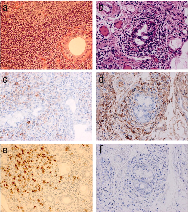
Histological specimens from patients with IgG4+MOLPS showed marked IgG4+ plasma cell infiltration with occasional lymphocyte follicular formation, but without lymphoepithelial lesions (Figure 1)7. This may explain the marked glandular swelling without severe dryness in patients with IgG4+ MOLPS. Importantly, treatment with glucocorticoids resulted in marked clinical improvements in almost all patients with IgG4+ MOLPS, while the effects of glucocorticoids on SS were not so dramatic10.

Figure 1.
  • Download figure
  • Open in new tab
  • Download powerpoint
Figure 1.

Histopathological findings of labial minor salivary gland biopsy in patients with IgG4+ MOLPS/Mikulicz’s disease (a, c, e) and Sjögren’s syndrome (b, d, f). (a, b) Hematoxylin and eosin staining; (c, d) IgG immunostaining; (e, f) IgG4 immunostaining. (a) Massive lymphocyte and plasmacyte infiltration and lymphoid follicle formation were seen in IgG4+ MOLPS. The ducts remained clear without lymphocytic infiltration. Both IgG+ and IgG4+ plasma cells were scattered in the periphery of the follicles (c, e). In contrast, there were few or no IgG4+ cells in typical SS (d, f), not even in patients with severe lymphocytic infiltration (b).

Autoimmune pancreatitis and IgG4

Autoimmune pancreatitis (AIP) is a unique form of chronic pancreatitis, first described by Sarles, et al in 196111 and characterized by infrequent attacks of abdominal pain, jaundice, irregular narrowing of the pancreatic duct, and swelling of the pancreatic parenchyma11–22. Kawaguchi, et al described cases complicated with similar pathological features in the common bile duct, gall bladder, and minor salivary glands, suggesting a systemic disorder12. Yoshida, et al described the typical features of AIP as hyper-γ-globulinemia, the presence of autoantibodies (RF and ANA), lymphocytic infiltration of pancreas tissue, coexistence of other manifestations such as sicca complex, and good responsiveness to glucocorticoids13. AIP is now known to be associated with types of sialadenitis and cholangitis distinct from SS and primary sclerosing cholangitis.

In 2001, Hamano, et al first reported high serum IgG4 concentrations in patients with sclerosing pancreatitis14. Further, massive IgG4+ plasmacytic infiltration in the pancreatic tissue was reported15. There have been many recent reports of AIP in Asia12–19 and in Western countries20,21.

Various diagnostic criteria for AIP have been proposed in Japan23, Korea17, and the United States (Mayo Clinic)21. In 2008, the Japan-Korea Symposium on AIP proposed Asian diagnostic criteria19. Further international criteria are currently under discussion.

IgG4 and other clinical conditions (Figure 2)

Figure 2.
  • Download figure
  • Open in new tab
  • Download powerpoint
Figure 2.

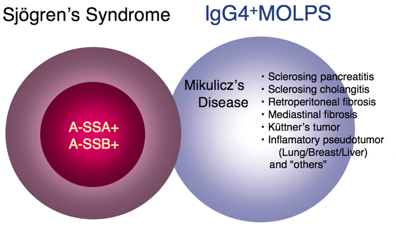
IgG4+ MOLPS should be defined as a distinct clinicopathological entity that includes Mikulicz’s disease (MD), autoimmune pancreatitis (AIP), sclerosing cholangitis, and other clinical conditions with good response to glucocorticoids. Although the diagnostic criteria of SS may include some patients with IgG4+ MOLPS/MD, typical SS and IgG4+ MOLPS/MD are different clinical conditions.

Hyper-IgG4-γ-globulinemia and IgG4+ plasma cell infiltration with sclerotic lesions, although first reported in patients with sclerosing pancreatitis, have also been reported in patients with many other disorders, including sclerosing cholangitis15,16; inflammatory pseudotumors of the lung24, liver16, and breast16,25; retroperitoneal or mediastinal fibrosis26; interstitial nephritis27; hypophysitis5; sclerosing dacryoadenitis28; sialadenitis (MD and Küttner’s tumor)4,5,29; inflammatory aortic aneurysm30,31; tumorous lesions of the coronary artery31; lymphadenopathy32; and many other inflammatory conditions in multiple organs.

In addition, various systemic involvements have been reported in each disorder. Kawaguchi, et al12 noted the same etiology between autoimmune pancreatitis and multifocal idiopathic fibrosclerosis (MIF) reported by Comings, et al33 because both conditions include occlusive phlebitis and sclerotic lesions.

DISCUSSION

Proposal of a new clinical entity, IgG4+ MOLPS, as a more generalized disorder

In addition to the term “IgG4+ MOLPS,” there are many synonyms, such as MIF, IgG4-related autoimmune disease15, IgG4-related plasmacytic disease6, and IgG4-related sclerosing disease18, all of which may refer to the same conditions.

Although various other disorders have been associated with hyper-IgG4-γ-globulinemia, including multicentric Castleman’s disease34, Wegener’s granulomatosis35, lymphoma36,37, and cancer38, IgG4+ MOLPS should be defined as a distinct clinicopathological entity, characterized by sclerosing sialadenitis and dacryoadenitis, AIP, sclerosing cholangitis, and other clinical conditions with good response to glucocorticoids.

Hypothetical mechanism of IgG4+ MOLPS

At present, the pathogenesis of IgG4+ MOLPS is not clear. Although some patients are positive for RF and ANA, these incidences are significantly lower than in SS, suggesting that RF and ANA positivity may be due to nonspecific immunoglobulin binding. Although IgG4+ MOLPS is accompanied by various immunological disorders, including AIP, there is little evidence that IgG4+ MOLPS is an autoimmune disorder because of the lack of disease-specific autoantibodies.

The role of IgG4 in IgG4+ MOLPS is still unknown. IgG4 represents the smallest population among IgG subclasses in the sera of normal subjects (3%–6% of total IgG), and is unique among the IgG subclasses in its inability to bind with the C1q complement39. IgG4 is associated with the pathogenicity of a small number of disorders, such as atopic dermatitis, parasitic disease, pemphigus vulgaris, and pemphigus foliaceus.

In clonality analysis, most tissue-infiltrating and circulating IgG4-positive cells are polyclonal40. These findings have suggested that IgG4 does not play a major pathological role in IgG4+ MOLPS, and that there may be other upstream regulators in its pathogenesis.

Zen, et al reported that the pathogenesis of IgG4-related AIP was characterized by the infiltration of T helper 2 and regulatory T cells (Treg), which secrete various cytokines such as interleukin 10 (IL-10) and tumor growth factor-ß (TGF-ß)41. Moreover, the level of Foxp3 messenger RNA expression was significantly increased in patients with AIP, and immunohistochemical staining revealed increases in the numbers of CD4+ CD25+ Foxp3+ cells. Treg may be involved in the in situ production of IL-10 and TGF-ß, which could be followed by IgG4 class switching and fibroplasia41.

The concentrations of IgG2, IgG4, and IgE have been shown to be significantly higher in patients with IgG4+ MOLPS than in those with typical SS, while the concentrations of IgG1, IgG3, IgA, and IgM were significantly higher in patients with typical SS than in those with IgG4+ MOLPS7. The immunoglobulin gene fragments Cμ, Cδ, Cγ3, Cγ1, Cα1, Cγ2, Cγ4, Cɛ, and Cα2, which encode IgM, IgD, IgG3, IgG1, IgA1, IgG2, IgG4, IgE, and IgA2, respectively, are arranged linearly in this order from upstream to downstream. Gene linkage and different class-switch mechanisms may cause the hyperproduction of the different immunoglobulin subclasses observed in these 2 diseases, which may contribute to the pathophysiology of IgG4+ MOLPS.

Future perspectives

Although IgG4+ MOLPS may be distributed worldwide, this disease entity has not been well recognized to date. Most reports on IgG4-related diseases have been from Japan, while many reports on AIP have come from Western countries, especially the Mayo Clinic21 in the United States. Therefore, we believe that an international consensus regarding IgG4-related diseases as new clinical entities is required.

In this regard, the Japanese IgG4 research group (Research Committee of Intractable Diseases, Health and Labor Sciences Research Grants, Ministry of Health, Labor and Welfare, Japan) has begun multicenter prospective clinical studies (UMIN: R000002820, R000002823) to formulate better diagnostic criteria, to identify novel diagnostic and prognostic factors, and to design better treatment strategies.

Acknowledgments

We thank all participants in the IgG4+ MOLPS/Mikulicz’s Disease Research Group and the researchers of the Autoimmune Pancreatitis Group for critical discussion.

Footnotes

  • Supported by grants from Intractable Diseases, the Health and Labor Sciences Research Grants from the Ministry of Health, Labor, and Welfare, and the Japanese Ministry of Education, Culture, Sports, Science, and Technology (13557160, 15024236, 15390313, and 13877075 to H. Umehara and 17591060 to Y. Masaki), Umehara Memorial Foundation (to H. Umehara), The Vehicle Racing Commemorative Foundation, and the Kanazawa Medical University Research Foundation (C2009-4 to H. Umehara and S2004-16 and S2007-5 to Y. Masaki).

  • Accepted for publication February 8, 2010.

REFERENCES

  1. 1.↵
    1. Mikulicz J.
    Über eine eigenartige symmetrische erkrankung der tränen und mundspeicheldrüsen. Stuttgart: Beitr.z.Chir.Fesrschr.f. Theodor Billroth; 1892:610–30.
  2. 2.↵
    1. Morgan WS,
    2. Castleman B
    . A clinicopathologic study of Mikulicz’s disease. Am J Pathol 1953;29:471–503.
    OpenUrlPubMed
  3. 3.↵
    1. Tsubota K,
    2. Fujita H,
    3. Tadano K,
    4. Onoda N,
    5. Tsuzaka K,
    6. Takeuchi T
    . Abnormal expression and function of Fas ligand of lacrimal glands and peripheral blood in Sjögren’s syndrome patients with enlarged exocrine glands. Clin Exp Immunol 2002;129:177–82.
    OpenUrlCrossRefPubMed
  4. 4.↵
    1. Yamamoto M,
    2. Ohara M,
    3. Suzuki C,
    4. Naishiro Y,
    5. Yamamoto H,
    6. Takahashi H,
    7. et al.
    Elevated IgG4 concentrations in serum of patients with Mikulicz’s disease. Scand J Rheumatol 2004;33:432–3.
    OpenUrlCrossRefPubMed
  5. 5.↵
    1. Yamamoto M,
    2. Ohara M,
    3. Suzuki C,
    4. Naishiro Y,
    5. Yamamoto H,
    6. Takahashi H,
    7. et al.
    A case of Mikulicz’s disease (IgG4-related plasmacytic disease) complicated by autoimmune hypophysitis. Scand J Rheumatol 2006;35:410–1.
    OpenUrlCrossRefPubMed
  6. 6.↵
    1. Yamamoto M,
    2. Takahashi H,
    3. Ohara M,
    4. Suzuki C,
    5. Naishiro Y,
    6. Yamamoto H,
    7. et al.
    A new conceptualization for Mikulicz’s disease as an IgG4-related plasmacytic disease. Mod Rheumatol 2006;16:335–40.
    OpenUrlCrossRefPubMed
  7. 7.↵
    1. Masaki Y,
    2. Dong L,
    3. Kurose N,
    4. Kitagawa K,
    5. Morikawa Y,
    6. Yamamoto M,
    7. et al.
    Proposal for a new clinical entity, IgG4-positive multiorgan lymphoproliferative syndrome: Analysis of 64 cases of IgG4-related disorders. Ann Rheum Dis 2009;68:1310–5.
    OpenUrlAbstract/FREE Full Text
  8. 8.↵
    1. Fujibayashi T,
    2. Sugai S,
    3. Tojo T,
    4. Miyawaki S,
    5. Miyasaka N,
    6. Ichikawa Y,
    7. et al.
    Revised Japanese criteria for Sjögren’s syndrome (1999): availability and validity. Mod Rheumatol 2004;14:425–34.
    OpenUrlCrossRef
  9. 9.↵
    1. Vitali C,
    2. Bombardieri S,
    3. Jonsson R,
    4. Moutsopoulos HM,
    5. Alexander EL,
    6. Carsons SE,
    7. et al.
    European Study Group on Classification Criteria for Sjögren’s Syndrome. Classification criteria for Sjögren’s syndrome: a revised version of the European criteria proposed by the American-European Consensus Group. Ann Rheum Dis 2002;61:554–8.
    OpenUrlAbstract/FREE Full Text
  10. 10.↵
    1. Miyawaki S,
    2. Nishiyama S,
    3. Matoba K
    . Efficacy of low-dose prednisolone maintenance for saliva production and serological abnormalities in patients with primary Sjögren’s syndrome. Intern Med 1999;38:938–43.
    OpenUrlPubMed
  11. 11.↵
    1. Sarles H,
    2. Sarles JC,
    3. Muratore R,
    4. Guien C
    . Chronic inflammatory sclerosis of the pancreas — an autonomous pancreatic disease? Am J Dig Dis 1961;6:688–98.
    OpenUrlCrossRefPubMed
  12. 12.↵
    1. Kawaguchi K,
    2. Koike M,
    3. Tsuruta K,
    4. Okamoto A,
    5. Tabata I,
    6. Fujita N
    . Lymphoplasmacytic sclerosing pancreatitis with cholangitis: a variant of primary sclerosing cholangitis extensively involving pancreas. Hum Pathol 1991;22:387–95.
    OpenUrlCrossRefPubMed
  13. 13.↵
    1. Yoshida K,
    2. Toki F,
    3. Takeuchi T,
    4. Watanabe S,
    5. Shiratori K,
    6. Hayashi N
    . Chronic pancreatitis caused by an autoimmune abnormality. Proposal of the concept of autoimmune pancreatitis. Dig Dis Sci 1995;40:1561–8.
    OpenUrlCrossRefPubMed
  14. 14.↵
    1. Hamano H,
    2. Kawa S,
    3. Horiuchi A,
    4. Unno H,
    5. Furuya N,
    6. Akamatsu T,
    7. et al.
    High serum IgG4 concentrations in patients with sclerosing pancreatitis. N Engl J Med 2001;344:732–8.
    OpenUrlCrossRefPubMed
  15. 15.↵
    1. Kamisawa T,
    2. Funata N,
    3. Hayashi Y,
    4. Eishi Y,
    5. Koike M,
    6. Tsuruta K,
    7. et al.
    A new clinicopathological entity of IgG4-related autoimmune disease. J Gastroenterol 2003;38:982–4.
    OpenUrlCrossRefPubMed
  16. 16.↵
    1. Zen Y,
    2. Harada K,
    3. Sasaki M,
    4. Sato Y,
    5. Tsuneyama K,
    6. Haratake J,
    7. et al.
    IgG4-related sclerosing cholangitis with and without hepatic inflammatory pseudotumor, and sclerosing pancreatitis-associated sclerosing cholangitis: do they belong to a spectrum of sclerosing pancreatitis? Am J Surg Pathol 2004;28:1193–203.
    OpenUrlCrossRefPubMed
  17. 17.↵
    1. Kim KP,
    2. Kim MH,
    3. Kim JC,
    4. Lee SS,
    5. Seo DW,
    6. Lee SK
    . Diagnostic criteria for autoimmune chronic pancreatitis revisited. World J Gastroenterol 2006;12:2487–96.
    OpenUrlPubMed
  18. 18.↵
    1. Kamisawa T,
    2. Okamoto A
    . Autoimmune pancreatitis: proposal of IgG4-related sclerosing disease. J Gastroenterol 2006;41:613–25.
    OpenUrlCrossRefPubMed
  19. 19.↵
    1. Otsuki M,
    2. Chung JB,
    3. Okazaki K,
    4. Kim MH,
    5. Kamisawa T,
    6. Kawa S,
    7. et al.
    Research Committee of Intractable Pancreatic Diseases provided by the Ministry of Health, Labor and Welfare of Japan and the Korean Society of Pancreatobiliary Diseases. Asian diagnostic criteria for autoimmune pancreatitis: consensus of the Japan-Korea Symposium on Autoimmune Pancreatitis. J Gastroenterol 2008;43:403–8.
    OpenUrlCrossRefPubMed
  20. 20.↵
    1. Notohara K,
    2. Burgart LJ,
    3. Yadav D,
    4. Chari S,
    5. Smyrk TC
    . Idiopathic chronic pancreatitis with periductal lymphoplasmacytic infiltration: clinicopathologic features of 35 cases. Am J Surg Pathol 2003;27:1119–27.
    OpenUrlCrossRefPubMed
  21. 21.↵
    1. Chari ST,
    2. Smyrk TC,
    3. Levy MJ,
    4. Topazian MD,
    5. Takahashi N,
    6. Zhang L,
    7. et al.
    Diagnosis of autoimmune pancreatitis: The Mayo Clinic experience. Clin Gastroenterol Hepatol 2006;4:1010–6.
    OpenUrlCrossRefPubMed
  22. 22.↵
    1. Okazaki K,
    2. Kawa S,
    3. Kamisawa T,
    4. Ito T,
    5. Inui K,
    6. Irie H,
    7. et al.
    Japanese clinical guidelines for autoimmune pancreatitis. Pancreas 2009;38:849–66.
    OpenUrlCrossRefPubMed
  23. 23.↵
    1. Okazaki K,
    2. Kawa S,
    3. Kamisawa T,
    4. Naruse S,
    5. Tanaka S,
    6. Nishimori I,
    7. et al.
    Research Committee of Intractable Diseases of the Pancreas. Clinical diagnostic criteria of autoimmune pancreatitis: revised proposal. J Gastroenterol 2006;41:626–31.
    OpenUrlCrossRefPubMed
  24. 24.↵
    1. Zen Y,
    2. Kitagawa S,
    3. Minato H,
    4. Kurumaya H,
    5. Katayanagi K,
    6. Masuda S,
    7. et al.
    IgG4-positive plasma cells in inflammatory pseudotumor (plasma cell granuloma) of the lung. Hum Pathol 2005;36:710–7.
    OpenUrlCrossRefPubMed
  25. 25.↵
    1. Zen Y,
    2. Kasahara Y,
    3. Horita K,
    4. Miyayama S,
    5. Miura S,
    6. Kitagawa S,
    7. et al.
    Inflammatory pseudotumor of the breast in a patient with a high serum IgG4 level: histologic similarity to sclerosing pancreatitis. Am J Surg Pathol 2005;29:275–8.
    OpenUrlCrossRefPubMed
  26. 26.↵
    1. Zen Y,
    2. Sawazaki A,
    3. Miyayama S,
    4. Notsumata K,
    5. Tanaka N,
    6. Nakanuma Y
    . A case of retroperitoneal and mediastinal fibrosis exhibiting elevated levels of IgG4 in the absence of sclerosing pancreatitis (autoimmune pancreatitis). Hum Pathol 2006;37:239–43.
    OpenUrlCrossRefPubMed
  27. 27.↵
    1. Saeki T,
    2. Saito A,
    3. Yamazaki H,
    4. Emura I,
    5. Imai N,
    6. Ueno M,
    7. et al.
    Tubulointerstitial nephritis associated with IgG4-related systemic disease. Clin Exp Nephrol 2007;11:168–73.
    OpenUrlCrossRefPubMed
  28. 28.↵
    1. Cheuk W,
    2. Yuen HK,
    3. Chan JK
    . Chronic sclerosing dacryoadenitis: part of the spectrum of IgG4-related sclerosing disease? Am J Surg Pathol 2007;31:643–5.
    OpenUrlCrossRefPubMed
  29. 29.↵
    1. Kitagawa S,
    2. Zen Y,
    3. Harada K,
    4. Sasaki M,
    5. Sato Y,
    6. Minato H,
    7. et al.
    Abundant IgG4-positive plasma cell infiltration characterizes chronic sclerosing sialadenitis (Küttner’s tumor). Am J Surg Pathol 2005;29:783–91.
    OpenUrlCrossRefPubMed
  30. 30.↵
    1. Kasashima S,
    2. Zen Y,
    3. Kawashima A,
    4. Konishi K,
    5. Sasaki H,
    6. Endo M,
    7. et al.
    Inflammatory abdominal aortic aneurysm: close relationship to IgG4-related periaortitis. Am J Surg Pathol 2008;32:197–204.
    OpenUrlCrossRefPubMed
  31. 31.↵
    1. Matsumoto Y,
    2. Kasashima S,
    3. Kawashima A,
    4. Sasaki H,
    5. Endo M,
    6. Kawakami K,
    7. et al.
    A case of multiple immunogloblin G4-related periarteritis: a tumorous lesion of the coronary artery and abdominal aortic aneurysm. Hum Pathol 2008;39:975–80.
    OpenUrlCrossRefPubMed
  32. 32.↵
    1. Cheuk W,
    2. Yuen HK,
    3. Chu SY,
    4. Chiu EK,
    5. Lam LK,
    6. Chan JK
    . Lymphadenopathy of IgG4-related sclerosing disease. Am J Surg Pathol 2008;32:671–81.
    OpenUrlCrossRefPubMed
  33. 33.↵
    1. Comings DE,
    2. Skubi KB,
    3. Van Eyes J,
    4. Motulsky AG
    . Familial multifocal fibrosclerosis. Findings suggesting that retroperitoneal fibrosis, mediastinal fibrosis, sclerosing cholangitis, Riedel’s thyroiditis, and pseudotumor of the orbit may be different manifestations of a single disease. Ann Intern Med 1967;66:884–92.
    OpenUrlCrossRefPubMed
  34. 34.↵
    1. Ishida F,
    2. Kitano K,
    3. Kobayashi H,
    4. Saito H,
    5. Kiyosawa K
    . Elevated IgG4 levels in a case with multicentric Castleman’s disease. Br J Haematol 1997;99:981–2.
    OpenUrlPubMed
  35. 35.↵
    1. Brouwer E,
    2. Tervaert JW,
    3. Horst G,
    4. Huitema MG,
    5. van der Giessen M,
    6. Limburg PC,
    7. et al.
    Predominance of IgG1 and IgG4 subclass of anti-neutrophil cytoplasmic autoantibodies (ANCA) in patients with Wegener’s granulomatosis and clinically related disorders. Clin Exp Immunol 1991;83:379–86.
    OpenUrlPubMed
  36. 36.↵
    1. Cheuk W,
    2. Yuen HK,
    3. Chan AC,
    4. Shih LY,
    5. Kuo TT,
    6. Ma MW,
    7. et al.
    Ocular adnexal lymphoma associated with IgG4+ chronic sclerosing dacryoadenitis: a previously undescribed complication of IgG4-related sclerosing disease. Am J Surg Pathol 2008;32:1159–67.
    OpenUrlCrossRefPubMed
  37. 37.↵
    1. Sato Y,
    2. Ohshima K,
    3. Ichimura K,
    4. Sato M,
    5. Yamadori I,
    6. Tanaka T,
    7. et al.
    Ocular adnexal IgG4-related disease has uniform clinicopathology. Pathol Int 2008;58:465–70.
    OpenUrlCrossRefPubMed
  38. 38.↵
    1. Oh HC,
    2. Kim JG,
    3. Kim JW,
    4. Lee KS,
    5. Kim MK,
    6. Chi KC,
    7. et al.
    Early bile duct cancer in a background of sclerosing cholangitis and autoimmune pancreatitis. Intern Med 2008;47:2025–8.
    OpenUrlCrossRefPubMed
  39. 39.↵
    1. van der Zee JS,
    2. van Swieten P,
    3. Aalberse RC
    . Inhibition of complement activation by IgG4 antibodies. Clin Exp Immunol 1986;64:415–22.
    OpenUrlPubMed
  40. 40.↵
    1. Yamada K,
    2. Kawano M,
    3. Inoue R,
    4. Hamano R,
    5. Kakuchi Y,
    6. Fujii H,
    7. et al.
    Clonal relationship between infiltrating immunoglobulin G4 (IgG4)-positive plasma cells in lacrimal glands and circulating IgG4-positive lymphocytes in Mikulicz’s disease. Clin Exp Immunol 2008;152:432–9.
    OpenUrlPubMed
  41. 41.↵
    1. Zen Y,
    2. Fujii T,
    3. Harada K,
    4. Kawano M,
    5. Yamada K,
    6. Takahira M,
    7. et al.
    Th2 and regulatory immune reactions are increased in immunoglobulin G4-related sclerosing pancreatitis and cholangitis. Hepatology 2007;45:1538–46.
    OpenUrlCrossRefPubMed
PreviousNext
Back to top

In this issue

The Journal of Rheumatology
Vol. 37, Issue 7
1 Jul 2010
  • Table of Contents
  • Table of Contents (PDF)
  • Index by Author
  • Editorial Board (PDF)
Print
Download PDF
Article Alerts
Sign In to Email Alerts with your Email Address
Email Article

Thank you for your interest in spreading the word about The Journal of Rheumatology.

NOTE: We only request your email address so that the person you are recommending the page to knows that you wanted them to see it, and that it is not junk mail. We do not capture any email address.

Enter multiple addresses on separate lines or separate them with commas.
IgG4-related Diseases Including Mikulicz’s Disease and Sclerosing Pancreatitis: Diagnostic Insights
(Your Name) has forwarded a page to you from The Journal of Rheumatology
(Your Name) thought you would like to see this page from the The Journal of Rheumatology web site.
CAPTCHA
This question is for testing whether or not you are a human visitor and to prevent automated spam submissions.
Citation Tools
IgG4-related Diseases Including Mikulicz’s Disease and Sclerosing Pancreatitis: Diagnostic Insights
YASUFUMI MASAKI, SUSUMU SUGAI, HISANORI UMEHARA
The Journal of Rheumatology Jul 2010, 37 (7) 1380-1385; DOI: 10.3899/jrheum.091153

Citation Manager Formats

  • BibTeX
  • Bookends
  • EasyBib
  • EndNote (tagged)
  • EndNote 8 (xml)
  • Medlars
  • Mendeley
  • Papers
  • RefWorks Tagged
  • Ref Manager
  • RIS
  • Zotero

 Request Permissions

Share
IgG4-related Diseases Including Mikulicz’s Disease and Sclerosing Pancreatitis: Diagnostic Insights
YASUFUMI MASAKI, SUSUMU SUGAI, HISANORI UMEHARA
The Journal of Rheumatology Jul 2010, 37 (7) 1380-1385; DOI: 10.3899/jrheum.091153
del.icio.us logo Twitter logo Facebook logo  logo Mendeley logo
  • Tweet Widget
  •  logo
Bookmark this article

Jump to section

  • Article
    • Abstract
    • MATERIALS AND METHODS
    • RESULTS
    • DISCUSSION
    • Acknowledgments
    • Footnotes
    • REFERENCES
  • Figures & Data
  • Info & Metrics
  • References
  • PDF

Related Articles

Cited By...

More in this TOC Section

  • Role of CD19 Chimeric Antigen Receptor T Cell Therapy in Idiopathic Inflammatory Myopathies
  • Canadian Rheumatology Association Guidance for Developing and Endorsing Quality Measures to Support Learning Health Systems
  • Historical Perspective on Antinuclear Antibody Testing
Show more Review

Similar Articles

Content

  • First Release
  • Current
  • Archives
  • Collections
  • Audiovisual Rheum
  • COVID-19 and Rheumatology

Resources

  • Guide for Authors
  • Submit Manuscript
  • Author Payment
  • Reviewers
  • Advertisers
  • Classified Ads
  • Reprints and Translations
  • Permissions
  • Meetings
  • FAQ
  • Policies

Subscribers

  • Subscription Information
  • Purchase Subscription
  • Your Account
  • Terms and Conditions

More

  • About Us
  • Contact Us
  • My Alerts
  • My Folders
  • Privacy/GDPR Policy
  • RSS Feeds
The Journal of Rheumatology
The content of this site is intended for health care professionals.
Copyright © 2025 by The Journal of Rheumatology Publishing Co. Ltd.
Print ISSN: 0315-162X; Online ISSN: 1499-2752
Powered by HighWire