
Main menu

  • Home
  • Content
    • First Release
    • Current
    • Archives
    • Collections
    • Audiovisual Rheum
    • COVID-19 and Rheumatology
  • Resources
    • Guide for Authors
    • Submit Manuscript
    • Payment
    • Reviewers
    • Advertisers
    • Classified Ads
    • Reprints and Translations
    • Permissions
    • Meetings
    • FAQ
    • Policies
  • Subscribers
    • Subscription Information
    • Purchase Subscription
    • Your Account
    • Terms and Conditions
  • About Us
    • About Us
    • Editorial Board
    • Letter from the Editor
    • Duncan A. Gordon Award
    • Privacy/GDPR Policy
    • Accessibility
  • Contact Us
  • JRheum Supplements
  • Services

User menu

  • My Cart
  • Log In

Search

  • Advanced search
The Journal of Rheumatology
  • JRheum Supplements
  • Services
  • My Cart
  • Log In
The Journal of Rheumatology

Advanced Search

  • Home
  • Content
    • First Release
    • Current
    • Archives
    • Collections
    • Audiovisual Rheum
    • COVID-19 and Rheumatology
  • Resources
    • Guide for Authors
    • Submit Manuscript
    • Payment
    • Reviewers
    • Advertisers
    • Classified Ads
    • Reprints and Translations
    • Permissions
    • Meetings
    • FAQ
    • Policies
  • Subscribers
    • Subscription Information
    • Purchase Subscription
    • Your Account
    • Terms and Conditions
  • About Us
    • About Us
    • Editorial Board
    • Letter from the Editor
    • Duncan A. Gordon Award
    • Privacy/GDPR Policy
    • Accessibility
  • Contact Us
  • Follow jrheum on Twitter
  • Visit jrheum on Facebook
  • Follow jrheum on LinkedIn
  • Follow jrheum on YouTube
  • Follow jrheum on Instagram
  • Follow jrheum on RSS
Research ArticleArticle

Visual Assessment of the Spine Bruckel Instrument, a Novel Status Tool to Reflect Appearance of the Spine in Patients with Ankylosing Spondylitis

DOMINIK W. PODBIELSKI, JANE BRUCKEL, EMMA POMEROY, ROBERT D. INMAN, ROBERT W. WARREN, LAURIE M. SAVAGE, JENNY RICHARDSON, ANGELO PAPACHRISTOS, REBECCA J. MOGG and MILLICENT A. STONE
The Journal of Rheumatology March 2010, 37 (3) 628-632; DOI: https://doi.org/10.3899/jrheum.090548
DOMINIK W. PODBIELSKI
  • Find this author on Google Scholar
  • Find this author on PubMed
  • Search for this author on this site
JANE BRUCKEL
  • Find this author on Google Scholar
  • Find this author on PubMed
  • Search for this author on this site
EMMA POMEROY
  • Find this author on Google Scholar
  • Find this author on PubMed
  • Search for this author on this site
ROBERT D. INMAN
  • Find this author on Google Scholar
  • Find this author on PubMed
  • Search for this author on this site
ROBERT W. WARREN
  • Find this author on Google Scholar
  • Find this author on PubMed
  • Search for this author on this site
LAURIE M. SAVAGE
  • Find this author on Google Scholar
  • Find this author on PubMed
  • Search for this author on this site
JENNY RICHARDSON
  • Find this author on Google Scholar
  • Find this author on PubMed
  • Search for this author on this site
ANGELO PAPACHRISTOS
  • Find this author on Google Scholar
  • Find this author on PubMed
  • Search for this author on this site
REBECCA J. MOGG
  • Find this author on Google Scholar
  • Find this author on PubMed
  • Search for this author on this site
MILLICENT A. STONE
  • Find this author on Google Scholar
  • Find this author on PubMed
  • Search for this author on this site
  • For correspondence: stonem@smh.toronto.on.ca
  • Article
  • Figures & Data
  • References
  • Info & Metrics
  • PDF
  • eLetters
PreviousNext
Loading

Abstract

Objective. The Visual Assessment of the Spine Bruckel Instrument (VASBI) is a new status tool developed by the Spondylitis Association of America and the University of Toronto to reflect spinal appearance in patients with ankylosing spondylitis (AS). Our objective was to validate the VASBI according to the Outcome Measures in Rheumatoid Arthritis Clinical Trials filter (truth, discrimination, and feasibility).

Methods. Three hundred patients with AS were asked to rate their degree of perceived spinal deformity using the VASBI. To evaluate construct validity, VASBI scores were compared with functional outcome, spinal mobility, and radiographic spinal damage. Test-retest reliability was evaluated using kappa statistic (κ).

Results. Patient VASBI demonstrated strong correlation with spinal mobility (r = 0.543) and moderate correlation with functional impairment (r = 0.490) and structural damage (r = 0.309). Reliability for VASBI was very good (κ = 0.973, p < 0.001).

Conclusion. The VASBI is a novel tool with practical applications in a busy clinical setting as it simplifies assessment of AS spinal deformity. Our study demonstrates that the VASBI has good feasibility, construct validity, and reliability.

  • SPINE BRUCKEL INSTRUMENT
  • ANKYLOSING SPONDYLITIS
  • SELF-ASSESSMENT

One of the earliest features of ankylosing spondylitis (AS) is loss of spinal mobility1. Evaluation of spinal mobility is an important part of patient subgroup stratification. Further, it provides a baseline at the commencement of appropriate therapy and determination of clinical outcomes2,3. To this end, the Assessment in AS (ASAS) Study Group has identified spinal mobility as one of the core domains for the evaluation of disease-controlling therapies and for clinical recordkeeping in AS4. Based on the ASAS Study Group recommendation, spinal assessment should be incorporated into routine clinical and research practice4,5. Spinal deformity is also a common feature of AS, and at present, rheumatologists rely on clinical and radiographic examination to assess the degree of spinal deformity in patients with AS. Measurements of spinal involvement (mobility/deformity) in the clinical setting can be assessed using the Bath Ankylosing Spondylitis Metrology Index (BASMI)6, which consists of 5 clinical measures to assess axial spine status in patients with AS. Radiographic damage is measured using the modified Stoke Ankylosing Spondylitis Spine Score (mSASSS), which is considered superior to the other radiographic scoring systems7,8. However, these measurements can be time-consuming, requiring a metrologist for accuracy and appropriate resources that are often lacking in physicians’ offices. Other spinal measurement tools have been developed9,10, but few have undergone validation according to the Outcome Measures in Rheumatoid Arthritis Clinical Trials (OMERACT) filter, which examines truth, discrimination, and feasibility11.

To date, no quick and easy tool has been developed for defining the status of spinal deformity in patients with AS. Thus the Visual Assessment of the Spine Bruckel Instrument (VASBI) was developed for use in AS by the Spondylitis Association of America (SAA) and the University of Toronto (Figure 1). It is named after Jane Bruckel, a former executive director of the SAA. It is a simple status tool reflecting a patient’s spinal appearance that can be used by anyone regardless of their education background. It is very feasible as it takes less than 10 seconds to complete, is of minimal cost, and is easily scored.

Figure 1.
  • Download figure
  • Open in new tab
  • Download powerpoint
Figure 1.

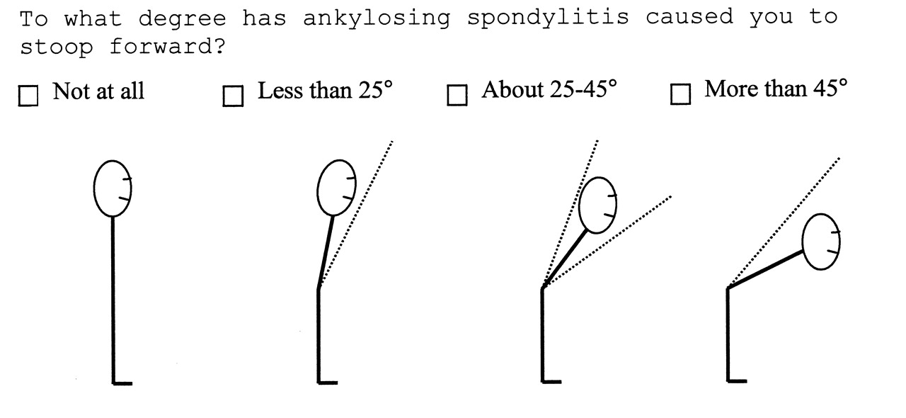
Patient version of the Visual Assessment of the Spine Bruckel Instrument (VASBI).

In this report, we aim to demonstrate construct validity and reliability of this tool that simplifies assessment of spinal deformity in patients with AS. Using a large and representative sample of patients with AS from the United Kingdom, we illustrate VASBI’s validity according to components of the OMERACT filter.

MATERIALS AND METHODS

Development of the VASBI

An illustration for assessment of spinal deformity was developed by Jane Bruckel and a group at the SAA consisting of health professionals, patients with AS, a methodologist, and a graphic designer. It was originally developed for patient use for self-assessment of the degree of perceived spinal deformity. The individual using the tool is asked to choose 1 of 4 diagrams depicting the forward stoop and representative of spinal curvature of the patient with AS. The score range is 1 (no stoop forward) to 4 (> 45° stoop forward). The authors were not involved in designing the illustration.

Patients

Three hundred individuals enrolled in the Spondyloarthropathy Methodology and Research Therapeutics (SMART) Study at the Royal National Hospital for Rheumatic Diseases (RNHRD) in Bath, England, were recruited after giving informed consent. This cohort was a combination of inpatients and outpatients followed prospectively by the attending rheumatologist. To identify the spectrum of disease in AS, the European Spondylarthropathy Study Group guidelines were used as the inclusion criteria12.

Methods

As part of the SMART clinic at RNHRD, patients are asked to complete a set of questionnaires that included the VASBI and demographic, socioeconomic, and clinical data. Patients were asked to rate the degree of perceived spinal deformity by scoring the VASBI (Figure 1).

The following clinical variables were collected: age, disease duration, gender, validated measures for axial spinal status using BASMI6, radiographic spinal damage using mSASSS7 (which is recommended by ASAS13), functional outcome using BASFI14, and the Ankylosing Spondylitis Quality of Life Questionnaire (ASQOL)15. Not all patients had complete assessment of each outcome measure because of the logistical time constraints of conducting the study in an ambulatory outpatient clinic.

Data from these tests were used to assess construct validity of the VASBI. The respective contribution to spinal mobility was determined according to individual items of the BASMI, the entire BASFI score, and items 1, 2, and 8 of the BASFI. We investigated independent questions from the BASFI because they represented activities (i.e., putting on socks, bending forward, looking over shoulder) in which spinal mobility was necessary. All the BASMI were scored by one AS physiotherapist and a member of the group that developed the BASMI instrument. All radiographs were scored by one observer who is a trained expert in mSASSS. Test-retest reliability of the VASBI was assessed by asking participants to repeat the assessment 1 day and 1 week after their hospital visit.

Members of the SAA and physicians from the University of Toronto performed a pilot study to evaluate construct validity of the VASBI in 200316. Self-perceived spinal deformity, using this instrument, was compared to functional outcome, as measured by the BASFI, in 2720 patients with AS who participated in the Life Impact Study, a nationwide survey conducted by the SAA in 2003. Based on these data, our study was planned and conducted, in which the instrument could be tested more rigorously in the clinical setting according to components of the OMERACT filter.

Face validity

Members of the SAA, patients with AS, and physicians from the University of Toronto with international reputations as experts in the management of AS (M.A. Stone and R.D. Inman), determined that VASBI had good face and content validity, according to OMERACT filter definition11. Based on the consultative process, it was felt that the VASBI had face validity because it appeared to an expert in the field to measure at least one aspect of AS (spinal appearance). Moreover, the VASBI achieved content validity because experts in the field felt that it determined the dimensionality of the manifestations of AS (spinal structure and mobility).

Truth: construct validity

Construct validity was gauged by analyzing correlations (using Spearman’s rho for nonparametric data) between the patient-scored VASBI and other indices of spinal deformity. These included spinal mobility using BASMI, radiographic spinal damage using mSASSS, and functional outcome using BASFI. A positive and moderate correlation of these indices and the VASBI was expected. The VASBI score was also compared with individual items on the BASMI and BASFI as described in the Methods section. The correlation coefficients were interpreted according to Cohen’s categories for strength of association, which are strong (r between 0.5 and 1.0), moderate (r between 0.3 and 0.5), small (r between 0.1 and 0.3), and insignificant (r < 0.1)17.

Discrimination: reliability

The agreement between test and retest was calculated using Kappa statistic, where a value of 0.2–0.4 was fair agreement beyond chance, 0.4–0.6 moderate agreement beyond chance, 0.6–0.8 substantial agreement beyond chance, and 0.8–1.0 almost perfect agreement beyond chance18.

Discrimination: responsiveness

Because of the small number of potential scores for the VASBI (1–4), it was not appropriate to evaluate common indicators of responsiveness such as the effect size or standardized response mean. Because the VASBI is a status tool, the authors did not assess its responsiveness.

The statistical package used was SPSS for Windows (version 12.0). A p value < 0.05 was considered significant in all analyses.

RESULTS

Patient characteristics

Of the 300 individuals enrolled in the study, 57% were experiencing extraarticular features and 80% were diagnosed with AS according to the modified New York criteria1. Of the 236 who were tested for HLA-B27, 89.4% were positive (Table 1). Most of the patients were male (75%) and the mean (± SD) duration of symptoms since onset was 27.1 (12.57) years. The mean ± SD for BASFI, BASMI, and mSASSS were 4.6 (2.54), 3.6 (1.91), and 14.2 (19.92), respectively (Table 1).

View this table:
  • View inline
  • View popup
Table 1.

Characteristics of patients with SpA in VASBI study.

Patient VASBI (Table 2) demonstrated moderate correlation with functional impairment (BASFI: r = 0.490), and structural damage (mSASSS: r = 0.309), and a strong correlation with spinal mobility (BASMI: r = 0.543). The patient-scored VASBI scores demonstrated moderate correlation with the 3 BASFI questions.

View this table:
  • View inline
  • View popup
Table 2.

Assessment of construct validity of the VASBI: Correlation between measures (BASFI, BASMI, mSASSS) and patient VASBI score.

Of the individual BASMI components, tragus-to-wall measurement showed a strong correlation with patient-scored VASBI; lumbar flexion, lumbar side flexion, and cervical rotation showed moderate correlations with patient-scored VASBI and weak correlation with intermalleolar distance. Correlations with individual mSASSS component scores were weak.

The VASBI was assessed in patients for reliability. The VASBI scores at baseline (clinic response) were found to have substantial agreement with VASBI results in the same patient 1 day and 1 week later [1 day: n = 67, Kappa = 0.739 (SE = 0.080); 1 week: n = 63, Kappa = 0.786 (SE = 0.076)]. One-day and 1-week responses were in near-perfect agreement [n = 75, Kappa = 0.973 (SE = 0.027)].

DISCUSSION

We have demonstrated that the VASBI, a new status tool to assess the appearance of the spine in AS, is very feasible and shows good construct and face validity, and reliability. It is a simple tool that relies on the visual assessment of the spine in patients with AS and can be used in a busy clinical setting to quickly assess the appearance of the spine, which in turn is a reflection of spinal damage and mobility.

Patient-assessed health instruments have become an important tool in determining different aspects of a patient’s health, particularly in rheumatology19–21. Currently in AS, the plain radiograph is the “gold standard” for assessing structural damage and the BASMI serves as a clinical measure of spinal mobility. However, plain radiographs are not always readily available and along with the BASMI require training to score appropriately. The present study was prompted by the lack of a quick and easy tool that can reflect spinal degeneration in patients with spondyloarthropathy (SpA). There is a need for a simple tool to measure spine involvement, especially in small communities with restricted resources.

The majority of self-assessment measures in SpA reveal the functional status of patients and some, like the BASDAI, describe disease activity22,23. Other self-report instruments like the Patient Acceptable Symptom State24 are used, but the VASBI is unique because it is an illustration meant to mirror a patient’s spinal appearance. When we compared this novel instrument to other indices of spinal function and structure (i.e., BASFI, BASMI, mSASSS), we achieved significant correlation, demonstrating good construct validity25. Reliability of the instrument was also highly significant on test-retest comparisons.

The VASBI’s categorical quality prevented the assessment of responsiveness using common statistical methods such as effect size or standardized response means. Further, since AS is a slowly progressive disease in terms of spinal damage and loss of mobility, transition between the relatively broad categories of the VASBI would be expected to occur over a number of years in most cases26–29. Therefore we would expect that the majority of patients show no change over the 2-week intensive rehabilitation course because major changes in spinal deformity resulting in transition from one VASBI score to another would be unlikely. Further refinement of the instrument for precision is required before this instrument can be used to detect a change in therapy.

Patient VASBI scores achieved high correlation with BASFI items, indicating that a patient’s view of disease status tends to be influenced by their experience of the illness, again consistent with an earlier study30. Previous work by our group demonstrated a strong correlation between BASFI and VASBI16. Personality traits and a patient’s point of view influence self-reported health status31,32. However, the VASBI contains only 4 items, which makes it unlikely for personality type to influence the score unless the person’s spinal deformity is on the border between 2 items of the instrument. The precision of the instrument may also play a role if it is used to monitor response to treatment.

A study in Canada by Bellamy, et al found that rheumatologists are more likely to longitudinally follow patients with rheumatoid arthritis (100%) and AS (96%) than osteoarthritis (58%–74%) or fibromyalgia (51%)33. It was also pointed out that physicians agree on certain qualities of assessment techniques such as brevity, simplicity, and validity. The VASBI may be employed in the absence of spinal measuring tools such as the inclinometer or goniometer, which are not routinely available or used in clinical practice. It may also serve as a status tool to follow patients longitudinally and with modification to improve precision, for the detection of changes with treatment. Further, VASBI might be a cost-effective visual assessment tool that can be scored by both patients and physicians. Because of its availability and simplicity, VASBI is an ideal tool for use outside large urban centers to give us a better understanding of the spectrum of disease in the population.

VASBI is a feasible and reliable tool with good construct and face validity that may be used for assessing the spine in large cohorts and for tracking disease progression. This measure can provide physicians with a quick and easy tool to follow patients with spinal deformities such as AS.

Footnotes

  • Supported by a joint award from the Spondylitis Association of America and the National Ankylosing Spondylitis Society of the UK; and by the donated funds award from the Royal National Hospital for Rheumatic Diseases, Bath. Dr. Podbielski is the recipient of a Canadian Arthritis Network Summer Research Program Studentship, the Spondylitis Association of America (SAA) Studentship, and the St. Michael’s Hospital Summer Research Student Scholarship. Dr. Stone is the recipient of the Cumming Visiting Professor Award and SAA award.

    • Accepted for publication October 9, 2009.

REFERENCES

  1. 1.↵
    1. van der Linden S,
    2. Valkenburg HA,
    3. Cats A
    . Evaluation of diagnostic criteria for ankylosing spondylitis. A proposal for modification of the New York Criteria. Arthritis Rheum 1984;27:361–8.
    OpenUrlCrossRefPubMed
  2. 2.↵
    1. Dawes PT
    . Stoke Ankylosing Spondylitis Spine Score. J Rheumatol 1999;26:993–6.
    OpenUrlPubMed
  3. 3.↵
    1. van der Heijde D,
    2. Spoorenberg A
    . Plain radiographs as an outcome measure in ankylosing spondylitis. J Rheumatol 1999;26:985–7.
    OpenUrlPubMed
  4. 4.↵
    1. van der Heijde D,
    2. Calin A,
    3. Dougados M,
    4. Khan MA,
    5. van der Linden S,
    6. Bellamy N
    . Selection of instruments in the core set for DC-ART, SMARD, physical therapy, and clinical record keeping in ankylosing spondylitis. Progress report of the ASAS Working Group. Assessments in Ankylosing Spondylitis. J Rheumatol 1999;26:951–4.
    OpenUrlPubMed
  5. 5.↵
    1. van der Heijde D,
    2. Bellamy N,
    3. Calin A,
    4. Dougados M,
    5. Khan MA,
    6. van der Linden S
    . Preliminary core sets for endpoints in ankylosing spondylitis. Assessments in Ankylosing Spondylitis Working Group. J Rheumatol 1997;24:2225–9.
    OpenUrlPubMed
  6. 6.↵
    1. Jenkinson TR,
    2. Mallorie PA,
    3. Whitelock HC,
    4. Kennedy LG,
    5. Garrett SL,
    6. Calin A
    . Defining spinal mobility in ankylosing spondylitis (AS). The Bath AS Metrology Index. J Rheumatol 1994;21:1694–8.
    OpenUrlPubMed
  7. 7.↵
    1. Creemers MC,
    2. Franssen MJ,
    3. van’t Hof MA,
    4. Gribnau FW,
    5. van de Putte LB,
    6. van Riel PL
    . Assessment of outcome in ankylosing spondylitis: An extended radiographic scoring system. Ann Rheum Dis 2005;64:127–9.
    OpenUrlAbstract/FREE Full Text
  8. 8.↵
    1. Salaffi F,
    2. Carotti M,
    3. Garofalo G,
    4. Giuseppetti GM,
    5. Grassi W
    . Radiological scoring methods for ankylosing spondylitis: A comparison between the Bath Ankylosing Spondylitis Radiology Index and the modified Stoke Ankylosing Spondylitis Spine Score. Clin Exp Rheumatol 2007;25:67–74.
    OpenUrlPubMed
  9. 9.↵
    1. Pile KD,
    2. Laurent MR,
    3. Salmond CE,
    4. Best MJ,
    5. Pyle EA,
    6. Moloney RO
    . Clinical assessment of ankylosing spondylitis: A study of observer variation in spinal measurements. Br J Rheumatol 1991;30:29–34.
    OpenUrlAbstract/FREE Full Text
  10. 10.↵
    1. Maksymowych WP,
    2. Mallon C,
    3. Richardson R,
    4. Conner-Spady B,
    5. Jauregui E,
    6. Chung C,
    7. et al.
    Development and validation of the Edmonton Ankylosing Spondylitis Metrology Index. Arthritis Rheum 2006;55:575–82.
    OpenUrlCrossRefPubMed
  11. 11.↵
    1. Bellamy N
    . Clinimetric concepts in outcome assessment: The OMERACT filter. J Rheumatol 1999;26:948–50.
    OpenUrlPubMed
  12. 12.↵
    1. Dougados M,
    2. van der Linden S,
    3. Juhlin R,
    4. Huitfeldt B,
    5. Amor B,
    6. Calin A,
    7. et al.
    The European Spondylarthropathy Study Group preliminary criteria for the classification of spondylarthropathy. Arthritis Rheum 1991;34:1218–27.
    OpenUrlCrossRefPubMed
  13. 13.↵
    1. van der Heijde D,
    2. Landewe R
    . Selection of a method for scoring radiographs for ankylosing spondylitis clinical trials, by the Assessment in Ankylosing Spondylitis Working Group and OMERACT. J Rheumatol 2005;32:2048–9.
    OpenUrlAbstract/FREE Full Text
  14. 14.↵
    1. Calin A,
    2. Garrett S,
    3. Whitelock H,
    4. Kennedy LG,
    5. O’Hea J,
    6. Mallorie P,
    7. et al.
    A new approach to defining functional ability in ankylosing spondylitis: The development of the Bath Ankylosing Spondylitis Functional Index. J Rheumatol 1994;21:2281–5.
    OpenUrlPubMed
  15. 15.↵
    1. Doward LC,
    2. Spoorenberg A,
    3. Cook SA,
    4. Whalley D,
    5. Helliwell PS,
    6. Kay LJ,
    7. et al.
    Development of the ASQoL: A Quality of Life instrument specific to ankylosing spondylitis. Ann Rheum Dis 2003;62:20–6.
    OpenUrlAbstract/FREE Full Text
  16. 16.↵
    1. Podbielski D,
    2. Bruckel J,
    3. Warren RW,
    4. Inman RD,
    5. Cortinovis D,
    6. Papachristos A,
    7. et al.
    Validation of a self assessment spinal deformity tool for ankylosing spondylitis [abstract]. Arthritis Rheum 2005;52 Suppl:S413–4.
    OpenUrl
  17. 17.↵
    1. Cohen J
    . Statistical power analysis for the behavioral sciences. 2nd ed. Hillsdale, New Jersey: Lawrence Erlbaum Associates; 1988.
  18. 18.↵
    1. McGinn T,
    2. Wyer PC,
    3. Newman TB,
    4. Keitz S,
    5. Leipzig R,
    6. For GG,
    7. et al.
    Tips for learners of evidence-based medicine: 3. Measures of observer variability (kappa statistic). CMAJ 2004;171:1369–73.
    OpenUrlFREE Full Text
  19. 19.↵
    1. Garratt A,
    2. Schmidt L,
    3. Mackintosh A,
    4. Fitzpatrick R
    . Quality of life measurement: Bibliographic study of patient assessed health outcome measures. BMJ 2002;324:1417.
    OpenUrlAbstract/FREE Full Text
  20. 20.↵
    1. Fitzpatrick R,
    2. Davey C,
    3. Buxton MJ,
    4. Jones DR
    . Evaluating patient-based outcome measures for use in clinical trials. Health Technol Assess 1998;2:i,iv,1–74.
    OpenUrlPubMed
  21. 21.↵
    1. Saag KG
    . OMERACT 6 brings new perspectives to rheumatology measurement research. J Rheumatol 2003;30:639–41.
    OpenUrlFREE Full Text
  22. 22.↵
    1. Garrett S,
    2. Jenkinson T,
    3. Kennedy LG,
    4. Whitelock H,
    5. Gaisford P,
    6. Calin A
    . A new approach to defining disease status in ankylosing spondylitis: The Bath Ankylosing Spondylitis Disease Activity Index. J Rheumatol 1994;21:2286–91.
    OpenUrlPubMed
  23. 23.↵
    1. Haywood KL,
    2. Garratt AM,
    3. Dawes PT
    . Patient-assessed health in ankylosing spondylitis: A structured review. Rheumatology 2005;44:577–86.
    OpenUrlAbstract/FREE Full Text
  24. 24.↵
    1. Tubach F,
    2. Ravaud P,
    3. Baron G,
    4. Falissard B,
    5. Logeart I,
    6. Bellamy N,
    7. et al.
    Evaluation of clinically relevant states in patient reported outcomes in knee and hip osteoarthritis: The patient acceptable symptom state. Ann Rheum Dis 2005;64:34–7.
    OpenUrlAbstract/FREE Full Text
  25. 25.↵
    1. Haywood KL,
    2. Garratt AM,
    3. Jordan K,
    4. Dziedzic K,
    5. Dawes PT
    . Spinal mobility in ankylosing spondylitis: Reliability, validity and responsiveness. Rheumatology 2004;43:750–7.
    OpenUrlAbstract/FREE Full Text
  26. 26.↵
    1. Carette S,
    2. Graham D,
    3. Little H,
    4. Rubenstein J,
    5. Rosen P
    . The natural disease course of ankylosing spondylitis. Arthritis Rheum 1983;26:186–90.
    OpenUrlCrossRefPubMed
  27. 27.↵
    1. Brophy S,
    2. Mackay K,
    3. Al-Saidi A,
    4. Taylor G,
    5. Calin A
    . The natural history of ankylosing spondylitis as defined by radiological progression. J Rheumatol 2002;29:1236–43.
    OpenUrlAbstract/FREE Full Text
  28. 28.↵
    1. Wanders AJ,
    2. Landewe RB,
    3. Spoorenberg A,
    4. Dougados M,
    5. van der Linden S,
    6. Mielants H,
    7. et al.
    What is the most appropriate radiologic scoring method for ankylosing spondylitis? A comparison of the available methods based on the outcome measures in rheumatology clinical trials filter. Arthritis Rheum 2004;50:2622–32.
    OpenUrlCrossRefPubMed
  29. 29.↵
    1. Sengupta R,
    2. Hunt LP,
    3. Richardson J,
    4. Gay L,
    5. Pomeroy E,
    6. Mogg R,
    7. et al.
    Analysis of longitudinal cervical spinal mobility measures over a 32 year period indicates that AS is a progressive disease with clear cut gender differences. Ann Rheum Dis 2007;66 Suppl II:409.
    OpenUrl
  30. 30.↵
    1. Spoorenberg A,
    2. van Tubergen A,
    3. Landewe R,
    4. Dougados M,
    5. van der Linden S,
    6. Mielants H,
    7. et al.
    Measuring disease activity in ankylosing spondylitis: Patient and physician have different perspectives. Rheumatology 2005;44:789–95.
    OpenUrlAbstract/FREE Full Text
  31. 31.↵
    1. Hidding A,
    2. de Witte L,
    3. van der Linden S
    . Determinants of self-reported health status in ankylosing spondylitis. J Rheumatol 1994;21:275–8.
    OpenUrlPubMed
  32. 32.↵
    1. Bakker C,
    2. Boers M,
    3. van der Linden S
    . Measures to assess ankylosing spondylitis: Taxonomy, review and recommendations. J Rheumatol 1993;20:1724–30.
    OpenUrlPubMed
  33. 33.↵
    1. Bellamy N,
    2. Kaloni S,
    3. Pope J,
    4. Coulter K,
    5. Campbell J
    . Quantitative rheumatology: A survey of outcome measurement procedures in routine rheumatology outpatient practice in Canada. J Rheumatol 1998;25:852–8.
    OpenUrlPubMed
PreviousNext
Back to top

In this issue

The Journal of Rheumatology
Vol. 37, Issue 3
1 Mar 2010
  • Table of Contents
  • Table of Contents (PDF)
  • Index by Author
  • Editorial Board (PDF)
Print
Download PDF
Article Alerts
Sign In to Email Alerts with your Email Address
Email Article

Thank you for your interest in spreading the word about The Journal of Rheumatology.

NOTE: We only request your email address so that the person you are recommending the page to knows that you wanted them to see it, and that it is not junk mail. We do not capture any email address.

Enter multiple addresses on separate lines or separate them with commas.
Visual Assessment of the Spine Bruckel Instrument, a Novel Status Tool to Reflect Appearance of the Spine in Patients with Ankylosing Spondylitis
(Your Name) has forwarded a page to you from The Journal of Rheumatology
(Your Name) thought you would like to see this page from the The Journal of Rheumatology web site.
CAPTCHA
This question is for testing whether or not you are a human visitor and to prevent automated spam submissions.
Citation Tools
Visual Assessment of the Spine Bruckel Instrument, a Novel Status Tool to Reflect Appearance of the Spine in Patients with Ankylosing Spondylitis
DOMINIK W. PODBIELSKI, JANE BRUCKEL, EMMA POMEROY, ROBERT D. INMAN, ROBERT W. WARREN, LAURIE M. SAVAGE, JENNY RICHARDSON, ANGELO PAPACHRISTOS, REBECCA J. MOGG, MILLICENT A. STONE
The Journal of Rheumatology Mar 2010, 37 (3) 628-632; DOI: 10.3899/jrheum.090548

Citation Manager Formats

  • BibTeX
  • Bookends
  • EasyBib
  • EndNote (tagged)
  • EndNote 8 (xml)
  • Medlars
  • Mendeley
  • Papers
  • RefWorks Tagged
  • Ref Manager
  • RIS
  • Zotero

 Request Permissions

Share
Visual Assessment of the Spine Bruckel Instrument, a Novel Status Tool to Reflect Appearance of the Spine in Patients with Ankylosing Spondylitis
DOMINIK W. PODBIELSKI, JANE BRUCKEL, EMMA POMEROY, ROBERT D. INMAN, ROBERT W. WARREN, LAURIE M. SAVAGE, JENNY RICHARDSON, ANGELO PAPACHRISTOS, REBECCA J. MOGG, MILLICENT A. STONE
The Journal of Rheumatology Mar 2010, 37 (3) 628-632; DOI: 10.3899/jrheum.090548
del.icio.us logo Digg logo Reddit logo Twitter logo CiteULike logo Facebook logo Google logo Mendeley logo
  • Tweet Widget
  • Facebook Like
  • Google Plus One
Bookmark this article

Jump to section

  • Article
    • Abstract
    • MATERIALS AND METHODS
    • RESULTS
    • DISCUSSION
    • Footnotes
    • REFERENCES
  • Figures & Data
  • References
  • Info & Metrics
  • PDF
  • eLetters

Related Articles

Cited By...

More in this TOC Section

  • Follow-up Contrast-Enhanced Ultrasonography of the Carotid Artery in Patients With Takayasu Arteritis: A Retrospective Study
  • Disease Flare of Systemic Lupus Erythematosus in Patients With Endstage Renal Disease on Dialysis
  • A Practical Guide for Assessment of Skin Burden in Patients With Psoriatic Arthritis
Show more Articles

Similar Articles

Content

  • First Release
  • Current
  • Archives
  • Collections
  • Audiovisual Rheum
  • COVID-19 and Rheumatology

Resources

  • Guide for Authors
  • Submit Manuscript
  • Author Payment
  • Reviewers
  • Advertisers
  • Classified Ads
  • Reprints and Translations
  • Permissions
  • Meetings
  • FAQ
  • Policies

Subscribers

  • Subscription Information
  • Purchase Subscription
  • Your Account
  • Terms and Conditions

More

  • About Us
  • Contact Us
  • My Alerts
  • My Folders
  • Privacy/GDPR Policy
  • RSS Feeds
The Journal of Rheumatology
The content of this site is intended for health care professionals.
Copyright © 2022 by The Journal of Rheumatology Publishing Co. Ltd.
Print ISSN: 0315-162X; Online ISSN: 1499-2752
Powered by HighWire