
Main menu

  • Home
  • Content
    • First Release
    • Current
    • Archives
    • Collections
    • Audiovisual Rheum
    • 50th Volume Reprints
  • Resources
    • Guide for Authors
    • Submit Manuscript
    • Payment
    • Reviewers
    • Advertisers
    • Classified Ads
    • Reprints and Translations
    • Permissions
    • Meetings
    • FAQ
    • Policies
  • Subscribers
    • Subscription Information
    • Purchase Subscription
    • Your Account
    • Terms and Conditions
  • About Us
    • About Us
    • Editorial Board
    • Letter from the Editor
    • Duncan A. Gordon Award
    • Privacy/GDPR Policy
    • Accessibility
  • Contact Us
  • JRheum Supplements
  • Services

User menu

  • My Cart
  • Log In

Search

  • Advanced search
The Journal of Rheumatology
  • JRheum Supplements
  • Services
  • My Cart
  • Log In
The Journal of Rheumatology

Advanced Search

  • Home
  • Content
    • First Release
    • Current
    • Archives
    • Collections
    • Audiovisual Rheum
    • 50th Volume Reprints
  • Resources
    • Guide for Authors
    • Submit Manuscript
    • Payment
    • Reviewers
    • Advertisers
    • Classified Ads
    • Reprints and Translations
    • Permissions
    • Meetings
    • FAQ
    • Policies
  • Subscribers
    • Subscription Information
    • Purchase Subscription
    • Your Account
    • Terms and Conditions
  • About Us
    • About Us
    • Editorial Board
    • Letter from the Editor
    • Duncan A. Gordon Award
    • Privacy/GDPR Policy
    • Accessibility
  • Contact Us
  • Follow Jrheum on BlueSky
  • Follow jrheum on Twitter
  • Visit jrheum on Facebook
  • Follow jrheum on LinkedIn
  • Follow jrheum on YouTube
  • Follow jrheum on Instagram
  • Follow jrheum on RSS
Case ReportImages in Rheumatology

Glomus Tumor

ALEJANDRO ESPINOSA-GUTIERREZ, ALDO IZAGUIRRE, LETICIA BAENA-OCAMPO, RAFAEL FEYNOSO-CAMPO and CARLOS PINEDA
The Journal of Rheumatology June 2009, 36 (6) 1343-1344; DOI: https://doi.org/10.3899/jrheum.081026
ALEJANDRO ESPINOSA-GUTIERREZ
  • Find this author on Google Scholar
  • Find this author on PubMed
  • Search for this author on this site
ALDO IZAGUIRRE
  • Find this author on Google Scholar
  • Find this author on PubMed
  • Search for this author on this site
LETICIA BAENA-OCAMPO
  • Find this author on Google Scholar
  • Find this author on PubMed
  • Search for this author on this site
RAFAEL FEYNOSO-CAMPO
  • Find this author on Google Scholar
  • Find this author on PubMed
  • Search for this author on this site
CARLOS PINEDA
  • Find this author on Google Scholar
  • Find this author on PubMed
  • Search for this author on this site
  • For correspondence: carpineda{at}yahoo.com
  • Article
  • Figures & Data
  • Info & Metrics
  • References
  • PDF
PreviousNext
Loading

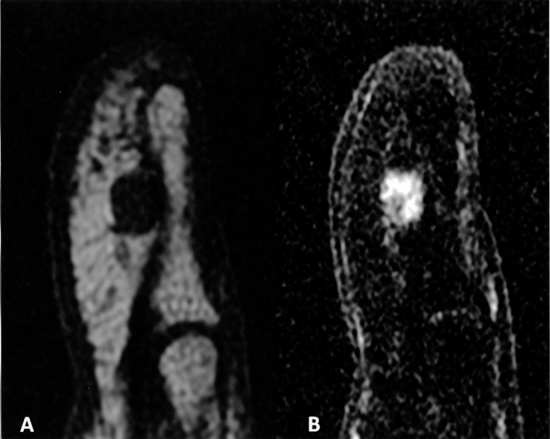
A 48-year-old man presented with a 3-year history of progressive, localized, paroxysmal pain, and swelling of the 5th digit pulp. The pain worsened with cold exposure, accidental tapping, and pressure. Prior consultations with an orthopedic surgeon, internal medicine specialist, and a neurologist failed to establish a diagnosis. Treatment with analgesics, nonsteroidal antiinflammatory drugs, physical therapy, and antibiotics was unsuccessful. Examination revealed a bulbous reddish deformity of the pulp. Love’s pin test (excruciating pain elicited by gentle pressure with a pin head), Hildreth’s test (a tourniquet is applied at the base of the digit, and by repeating Love’s pin test, the pain is abolished), and cold sensitivity test (increased pain when exposed to cold water) were positive. Plain radiographs displayed subtle erosion of the volar cortex of the distal phalanx. High-resolution ultrasonography (US) showed a globular hypoechoic area of 8 mm on long axis view. Color Doppler and power Doppler US displayed marked hypervascularity of the lesion (Figure 1). Magnetic resonance imaging (MRI) showed a hypointense well defined nodule on T1-weighted images, which became hyperintense on T2-weighted images (Figure 2). Nodule enhancement appeared with gadolinium. Surgical excision was performed. Gross pathology revealed a well defined red-blue nodule. Histology revealed a glomus tumor composed of compact nests of monotonous polygonal cells with rounded nuclei and eosinophilic cytoplasm (Figure 3). On followup the patient is free of symptoms with no evidence of recurrence.

Longitudinal color Doppler 13-5MHz US image obtained over pulp of the 5th finger shows marked intratumor vasculature related to high-velocity shunts of the glomus tumor.
  • Download figure
  • Open in new tab
  • Download powerpoint
Figure 1.

Longitudinal color Doppler 13-5MHz US image obtained over pulp of the 5th finger shows marked intratumor vasculature related to high-velocity shunts of the glomus tumor.

Sagittal T1 (A), and T1-weighted fat suppression with gadolinium (B) images of distal phalanx of the 5th finger, showing low signal intensity, well defined nodule that enhances with contrast administration.
  • Download figure
  • Open in new tab
  • Download powerpoint
Figure 2.

Sagittal T1 (A), and T1-weighted fat suppression with gadolinium (B) images of distal phalanx of the 5th finger, showing low signal intensity, well defined nodule that enhances with contrast administration.

(A) Histology revealed glomus cells with typical centrally located rounded nucleus, and well defined cell membranes with eosinophilic cytoplasm surrounding endothelial-lined vascular spaces. (B) Cells displayed consistent strong immunoreactivity for smooth-muscle actin.
  • Download figure
  • Open in new tab
  • Download powerpoint
Figure 3.

(A) Histology revealed glomus cells with typical centrally located rounded nucleus, and well defined cell membranes with eosinophilic cytoplasm surrounding endothelial-lined vascular spaces. (B) Cells displayed consistent strong immunoreactivity for smooth-muscle actin.

Glomus tumors are benign hamartomas that account for 1% to 5% of all soft-tissue tumors of the hand. They arise from the normal glomus apparatus, located in the subcutaneous tissue. Glomus tumors are encountered most frequently in the subungual region of digits, but can be found almost anywhere in the body1. Glomus tumors present with the classic and virtually diagnostic triad of limb pain out of proportion to the size of the lesion, exquisite tenderness to even the lightest touch, and exacerbation of symptoms on exposure to temperature changes, especially cold2,3. Scintigraphy is a useful but nonspecific diagnostic tool4. MRI provides an excellent contrast between the neoplasm and normal tissue. On the other hand, US is able to demonstrate the size, site, and shape of the tumor, even if the tumor is not palpable5. The combination of clinical testing, US, and MRI allows for early and accurate diagnosis of glomus tumors. Glomus tumors are unusual lesions, which can present a diagnostic enigma to an unaware rheumatologist.

Footnotes

REFERENCES

  1. 1.↵
    1. Tuncali D,
    2. Yilmaz AC,
    3. Terzioglu A,
    4. Aslan G
    . Multiple occurrences of different histologic types of the glomus tumor. J Hand Surg 2005;30A:161–4.
    OpenUrl
  2. 2.↵
    1. McDermott EM,
    2. Weiss AP
    . Glomus tumors. J Hand Surg 2006;31A:1397–400.
    OpenUrl
  3. 3.↵
    1. Bhaskaranand K,
    2. Navadgi BC
    . Glomus tumor of the hand. J Hand Surg 2002; 27B:3:229–31.
    OpenUrlAbstract/FREE Full Text
  4. 4.↵
    1. Geertruyden JV,
    2. Lorea P,
    3. Goldschmidt D,
    4. et al
    . Glomus tumours of the hand. A retrospective study of 51 cases. J Hand Surg 1996;21B:257–60.
    OpenUrlAbstract/FREE Full Text
  5. 5.↵
    1. Chen SH,
    2. Chen YL,
    3. Cheng MH,
    4. Yeow KM,
    5. Chen HC,
    6. Wei FC
    . The use of ultrasonography in preoperative localization of digital glomus tumors. Plast Reconstr Surg 2003;112:115–9.
    OpenUrlCrossRefPubMed
PreviousNext
Back to top

In this issue

The Journal of Rheumatology
Vol. 36, Issue 6
1 Jun 2009
  • Table of Contents
  • Table of Contents (PDF)
  • Index by Author
  • Editorial Board (PDF)
Print
Download PDF
Article Alerts
Sign In to Email Alerts with your Email Address
Email Article

Thank you for your interest in spreading the word about The Journal of Rheumatology.

NOTE: We only request your email address so that the person you are recommending the page to knows that you wanted them to see it, and that it is not junk mail. We do not capture any email address.

Enter multiple addresses on separate lines or separate them with commas.
Glomus Tumor
(Your Name) has forwarded a page to you from The Journal of Rheumatology
(Your Name) thought you would like to see this page from the The Journal of Rheumatology web site.
CAPTCHA
This question is for testing whether or not you are a human visitor and to prevent automated spam submissions.
Citation Tools
Glomus Tumor
ALEJANDRO ESPINOSA-GUTIERREZ, ALDO IZAGUIRRE, LETICIA BAENA-OCAMPO, RAFAEL FEYNOSO-CAMPO, CARLOS PINEDA
The Journal of Rheumatology Jun 2009, 36 (6) 1343-1344; DOI: 10.3899/jrheum.081026

Citation Manager Formats

  • BibTeX
  • Bookends
  • EasyBib
  • EndNote (tagged)
  • EndNote 8 (xml)
  • Medlars
  • Mendeley
  • Papers
  • RefWorks Tagged
  • Ref Manager
  • RIS
  • Zotero

 Request Permissions

Share
Glomus Tumor
ALEJANDRO ESPINOSA-GUTIERREZ, ALDO IZAGUIRRE, LETICIA BAENA-OCAMPO, RAFAEL FEYNOSO-CAMPO, CARLOS PINEDA
The Journal of Rheumatology Jun 2009, 36 (6) 1343-1344; DOI: 10.3899/jrheum.081026
del.icio.us logo Twitter logo Facebook logo  logo Mendeley logo
  • Tweet Widget
  •  logo
Bookmark this article

Jump to section

  • Article
    • Footnotes
    • REFERENCES
  • Figures & Data
  • Info & Metrics
  • References
  • PDF

Related Articles

Cited By...

More in this TOC Section

  • When Infection Leaves Its Mark: Calcinosis in Systemic Sclerosis
  • Colonic Telangiectasias Associated With Systemic Sclerosis
  • Peripheral Ulcerative Keratitis in VEXAS Syndrome
Show more Images in Rheumatology

Similar Articles

Content

  • First Release
  • Current
  • Archives
  • Collections
  • Audiovisual Rheum
  • COVID-19 and Rheumatology

Resources

  • Guide for Authors
  • Submit Manuscript
  • Author Payment
  • Reviewers
  • Advertisers
  • Classified Ads
  • Reprints and Translations
  • Permissions
  • Meetings
  • FAQ
  • Policies

Subscribers

  • Subscription Information
  • Purchase Subscription
  • Your Account
  • Terms and Conditions

More

  • About Us
  • Contact Us
  • My Alerts
  • My Folders
  • Privacy/GDPR Policy
  • RSS Feeds
The Journal of Rheumatology
The content of this site is intended for health care professionals.
Copyright © 2025 by The Journal of Rheumatology Publishing Co. Ltd.
Print ISSN: 0315-162X; Online ISSN: 1499-2752
Powered by HighWire