
Main menu

  • Home
  • Content
    • First Release
    • Current
    • Archives
    • Collections
    • Audiovisual Rheum
    • 50th Volume Reprints
  • Resources
    • Guide for Authors
    • Submit Manuscript
    • Payment
    • Reviewers
    • Advertisers
    • Classified Ads
    • Reprints and Translations
    • Permissions
    • Meetings
    • FAQ
    • Policies
  • Subscribers
    • Subscription Information
    • Purchase Subscription
    • Your Account
    • Terms and Conditions
  • About Us
    • About Us
    • Editorial Board
    • Letter from the Editor
    • Duncan A. Gordon Award
    • Privacy/GDPR Policy
    • Accessibility
  • Contact Us
  • JRheum Supplements
  • Services

User menu

  • My Cart
  • Log In

Search

  • Advanced search
The Journal of Rheumatology
  • JRheum Supplements
  • Services
  • My Cart
  • Log In
The Journal of Rheumatology

Advanced Search

  • Home
  • Content
    • First Release
    • Current
    • Archives
    • Collections
    • Audiovisual Rheum
    • 50th Volume Reprints
  • Resources
    • Guide for Authors
    • Submit Manuscript
    • Payment
    • Reviewers
    • Advertisers
    • Classified Ads
    • Reprints and Translations
    • Permissions
    • Meetings
    • FAQ
    • Policies
  • Subscribers
    • Subscription Information
    • Purchase Subscription
    • Your Account
    • Terms and Conditions
  • About Us
    • About Us
    • Editorial Board
    • Letter from the Editor
    • Duncan A. Gordon Award
    • Privacy/GDPR Policy
    • Accessibility
  • Contact Us
  • Follow Jrheum on BlueSky
  • Follow jrheum on Twitter
  • Visit jrheum on Facebook
  • Follow jrheum on LinkedIn
  • Follow jrheum on YouTube
  • Follow jrheum on Instagram
  • Follow jrheum on RSS
LetterLetter

A Unique Articular Manifestation in a Child with Relapsing Polychondritis

SHEILA KNUPP FEITOSA de OLIVEIRA, ADRIANA RODRIGUES FONSECA, ROMEU CORTES DOMINGUES and IERECÊ LINS AYMORÉ
The Journal of Rheumatology March 2009, 36 (3) 659-660; DOI: https://doi.org/10.3899/jrheum.080672
SHEILA KNUPP FEITOSA de OLIVEIRA
  • Find this author on Google Scholar
  • Find this author on PubMed
  • Search for this author on this site
  • For correspondence: sheila_knupp@hotmail.com
ADRIANA RODRIGUES FONSECA
  • Find this author on Google Scholar
  • Find this author on PubMed
  • Search for this author on this site
ROMEU CORTES DOMINGUES
  • Find this author on Google Scholar
  • Find this author on PubMed
  • Search for this author on this site
IERECÊ LINS AYMORÉ
  • Find this author on Google Scholar
  • Find this author on PubMed
  • Search for this author on this site
  • Article
  • Figures & Data
  • Info & Metrics
  • References
  • PDF
PreviousNext
Loading

To the Editor:

Relapsing polychondritis (RP) is a rare multisystem disease1 for which the etiopathogenesis is probably autoimmune. RP predominantly affects middle-aged Caucasians1, but it has been reported in 31 children and adolescents under 18 years of age1–3. It is mainly characterized by recurrent destructive inflammatory lesions of cartilaginous structures and proteoglycan-rich tissues1,3 such as outer and inner ears, nose, the laryngotracheobronchial tree, hyaline cartilage of joints, eyes, heart, and blood vessels; kidneys and skin may also be involved. We describe a rare and unique childhood articular manifestation of RP.

A 4-year-old girl of African descent was diagnosed with RP after a second episode of left-ear chondritis. Conjunctivitis and arthritis began 14 months later, involving proximal interphalangeal (PIP) joints, wrists, ankles, and knees. She developed a chronic arthritis course with some episodic flares. Nasal chondritis started 2 years later. Laboratory tests showed negative antinuclear antibody and rheumatoid factor, and during flares a moderate to high erythrocyte sedimentation rate, leukocytosis, and thrombocytosis. At the age of 10 years, she presented with enlargement of distal forearms and legs (Figure 1A) associated with peculiar radiological changes around the growth plate: metaphyseal widening and irregularities in the distal epiphysis of the radius, tibias, and fibulas (Figure 1B). Magnetic resonance imaging (MRI) showed edema in the tarsal bones and also in the metaphyseal regions of metatarsal bones and irregularities in the distal epiphysis of tibia, fibulas, and radius (Figures 1C, 1D).A biopsy performed in the distal growth plate of the radius revealed a loss of basophilic staining, inflammatory infiltrates, decreased number and pyknosis of chondrocytes, in addition to areas of growth cartilage replacement with fibroblastic granulation tissue.

Figure 1.
  • Download figure
  • Open in new tab
  • Download powerpoint
Figure 1.

The patient at age 10 years. A. Widening of wrists and ankles; B. conventional radiograph of the right wrist shows metaphyseal widening and irregularities in the distal growth plate of the radius; C. coronal T1-weighted MRI of right wrist shows irregularity of the distal physis and epiphysis of the radius; D. sagittal T1-weighted MRI of the right ankle demonstrates, in the tibia, irregularity of the distal physis and anterior portion of the metaphysis, with some marrow edema. Edema of the navicular bone can also be seen.

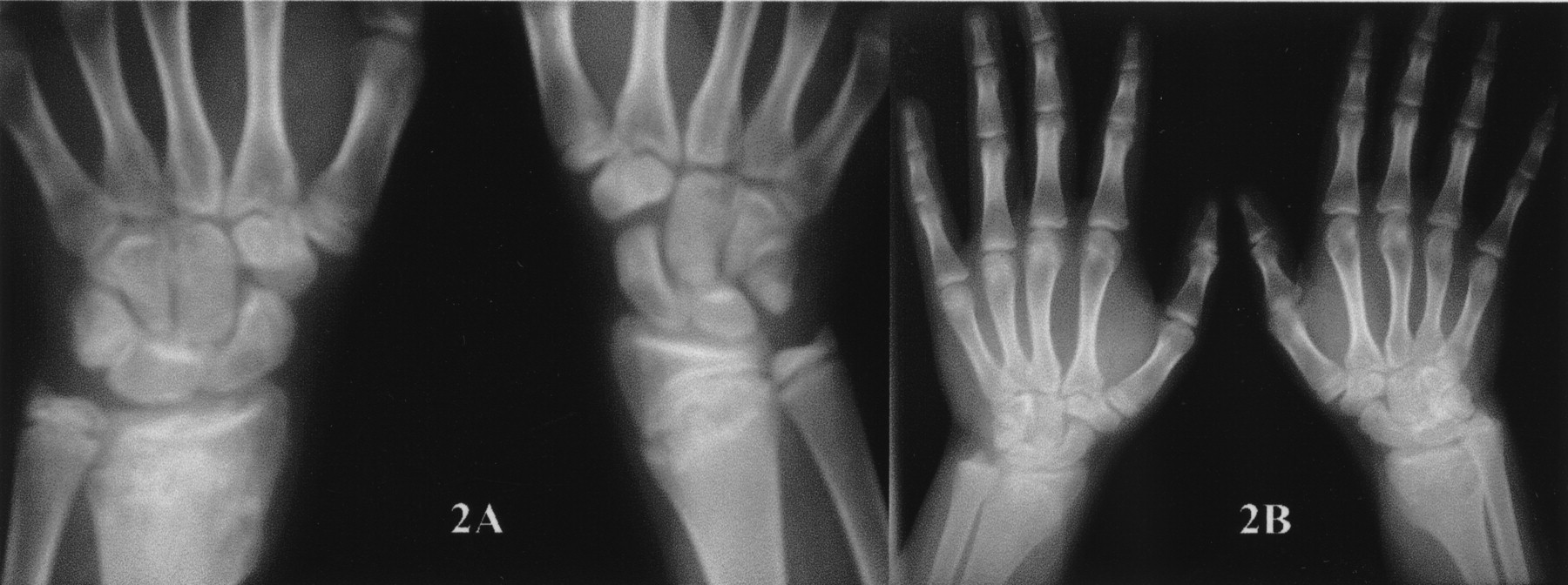
During 10 years of follow-up, no other organ has been involved (including airways) and she has been treated with corticosteroids, cyclosporine, azathioprine, infliximab, and recently with etanercept, which have yielded partial responses. There have been persistent bilateral deforming ear chondritis, deformities of the distal epiphysis, and laboratory markers of inflammation. However, it should be noted that linear growth and functional ability were not impaired, and also that erosive radiological changes resolved after epiphyseal fusion (Figures 2A, 2B).

Figure 2.
  • Download figure
  • Open in new tab
  • Download powerpoint
Figure 2.

Conventional radiographs at age 14 years (A) and 16 years (B) show that erosive radiological changes have not affected epiphyseal growth and fusion.

Articular manifestations (arthralgia, arthritis, and costochondritis) may occur in 70%–80% of adult and pediatric patients1–3. The most common pattern is episodic, asymmetric, nonerosive, nondeforming peripheral arthritis, lasting for weeks or months, commonly affecting ankles, wrists, PIP joints, metacarpophalangeal joints, elbows, and metatarsophalangeal joints3–5. Plain radiographs, in the absence of an associated disease, classically show only juxtaarticular osteopenia and minor uniform joint space narrowing, typically without erosive changes or deformities6,7. However, in this case, arthritis was persistent and destructive, with widening of distal forearms and legs, in addition to radiological and histopathological changes in the distal growth plate of the radius, fibulas, and tibias. There is a somewhat similar report8 in an 8-year-old boy, who developed a chronic polyarthritis with roentgenographic changes described as metaphyseal radiolucent defects of ulnas, radius, fibulas, and tibia, and also irregularities in the ossification centers of the fibulas, metacarpals, and proximal phalanges. Those rare articular and radiological findings may be considered unique to childhood RP.

Footnotes

REFERENCES

  1. 1.↵
    1. Damiani JM,
    2. Levine HL
    . Relapsing polychondritis — report of ten cases. Laryngoscope 1979;89:929–46.
    OpenUrlPubMed
  2. 2.
    1. Knipp S,
    2. Bier H,
    3. Horneff G,
    4. et al
    . Relapsing polychondritis in childhood — case report and short review. Rheumatol Int 2000;19:231–4.
    OpenUrlCrossRefPubMed
  3. 3.↵
    1. Gergely P,
    2. Poór G
    . Relapsing polychondritis. Best Pract Clin Res 2004;18:723–38.
    OpenUrl
  4. 4.
    1. O’Hanlan M,
    2. McAdam LP,
    3. Bluestone R,
    4. Pearson CM
    . The arthropathy of relapsing polychondritis. Arthritis Rheum 1976;19:191–4.
    OpenUrlPubMed
  5. 5.↵
    1. Balsa A,
    2. Espinosa A,
    3. Cuesta M,
    4. MacLeod TI,
    5. Gijon-Banos J,
    6. Maddison PJ
    . Joint symptoms in relapsing polychondritis. Clin Exp Rheumatol 1995;13:425–30.
    OpenUrlPubMed
  6. 6.↵
    1. Sartoris DJ,
    2. Resnick D
    . Radiologic vignette: primary disorders of articular cartilage in childhood. J Rheumatol 1988;15:812–9.
    OpenUrlPubMed
  7. 7.↵
    1. Johnson TH,
    2. Mital N,
    3. Rodnan GP,
    4. Wilson RJ
    . Relapsing polychondritis. Radiology 1973;106:313–5.
    OpenUrlCrossRefPubMed
  8. 8.↵
    1. Blau EB
    . Relapsing polychondritis and retroperitoneal fibrosis in an 8-year-old boy. Am J Dis Child 1976;130:1149–52.
    OpenUrlCrossRefPubMed
PreviousNext
Back to top

In this issue

The Journal of Rheumatology
Vol. 36, Issue 3
1 Mar 2009
  • Table of Contents
  • Table of Contents (PDF)
  • Index by Author
  • Editorial Board (PDF)
Print
Download PDF
Article Alerts
Sign In to Email Alerts with your Email Address
Email Article

Thank you for your interest in spreading the word about The Journal of Rheumatology.

NOTE: We only request your email address so that the person you are recommending the page to knows that you wanted them to see it, and that it is not junk mail. We do not capture any email address.

Enter multiple addresses on separate lines or separate them with commas.
A Unique Articular Manifestation in a Child with Relapsing Polychondritis
(Your Name) has forwarded a page to you from The Journal of Rheumatology
(Your Name) thought you would like to see this page from the The Journal of Rheumatology web site.
CAPTCHA
This question is for testing whether or not you are a human visitor and to prevent automated spam submissions.
Citation Tools
A Unique Articular Manifestation in a Child with Relapsing Polychondritis
SHEILA KNUPP FEITOSA de OLIVEIRA, ADRIANA RODRIGUES FONSECA, ROMEU CORTES DOMINGUES, IERECÊ LINS AYMORÉ
The Journal of Rheumatology Mar 2009, 36 (3) 659-660; DOI: 10.3899/jrheum.080672

Citation Manager Formats

  • BibTeX
  • Bookends
  • EasyBib
  • EndNote (tagged)
  • EndNote 8 (xml)
  • Medlars
  • Mendeley
  • Papers
  • RefWorks Tagged
  • Ref Manager
  • RIS
  • Zotero

 Request Permissions

Share
A Unique Articular Manifestation in a Child with Relapsing Polychondritis
SHEILA KNUPP FEITOSA de OLIVEIRA, ADRIANA RODRIGUES FONSECA, ROMEU CORTES DOMINGUES, IERECÊ LINS AYMORÉ
The Journal of Rheumatology Mar 2009, 36 (3) 659-660; DOI: 10.3899/jrheum.080672
del.icio.us logo Twitter logo Facebook logo  logo Mendeley logo
  • Tweet Widget
  •  logo
Bookmark this article

Jump to section

  • Article
    • Footnotes
    • REFERENCES
  • Figures & Data
  • Info & Metrics
  • References
  • PDF

Related Articles

Cited By...

More in this TOC Section

  • Seasonal Variations and Their Influence on Antineutrophil Cytoplasmic Antibody–Associated Vasculitis Relapse
  • Survival After Lung Transplantation in Patients With Rheumatoid Arthritis-Associated Lung Disease
  • High-Sensitivity Cardiac Troponin T as a Marker to Improve Cardiovascular Disease Risk Assessment Among Patients With Rheumatoid Arthritis
Show more Letter

Similar Articles

Content

  • First Release
  • Current
  • Archives
  • Collections
  • Audiovisual Rheum
  • COVID-19 and Rheumatology

Resources

  • Guide for Authors
  • Submit Manuscript
  • Author Payment
  • Reviewers
  • Advertisers
  • Classified Ads
  • Reprints and Translations
  • Permissions
  • Meetings
  • FAQ
  • Policies

Subscribers

  • Subscription Information
  • Purchase Subscription
  • Your Account
  • Terms and Conditions

More

  • About Us
  • Contact Us
  • My Alerts
  • My Folders
  • Privacy/GDPR Policy
  • RSS Feeds
The Journal of Rheumatology
The content of this site is intended for health care professionals.
Copyright © 2025 by The Journal of Rheumatology Publishing Co. Ltd.
Print ISSN: 0315-162X; Online ISSN: 1499-2752
Powered by HighWire