Article Text

Statistics from Altmetric.com
The diagnosis of enthesitis in clinical practice is difficult and usually based on conventional radiographic findings, which are not helpful in most cases.1 We previously reported that ultrasound (US) was sensitive in detecting peculiar pathological features of enthesitis around the heel.2 Furthermore, we have continued to study the efficacy of ultrasonographic diagnosis of enthesitis of other tendon and ligament insertion sites.
METHODS AND RESULTS
Sixteen patients (10 male, six female, mean age 45.6 years) with a diagnosis of seronegative arthropathy were recruited from the population for the study. Their mean disease duration was 6.3 years. They had seronegative arthropathy and knee enthesopathy without typical conventional radiographic evidence. An HDI 3000 ATL US machine (Advanced Technology Laboratories, USA), equipped with a 12 MHz linear transducer, was used to examine the knee patellar enthesis.
The present study produced interesting findings (figs 1A and B). The US images of the knee patellar enthesis showed loss of the normal fibrillar echo texture of the patellar tendon, no homogeneous pattern, blurring of the patellar tendon margins, irregular focal or generalised increased tendon thickness, and focal ill-defined tendon defects, with loss of their tightly packed echogenic dots. The US images clearly showed the definition of the patellar tendon margins, which were more precise and anatomically defined than the magnetic resonance (MR) images (figs 1C and D).

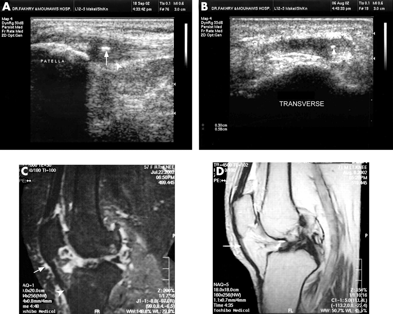
(A) A sagittal US scan shows thickened proximal entheses of the patellar ligament with loss of its fibrillar echo pattern, loss of the sharp definition of its posterior aspect compared with the distal portion (arrow head), calcific foci (arrow). (B) A transverse US scan of the same patient shows the thickened medial part of the patellar ligament with calcific focus (arrow). (C) A sagittal T2 fat suppression image shows the thickened distal part of the patellar tendon with altered signal intensity (arrow head) and prepatellar bursitis (arrow). (D) A sagittal Pd weighted image shows high intensity signals of the proximal patellar tendon.
DISCUSSION
The US examination of the knee joint clearly detected the early calcification foci of the patellar tendons. However, the calcification process of the knee patellar ligament developed less often in the patients than the calcification of the Achilles tendon found in a previous report.2,3
The process of fatty degeneration of the patellar tendon was detected early in US images, and appears as hyperechoic intratendinous lesions.4,5
This study detected a significant thickening of the patellar tendon, which can be measured by US. We believe that this US feature is more sensitive and reliable in diagnosing early enthesitis than a classical MR high signal intensity image within the superior medial and central aspects of the patellar tendon at its proximal attachment. This interesting observation has been confirmed in other related studies.6,7
In conclusion, we found several pathological differences between the pattern of patellar enthesitis and that of Achilles tendon and plantar fascia of the heel. The entheseal changes of the patellar tendon occurred at the tibial or patellar insertion either on its medial or lateral aspect, but in the case of the Achilles tendon, the entheseal changes were detected only in the calcaneal insertion. The presences of Calcific foci were found more often in the Achilles tendon than in the patellar tendon. US is a valuable and sensitive diagnostic method in patients with seronegative spondyloarthropathy and knee enthesopathy who have normal findings with conventional radiological images of the knee joint.