
Around 30 cases of bilateral or monomelic hypertrophic osteoarthropathy (HOA) of the lower limbs (or isolated clubbing of the toes) revealing an aortic prosthesis infection have been reported in the last 40 years1,2,3,4,5. Development of HOA localized to areas distal to a vascular prosthesis may allow early diagnosis of graft infection.
A 60-year-old man was admitted for septic shock. Blood cultures grew with Escherichia coli, Streptococcus anginosus, Lactobacillus rhamnosus, and Candida albicans. He had received an aorto-bifemoral prosthesis for atherosclerotic arterial disease 9 years before. He had complained for 6 months of painful legs, edema, and episodic fever. Examination showed clubbing and vasculitis of the toes (Figure 1). A 99mTc bone scan had previously shown a characteristic scintigraphic pattern of HOA of the lower limbs (Figure 2). Based on these findings, an infection of the aortic graft likely due to an intestinal fistula was suspected. Abdominal computed tomography scan showed periprosthetic inflammation. Indeed, gastroscopy revealed the presence of prosthetic material in the second duodenum (Figure 3). The patient was treated with parenteral antibiotics and the aortic prosthesis was replaced by an arterial homograft. The outcome was favorable.
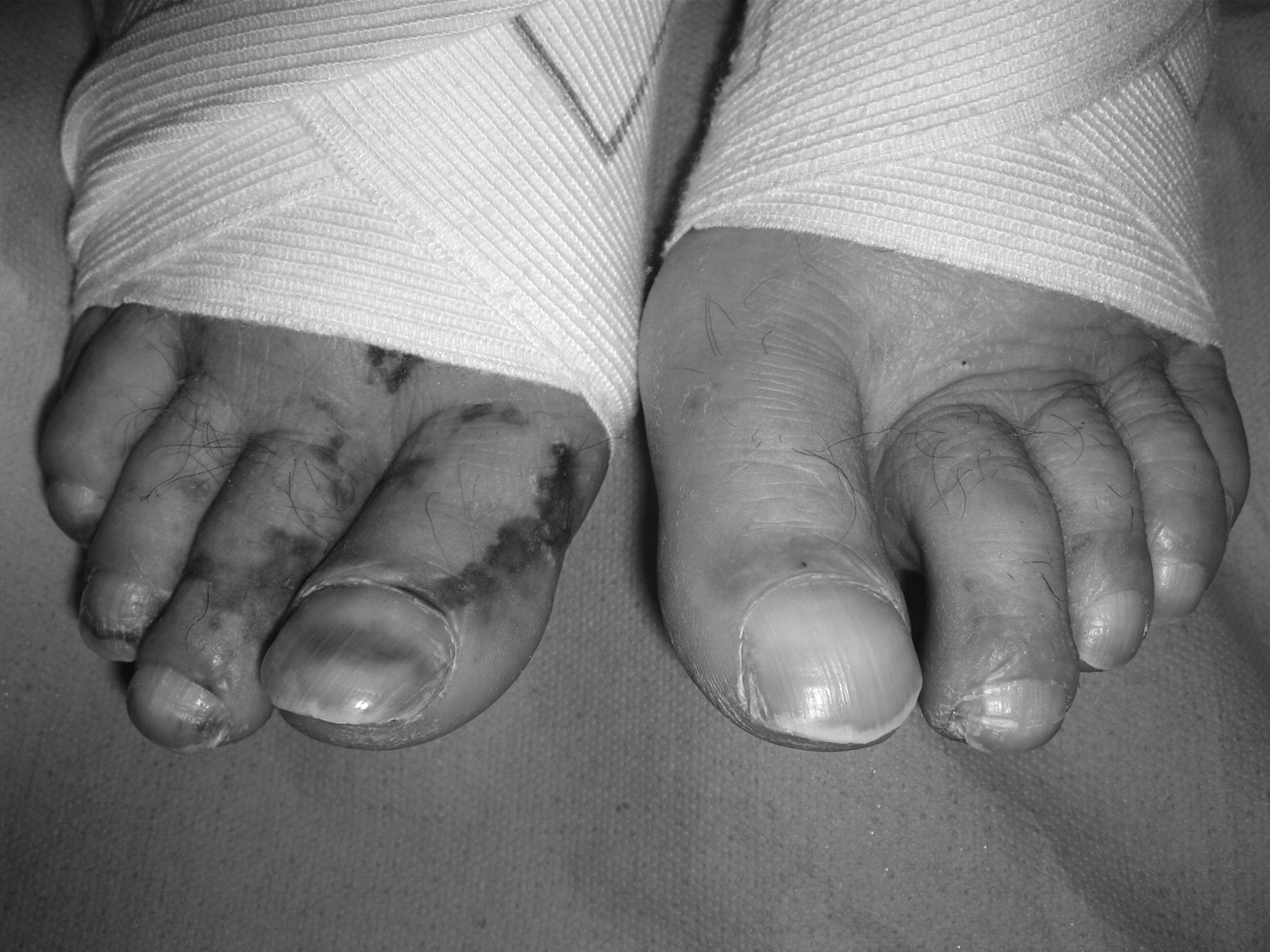
Clubbing and vasculitis of the toes.
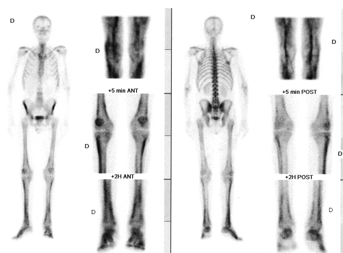
Scintigraphy scan shows characteristic pattern of HOA of the lower limbs.

Gastroscopy scan revealed prosthetic material in the second duodenum.
REFERENCES
- 1.
- 2.
- 3.
- 4.
- 5.