

Accessory sacroiliac joint (SIJ) is described as a common anatomical variant, identified in 13–18% and up to 40% of the general population1,2. It can be unilateral or bilateral and is related to aging, obesity, or women with multiple deliveries2,3.
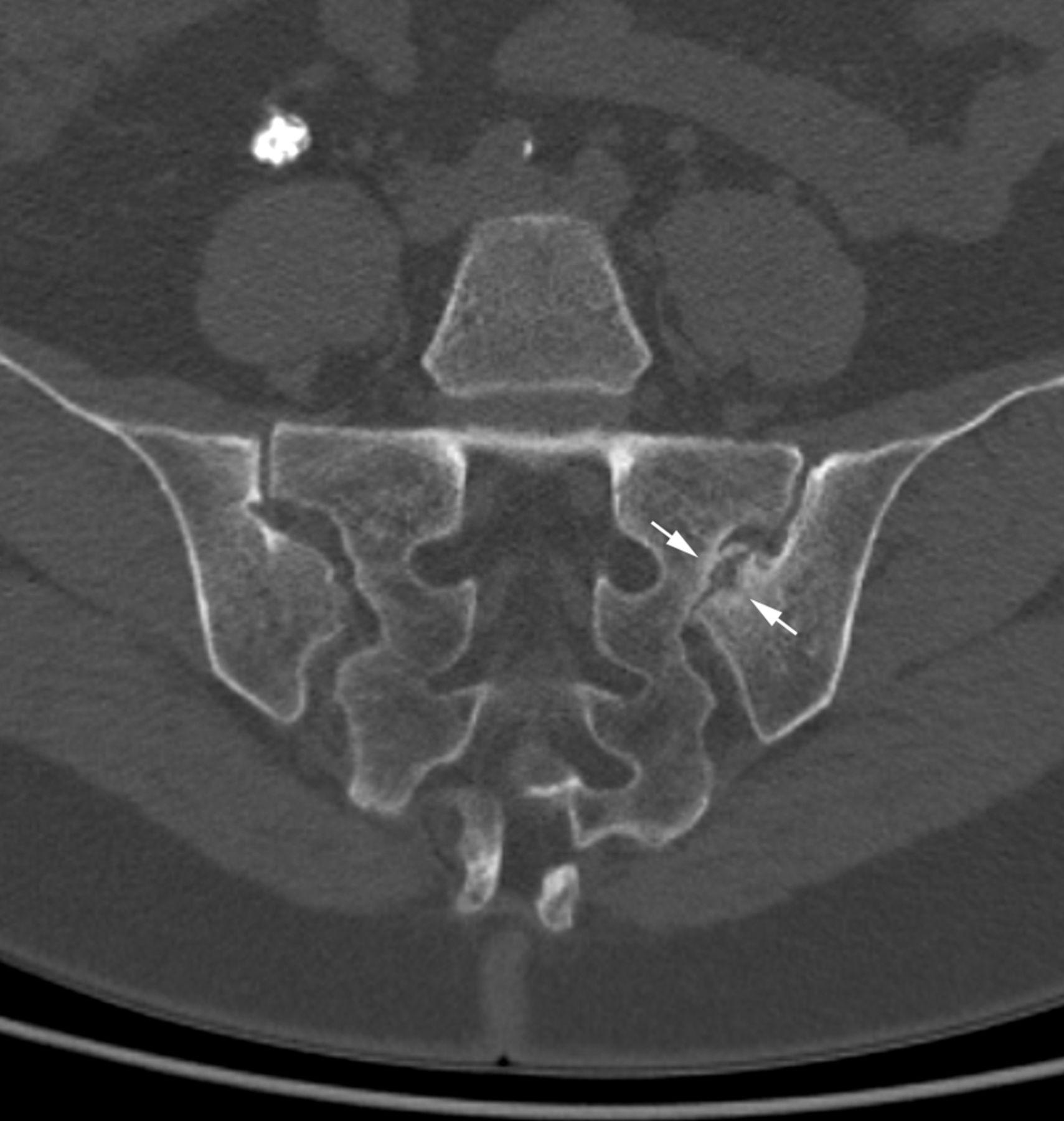
The patient, a 53-year-old white woman, presented with low back pain and left buttock pain for 6 months. She had no fever and local pressure on left SIJ reproduced the pain. Acute-phase reactants were normal and HLA-B27 was negative. Pelvic plain radiograph showed sclerosis of SIJ, predominantly on the left side. Bone marrow edema surrounding both sides of the left SIJ was observed on SIJ magnetic resonance imaging (MRI; Figure 1). Computed tomography scan of SIJ showed degenerative changes of the left SIJ with sclerosis and osteophytes but without erosions or ankylosis. An accessory SIJ was evidenced at the posterior part of the left SIJ (Figure 2).
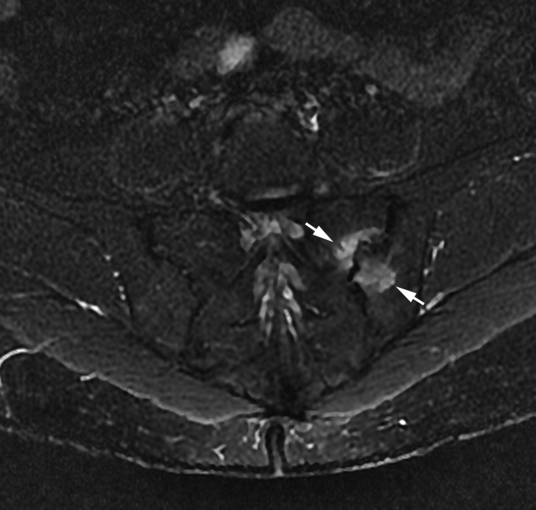
Sacroiliac joint MRI of the patient. On this T2-weighted STIR semicoronal image, subchondral bone marrow edema is visible on both sides of the left SIJ (arrows). MRI: magnetic resonance imaging; STIR: short-tau inversion recovery; SIJ: sacroiliac joint.

Semicoronal CT reconstruction showing a left accessory SIJ at the posterior part of the SIJ (arrows). CT: computed tomography; SIJ: sacroiliac joint.
The accessory SIJ is usually located at the posterior portion of the SIJ, from the level of the first to the second sacral foramen1,3. Degenerative changes of the accessory SIJ have been well described, including subchondral sclerosis, cysts, osteophytes, and even ankylosis (Figure 3). In patients with chronic or recurrent sacroiliac pain, bone marrow edema may be observed3. Accessory SIJ is common in the general population and may be associated with common back/SIJ pain and lead to inflammatory changes on MRI. Thus, rheumatologists must be aware of this anatomical variant and imaging mimicker of sacroiliitis that may be suggestive of axial spondyloarthritis.

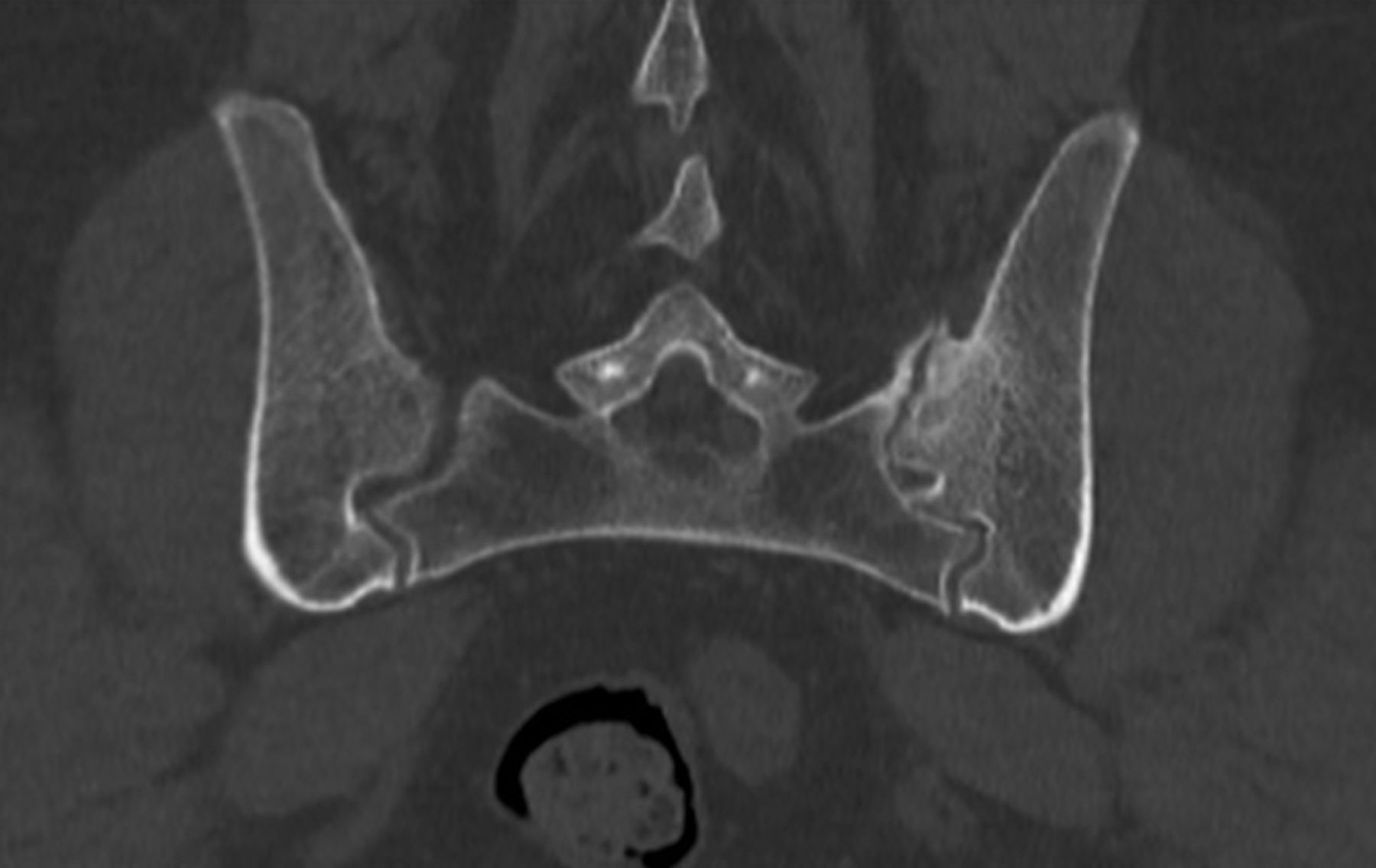
Axial CT reconstruction centered on the SIJ. Osteophytes and subchondral sclerosis of the left SIJ are consistent with degenerative changes. CT: computed tomography; SIJ: sacroiliac joint.
Footnotes
The patient gave her written informed consent to publish her case and imaging material. An approval statement from an Ethics Committee for case reports is not mandatory according to French law (loi Jardé).


