

Abstract
Objective. We investigated the association between objectively measured daily walking and knee structural change, defined either as radiographic worsening or as cartilage loss, in people at risk of or with knee osteoarthritis (OA).
Methods. Participants from the Multicenter Osteoarthritis Study (MOST) with Kellgren-Lawrence grades 0–2 and daily walking (measured with the StepWatch) at the 60-month visit were included. Participants had fixed-flexion, weight-bearing radiographs and knee magnetic resonance images (MRI) at 60 and 84 months. Radiographic worsening was read in both knees using the Osteoarthritis Research Society International grading, and MRI were read for 1 knee using the Whole-Organ MRI Score semiquantitative scoring. OR and 95% CI were calculated comparing those in the middle tertile against the lowest and highest tertiles of daily walking using logistic regression models and generalized estimating equations. Data on walking with moderate to vigorous intensity (min with > 100 steps/min/day) were associated to structural change using multivariate and logistic regression models.
Results. The 1179 study participants (59% women) were 67.0 years old (± 7.6), with a mean (± SD) body mass index of 29.8 kg/m2 (± 5.3) who walked 6981 (± 2630) steps/day. After adjusting for confounders, we found no significant associations between daily walking and radiographic worsening or cartilage loss. More time spent walking at a moderate to vigorous intensity was not associated with either radiographic worsening or cartilage loss.
Conclusion. Results from the MOST study indicated no association between daily walking and structural changes over 2 years in the knees of people at risk of or with mild knee OA.
The role of physical activity in the development and progression of knee osteoarthritis (OA) is controversial. Some studies suggest that physical activity at both high and low levels increase the risk of developing structural disease from repetitive compressive forces1,2. Others conclude that physical activity is protective against disease by promoting joint health3,4,5.
One reason for the conflicting findings of prior studies examining physical activity and structural change in the knee joint may be the assumption of a unidirectional effect of physical activity on risk of structural change rather than adverse effects of high and low levels of activity. Very low levels of walking may result in limited joint compression that may provide insufficient stimulation of cartilage cellular and matrix components. For instance, a sedentary lifestyle in animal models has been shown to lower proteoglycan content and synovial fluid volume6, which further led to cartilage fibrillation, ulceration, and erosion of the extracellular matrix7. At the extreme opposite, high levels of walking may lead to detrimental mechanical stress on joint tissues. Excessive mechanical stress may shift the chondrocyte to more catabolic activity7. Also, little is known about the risk of moderate to vigorous physical activity (MVPA), often characterized as walking at a frequency of > 100 steps/min8,9, on cartilage loss in people at risk of or with mild knee OA10. Walking is the most common type of physical activity for older adults11. However, the association of walking with structural changes in the knee joint as seen on radiograph and magnetic resonance imaging (MRI) is not well understood.
The aim of our present study was to examine the association of objectively measured daily walking with structural change 2 years later in people at risk of or with mild knee OA. We hypothesized that individuals in the lowest and highest tertiles of daily walking would have a higher degree of radiographic worsening than those in the middle tertile. Further, we hypothesized that individuals in the lowest and highest tertiles of daily walking would have a higher degree of cartilage loss than people in the middle tertile. In addition, we hypothesized that no time spent in walking at MVPA intensity (> 100 steps/min) would be associated with a higher risk of structural change.
MATERIALS AND METHODS
The Multicenter Osteoarthritis Study (MOST) is a prospective cohort study of men and women between 50–79 years of age at baseline, at risk of knee OA (i.e., overweight, obese, a history of knee injury, or with frequent knee pain), or with established knee OA. The study participants were from Birmingham, Alabama, and Iowa City, Iowa, USA, and were contacted by advertisements and mass mailings of study brochures in the first step and screened by telephone for eligibility in the next step12. The study started in 2003 and the study participants were interviewed by telephone and attended clinical visits at the timepoints 0 (baseline), 30, 60, and 84 months12,13. The visits included examination of background variables such as age, height, weight, self-reported physical activity level, knee extensor muscle strength, self-reported physical function and pain, radiographic evaluation, and MRI.
For inclusion in our study, we required that participants attended the 60-month visit when daily walking was measured for the first time using a StepWatch (Orthocare Innovations), and the 84-month visit, the first followup visit after StepWatch data were collected. Subjects had to have radiographic and/or MRI data at the 60- and 84-month visits and at least 3 days of step data. Also, we included those who at the 60-month visit had knee radiographs that showed Kellgren-Lawrence (KL) grades < 3 in at least 1 knee. Study participants who had KL grade 3 or 4 or who had undergone total knee replacement at the 30- or 60-month visits were excluded. We have previously written that identifying risk factors for progressive OA among those with more advanced disease was formidably difficult because of collider selection bias and other methodological challenges14,15.
Radiographic assessment
At the 60- and 84-month visits, standing posterior-anterior and lateral radiographs were taken using SynaFlexer frame for standardized positioning (Synarc Inc.). The radiographs were graded according to the KL classification system (grade 0–416; posteroanterior view), and the Osteoarthritis Research Society International (OARSI) atlas17 (posteroanterior and lateral view) by a musculoskeletal radiologist and a rheumatologist at Boston University blinded to clinical and MRI data. Discrepancies were solved in a group of 3 readers (including DTF). Interrater reliability results between κ = 0.77–0.80 have previously been published18 for radiographic scoring of the MOST data. Radiographic worsening in the tibiofemoral joint from 60 to 84 months was defined as any increase in the OARSI grade, e.g., increase within grade (0.5 grade) or a 1 or more grade increase19,20. The 60- and 84-month radiographs were read paired and compared for radiographic change19. Full limb radiographs of both legs were obtained at the 60-month visit18.
MRI assessment
MRI were obtained of both knees at the 60- and 84-month visits using a 1.0 Tesla MRI system (OrthOne, GE Medical Systems) and read for 1 randomly selected knee per participant. A circumferential transmit-receive extremity coil was used. Sagittal and axial fat-suppressed fast spin-echo proton density-weighted sequences (repetition time = 5800/2500 ms, time to echo = 35 ms, slice thickness = 3 mm, field of view = 14 cm, matrix = 288 × 192 pixels) and coronal short-tau inversion recovery sequence (repetition time = 7820 ms, time to echo = 15 ms, slice thickness = 3 mm, field of view = 14 cm, matrix = 256 × 256 pixels) were used for the MRI examination21. The MRI were read paired by 2 experienced readers (AG, FWR) for 1 randomly selected knee for each participant using the Whole-organ MRI Score (WORMS), including a total of 14 subregions22. Cartilage loss was graded using the WORMS from 0–622: 0 = normal thickness and no signal; 1 = normal thickness, but increased signal on proton density-weighted images; 2 = partial thickness focal defect < 1 cm in greatest width; 2.5 = full thickness focal defect < 1 cm in greatest width; 3 = multiple areas of partial thickness, intermixed with areas of normal thickness, or a grade 2.0 wider than 1 cm, but > 75% of the region; 4 = diffuse (> 75% of the region) partial thickness loss; 5 = multiple areas of full thickness loss or a grade 2.5 lesion wider than 1 cm, but < 75% of the region; 6 = diffuse (≥ 75% of the region). Cartilage loss versus no change between 60–84 months was defined on the basis of a comparison between the 60 and 84 months images of 5 subregions in the lateral and medial regions. The 5 medial and lateral subregions included femur central and posterior, and tibia anterior, central, and posterior. The change variables were defined as no change in the WORMS score, half-grade change, 1 grade change, and more than 1 grade change. We considered half-grade change or greater in any subregion as change between 60–84 months. Bone marrow lesion (BML) and meniscal tears and extrusions at the 60-month visit were included as covariates in sensitivity analyses. BML was graded in volume based on the WORMS score (grade 0–3)22,23.
Assessment of daily walking
Daily walking steps and number of minutes walking at MVPA were measured using a StepWatch Activity Monitor (Orthocare Innovations) at the 60-month visit. The StepWatch is a small waterproof accelerometer attached to the ankle. MOST participants were given written and verbal instructions for attaching the monitor each morning and removing it at bedtime for 7 consecutive days. No feedback was given to the users on the number of steps they had walked during the day. The StepWatch had to be worn for 3 valid days because it has been shown that 3 days provides an acceptable estimate of weekly activity using this monitor24,25. A valid day included wearing the StepWatch for 10 h per day, but not necessarily continuously26. The 10-h requirement has been shown to represent more than 66% of walking hours26. Time with no steps for ≥ 180 consecutive min did not count toward the 10-h requirement26,27,28. Because the StepWatch identified steps from only 1 leg, we doubled the number of steps recorded. The step data were converted as described by White, et al27: the number of steps were reduced by 25% for all the participants so that this number could be comparable to pedometer data that were reported in other studies29. The data were reported as average steps/day and tertiles. Further, we reported number of minutes per day with walking at MVPA8. High test-retest reliability has been reported for the measurement of daily walking using the monitor24.
Other variables
Data on weight, height, age, alignment, and history of knee injuries were collected at the 60-month visit. Calculation of body mass index (BMI) was based on weight ÷ (height2) in meters. Knee pain was examined using the Western Ontario McMaster Universities Osteoarthritis Index (WOMAC) pain subscale30. The subscale ranges from 0–20, in which 0 represented no pain and 20 represented extreme pain.
Statistical analyses
To examine the association of daily walking with structural change, we first classified the participants’ daily steps into tertiles that we named low, moderate, and high levels of daily walking. Next, we calculated the association of low, moderate (referent), and high levels of daily walking with radiographic worsening (coded as no change vs any change) using logistic regression models, reporting OR and 95% CI. In addition, we analyzed the association between daily walking as a continuous variable and radiographic worsening. In separate models, we carried out similar analyses for medial and lateral radiographic worsening. We repeated the analyses with MVPA as the independent variable to assess for an association with radiographic worsening. Potential confounders included age, sex, BMI, WOMAC knee pain, KL grade, alignment, and history of knee injury at the 60-month visit. Generalized estimation equations (GEE) were used to account for correlation between 2 knees within an individual31.
Logistic regression models were used to estimate the risk of cartilage loss in medial and lateral tibiofemoral compartments, respectively, with low or high compared with moderate levels of daily walking, and with daily walking as a continuous variable. Our analyses of MRI data were specific to the 5 subregions in each compartment (e.g., for medial, 1 of them is central medial femur) and we used GEE to account for the correlations of change in these 5 subregions. We also carried out analyses using MVPA as a continuous variable, and with a cutoff between those walking no minutes of MVPA versus those walking any minutes of MVPA. The analysis on cartilage loss was not adjusted for cartilage morphology at 60 months because this could introduce biased results32.
Sensitivity analyses were conducted, including the primary analysis for radiographic worsening and cartilage loss, but with an additional interaction term between the 60-month cartilage morphology status and daily walking incorporated. We also adjusted for 60-month BML and meniscus status (tear and extrusion). Further, we conducted analyses between cartilage loss and daily walking divided into < or ≥ 10,000 steps/day. Sensitivity analyses were additionally performed on those without a history of knee injury at the 60-month visit because of possible residual confounding by knee injuries33. Analyses were conducted to assess differences in sex, age, knee injury, BMI, and WOMAC pain between those included in the radiographic and MRI models (with KL grade 0–2) and the remainder of the MOST cohort (with KL grade 0–2). Statistical significance was set for p values < 0.05. Analyses were conducted using SAS and STATA 13.
RESULTS
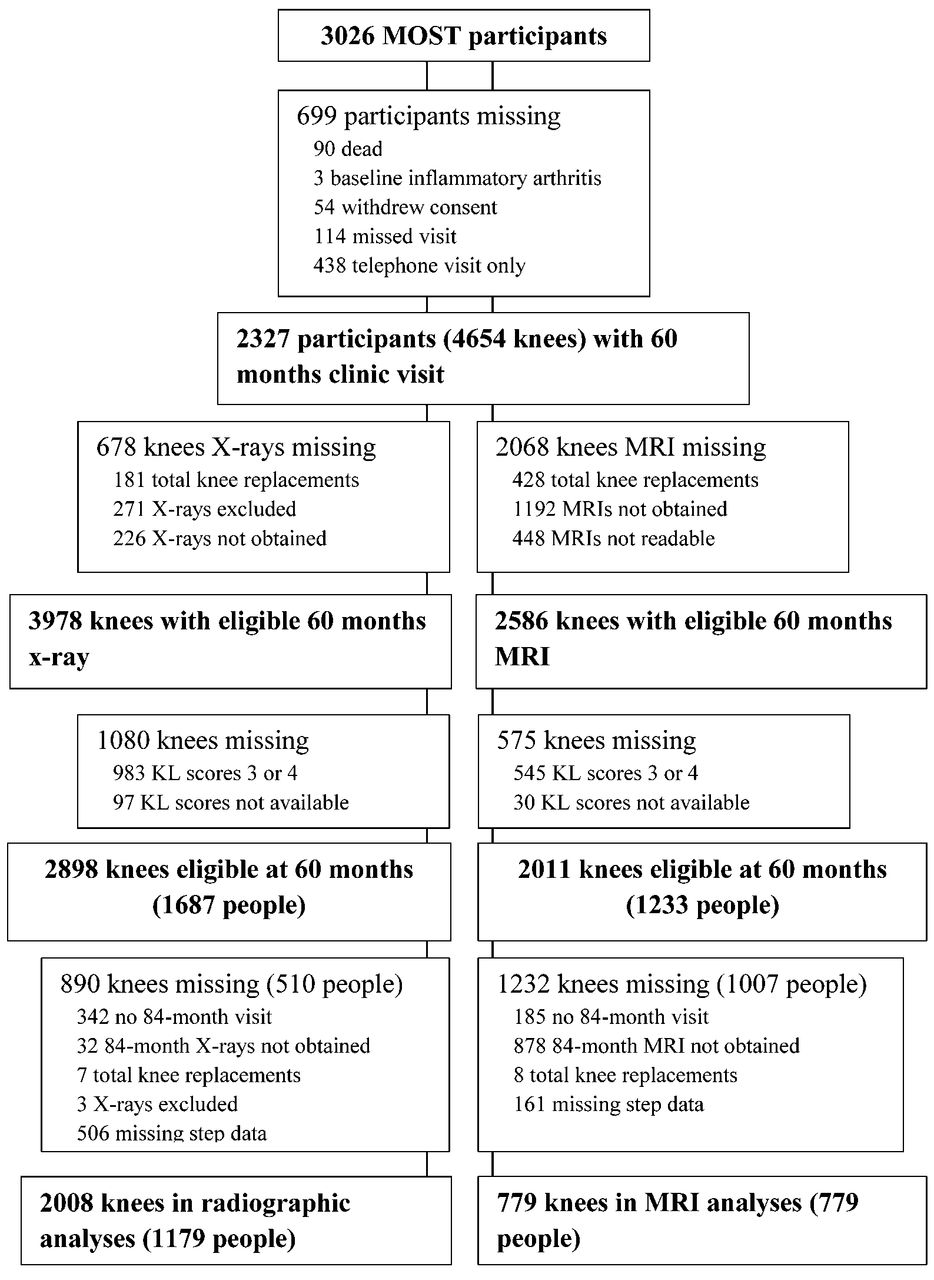
Of the 2898 knees (1687 people) with radiographs at the 60-month visit that were not endstage or already replaced, and 2011 knees (1233 people) with MRI, 890 knees (510 people) were missing for joint space narrowing (JSN) analyses and 1232 knees (1007 people) were missing for MRI analyses (Figure 1). The 1179 study participants (2008 knees: 1019 left and 989 right) included in the JSN analyses were on average (± SD) 67.0 (± 7.6) years old (range 55–84) and had a mean BMI of 29.8 (± 5.3, range 18–57). Women were 59% of the group. The mean WOMAC pain score was 1.9 (± 2.5, range 0–15). Mean steps/day were 6981 (± 2630; min–max 774–19,450), and the average minutes per day of MVPA were 8.8 (± 11.2, range 0–80). Corresponding numbers for those 779 included in the analyses for cartilage loss were mean age 67.0 years (± 7.6), mean BMI 29.5 (± 4.7); 61% were women, and there were 7185 mean steps/day (± 2565). Table 1 shows the characteristics of study participants across knees divided into tertiles of steps/day. Sixteen percent (calculated from 2008 knees) had no minutes of MVPA per day. At 60 months, 51.6%, 20.5%, and 27.9% of knees had KL grade 0, 1, and 2, respectively. Distribution of cartilage loss between 60–84 months across tertiles of daily walking is given in Table 2.

Flow chart of the study participants. MOST: Multicenter Osteoarthritis study; MRI: magnetic resonance imaging; KL: Kellgren-Lawrence.
Characteristics of study participants at the 60-month visit (n = 2008 knees). Analyses for continuous variables are performed using ANOVA. Analyses for categorical variables are conducted using chi-square test. Values are mean ± SD unless otherwise specified.
Number of knees with any cartilage loss (≥ half a grade change) between 60 to 84 months by tertiles of daily walking (n = 779). Values are numbers of knees followed by % of 260 knees.
Analyses showed no significant differences for those with KL grade 0–2 who were included in the regression model (n = 1179) versus those with KL grades 0–2 who were not included (n = 510) for BMI, sex, age, or knee injury. Those included in our analyses had significantly less WOMAC pain than those not included (1.9 vs 2.9, p < 0.001). Correspondingly, no differences were found for BMI, sex, or knee injury between those 779 who were included in the analyses on cartilage loss compared with the 454 who were not included. However, those not included were 1 year older (p = 0.03) and had 0.6 points higher WOMAC pain score (p = 0.002).
Associations between structural changes and daily walking
No significant associations were found between radiographic worsening and levels of daily walking (Table 3). Further, no significant associations were found between medial radiographic worsening and low (adjusted OR 0.86, 95% CI 0.59–1.27) and high (adjusted OR 1.21, 95% CI 0.82–1.79) versus moderate levels of daily walking, or lateral radiographic worsening and low (adjusted OR 1.10, 95% CI 0.66–1.83) and high (adjusted OR 0.62, 95% CI 0.33–1.16) versus moderate levels of daily walking. Further, no significant associations were found between walking with MVPA and radiographic worsening (Table 3).
The association between activity levels and structural worsening. The analyses are adjusted for the following confounders measured at the 60-month visit: age, sex, BMI, WOMAC knee pain, KL grade, alignment, and history of knee injury.
Significantly increased odds for cartilage loss in the medial compartment were seen for the lowest level of daily walking (< 6078 steps/day) versus those in the moderate level (6078–7938 steps/day) for unadjusted analysis (OR 1.70, 95% OR 1.06–2.73), but not for adjusted analyses. Figure 2 shows the adjusted association between tertiles of daily walking and proportion of medial cartilage loss. Analyzing daily walking as a continuous variable did not reveal a significant relationship with cartilage loss, and likewise, comparing those walking ≥ 10,000 versus < 10,000 steps/day did not reveal a statistical association in the adjusted model. No significant interactions were detected for daily walking and cartilage morphology status at the 60-month visit for either lateral (p = 0.106) or medial (p = 0.274) compartments. No significant association was found between walking at MVPA and medial cartilage loss (Figure 3) or lateral cartilage loss. Further, omitting those with a history of knee injury at the 60-month visit did not change the relationship between daily walking and medial or lateral cartilage loss.

Proportion of medial cartilage worsening for tertiles of daily walking (dashed lines).

Proportion of medial cartilage worsening for minutes of MVPA. MVPA: moderate to vigorous physical activity.
DISCUSSION
The results of our study did not confirm our hypotheses that those in the lowest and highest levels of daily walking had higher risk of structural changes compared with those with a moderate level of walking. Further, we did not find an association of walking with MVPA and structural changes over 2 years.
Even though not directly comparable, our results are in contrast to the literature suggesting associations between physical activity and structural changes in the knee joint2,34,35. Animal studies36,37,38, as well as studies on patients with injured knees39,40, and population-based cohort studies have suggested that high activity level may increase the risk of development and progression of knee OA1, although conflicting results have been reported41,42. No universal definitions of high, moderate, and low activity levels exist for walking, even though suggestions exist43. The MOST cohort does not represent a population with a high activity level, such as those in which an association of activity and structural worsening have been found (> 10,000 steps/day)2, and our tertiles may not represent the low, medium, and high activity levels for other populations. Tudor-Locke, et al43 have recommended this walking index: < 5000 steps/day reflects a sedentary lifestyle, 5000–7499 steps/day reflects low activity, 7500–9999 is somewhat active, and > 10,000 reflects a highly active level. The MOST data showed that the participants walked on average 6981 (± 2630, radiographic group) and 7185 (± 2565, MRI group) steps/day, with tertiles lower than the suggested classifications, but with mean values similar to the recommended 7000 steps in the American College of Sports Medicine Position Stand44. Our third tertile walked > 7950 steps/day, reflecting “somewhat active” according to Tudor-Locke, et al’s suggestion. Further, 16% of the study participants walked less than 1 min with MVPA, indicating a general low activity level in the MOST cohort45.
Doré, et al2 followed community-dwelling adults over 60 years of age for 2.7 years with measurements of daily walking at baseline using a pedometer and MRI findings both at baseline and after 2.7 years. They found that walking more than 10,000 steps/day was associated with increase in BML, meniscal pathology, and cartilage defect score. Further, they found an interaction effect between daily walking and cartilage volume, indicating that activity level was protective against volume loss in individuals with more baseline cartilage volume, but led to increased cartilage loss in those with less baseline cartilage volume. The authors concluded that individuals with knee abnormalities should not walk more than 10,000 steps/day. Doré, et al did not exclude those with moderate and severe radiographic OA and reported a prevalence of OA of 57% as compared with about 28% mild radiographic OA in our cohort. This may be the main reason for the different results across these 2 studies, in addition to the number of people walking > 10,000 steps/day. In another study, Lin, et al1 investigated physical activity in relation to change in T2 values in cartilage over 4 years using data from the Osteoarthritis Initiative. They found that both high and very low activity scores as measured with the Physical Activity for the Elderly Scale were associated with greater progression of T2 relaxation time in asymptomatic people without radiographic OA. They did not control for knee injury in their analyses, but according to their results and like ours, knee injury did not differ between tertiles of activity level. Currently, it is not known whether progression in T2 values leads to cartilage morphology changes. Our analyses showed that a history of knee injury measured at the 60-month visit was significantly associated to medial cartilage loss (results not shown). Thus, knee injury seems to be more important for medial structural worsening than activity level, at least when average activity level is relatively low as seen in the MOST cohort.
Our data showed no relation between structural change and minutes per day of walking with MVPA, but this is limited by a narrow range of step frequency in our subjects. Future studies should examine whether prolonged bouts of activity intensity as would be encouraged in exercise programs might influence joint structures.
Our study had weaknesses. Only 1 timepoint of physical activity measurement may not be optimal to assess daily walking levels as predictors of structural change. However, activity and step monitors have shown to be valid and reliable in measuring peoples’ physical activity levels24. StepWatch-recorded steps/day has high concurrent validity in comparison with several reference standard measures of step frequency46,47 and is 96% accurate for counting steps in older adults48. Further, following a prospective cohort of people over many years, high dropout rates may be expected. After selection of study participants according to our inclusion and exclusion criteria, we still included as many as 1179 people (2008 knees) for the radiographic worsening analyses and 779 people for the MRI analyses. As a result of the challenge with studying risk factors for progression of OA in knees with advanced disease, we narrowed our sample to people at risk of developing OA or those with mild OA. Studying cartilage loss in this population with no or mild OA using MRI with 1.0T magnet may not be optimal compared with newer MRI techniques49. Finally, even though our data included a substantial number of knees with worsening as seen in Table 3, a longer followup would have been better to detect radiographic structural changes in the knee joint.
Results from the MOST Study indicated no association between daily walking and structural changes over 2 years in people at risk of or with mild knee OA.
Footnotes
Multicenter Osteoarthritis study funded by US National Institutes of Health and National Institute on Aging grants (AG18820, AG18832, AG18947, and AG19069). The Research Council of Norway funds a postdoctoral position for the corresponding author.
- Accepted for publication April 13, 2015.


