

Ankylosing spondylitis (AS) is the prototype of diseases that belong to the category of spondyloarthropathies. AS usually affects the sacroiliac joints and invariably involves the axial skeleton. Peripheral joint involvement, enthesitis, and extraskeletal manifestations are also important clinical and radiographic features of the disease1,2.
A 55-year-old man was diagnosed with AS at the age of 18 years. He initially presented with both axial and peripheral involvement, including heel enthesitis and acute anterior uveitis. He had been doing well with nonsteroidal antiinflammatory drugs and sulfasalazine. At his most recent regular examination, he had experienced moderate pain at the proximal and lateral aspects of both thighs for several weeks. Clinical examination revealed signs of spinal ankylosis and tenderness over the trochanters.
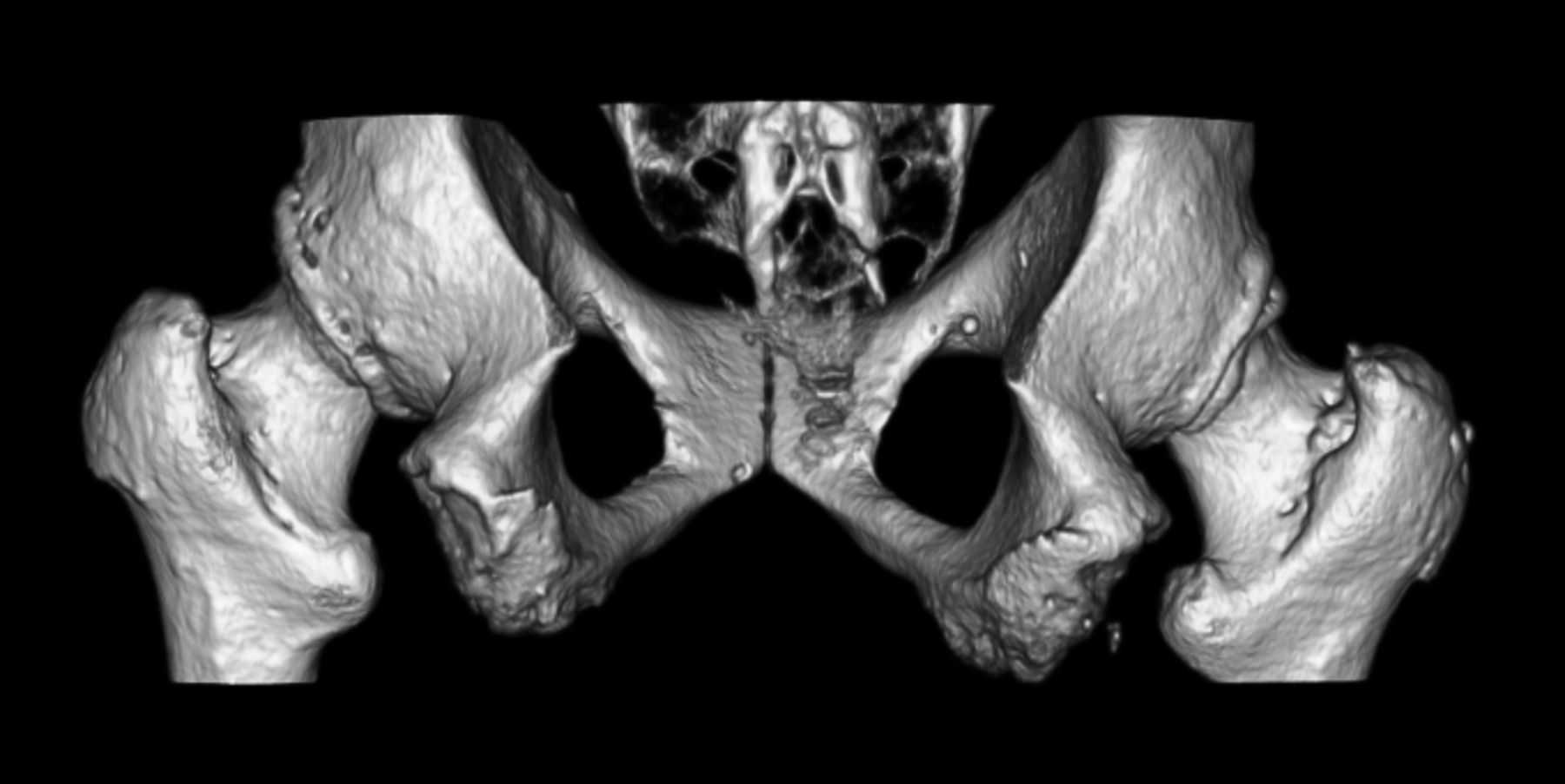
The patient was injected into the left greater trochanter twice, but without clinical improvement. Radiographs (Figure 1) showed sacroiliac fusion, whiskering at both ischial tuberosities, degenerative changes of both hips, and an uncertain lytic image at the right trochanter (possibly due to overpenetration of the radiograph). A computerized tomography (CT) scan was subsequently obtained, which revealed irregularity of the right trochanter, representing a possible avulsion of the tendinous insertion, osteoarthritic changes in both hips, and exuberant enthesopathy and bone proliferation at the ischial tuberosities. A 3-D volume rendering of the CT was performed (Figures 2 and 3), highlighting these findings. Eventually the patient was referred to the rehabilitation service and is doing well with ultrasound and laser therapy.

Anteroposterior view of whiskering.
Posteroanterior view of whiskering.

Radiograph showing whiskering.
The 3-D volume renderings from the CT scan show how dramatic enthesitis can be in these patients.


