

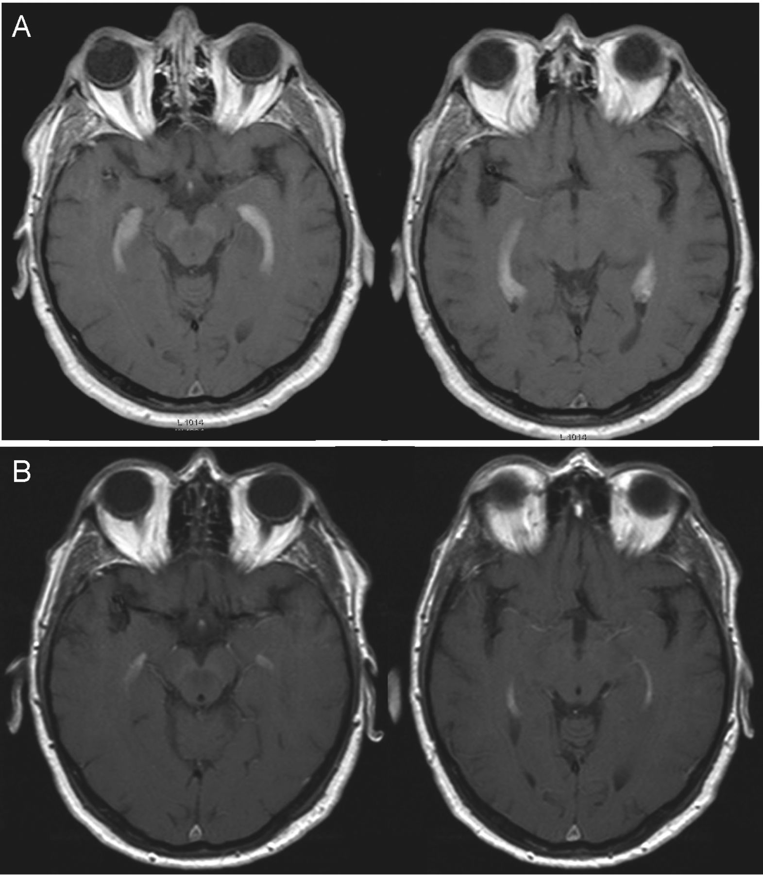
A 70-year-old man presented with a 3-day history of nausea, vomiting and constitutional symptoms (fatigue and anorexia). During hospitalization he developed diplopia. Cranial magnetic resonance imaging with gadolinium (Figure 1a) showed inflammation of the entire choroid plexus. A diagnosis of granulomatosis with polyangiitis (GPA) was made based on: presence of perinuclear antineutrophil cytoplasmic antibody at titer 1/40 with elevated anti-proteinase 3 antibodies; sinus radiography showing a mucosal thickening of the left maxillary sinus; and a lung biopsy demonstrating chronic granulomatous inflammation with multinucleated giant cells. The patient was treated with methylprednisolone [1 g/day intravenously (IV) for 3 days], cyclophosphamide (0.6 mg/m2/dose IV, monthly pulses for 6 mos) and mycophenolate mofetil (2 g/day PO, maintenance therapy). He made a full recovery. A followup MRI showed partial improvement of the inflammation (Figure 1B).

Axial contrast-enhanced T1-weighted images. Homogenous marked enhancement of enlarged choroid plexus within the 2 temporal horns (a); and followup imaging 3 months after immunosuppression therapy showing a prominent reduction in size and enhancement (b).
Although not commonly the initial symptom1,2, the most frequent pattern of central nervous system involvement is granulomatous inflammation that leads to compression of usually cranial nerves II–III, V–VIII, meningitis and pituitary gland inflammation3. Our literature review of choroid plexus involvement in GPA cases yielded no reports.
Acknowledgments
The authors thank Mireia Tomás for assistance in acquiring patient data for this report.


