

Amicrobial pustulosis of the folds (APF) is a rare, chronic, relapsing cutaneous disease seen almost exclusively in young women with a history of autoimmune disease, most commonly systemic lupus erythematosus (SLE), or who simply have circulating autoantibodies1,2,3. APF is characterized by recurrent crops of pustules primarily in the skin folds and periorificial regions that eventually coalesce into plaques. To our knowledge there are fewer than 30 cases in the literature to date.
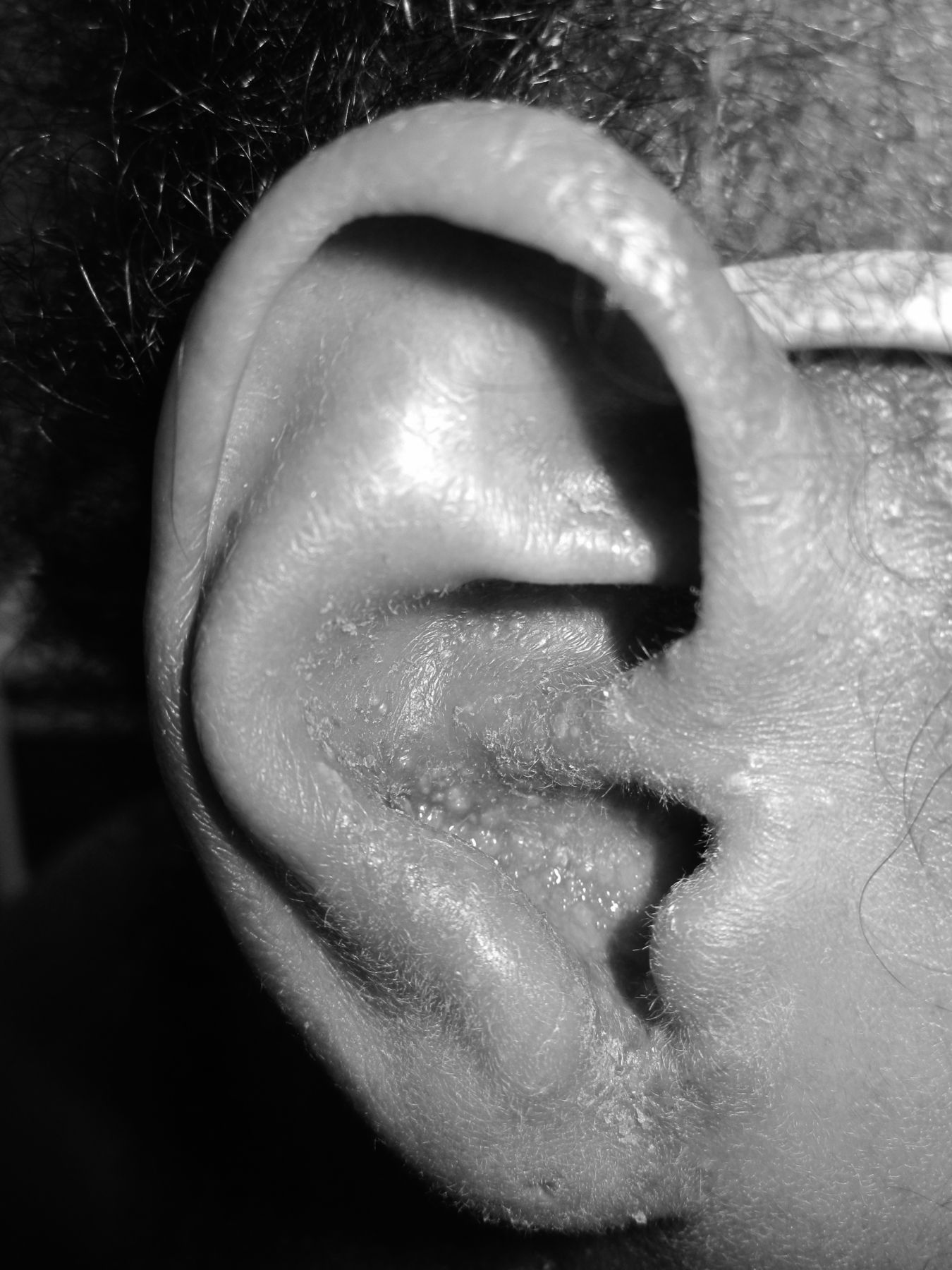
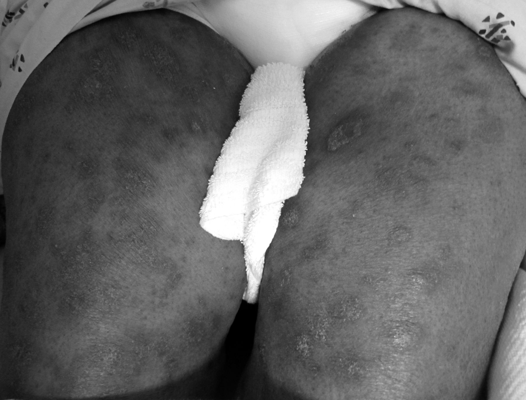
A 22-year-old African American woman with a history of SLE and taking hydroxychloroquine presented with a 1-year history of a recurrent generalized pruritic pustular eruption. She had multiple hospitalizations for this cutaneous eruption, which was originally thought to be superinfected atopic dermatitis, but had no resolution with topical steroids or oral antibiotics. Examination revealed multiple pustules with underlying erythema throughout the scalp, bilateral conchal bowls (Figure 1), forehead, eyelids, nasal ala, bilateral inframammary folds, and flanks. On the bilateral thighs and groin there were erythematous papules and pustules coalescing into plaques (Figure 2). Skin biopsy and clinical presentation was consistent with APF.

Small pustules with underlying erythema are seen in the right conchal bowl.

Erythematous papules and pustules have coalesced into plaques on the bilateral thighs.
The cause of APF is unknown, but is thought to be related to immune-complex activation of complement leading to neutrophil chemotaxis1,4. Recently, APF has been classified within the spectrum of neutrophilic dermatoses, which are now considered autoinflammatory diseases4,5. Flares of autoimmune disease have not been shown to correlate with the severity of APF1,2. There is no standard therapy established for APF, because multiple immune-modulating therapies have failed to show consistent longterm success1,2. Despite treatment with potent topical steroids and azathioprine, our patient has had no significant improvement to date.


