

Abstract
Objective. To examine the pain threshold in children with juvenile idiopathic arthritis (JIA) compared with healthy children by using a digital pressure algometer.
Methods. Fifty-eight children with JIA born between 1995 and 2000 and 91 age-related healthy children participated in the study. We used a digital pressure algometer to measure the pain threshold on 17 symmetric, anatomically predefined joint-related or bone-related areas. All children were asked to rate their current pain on a Faces Pain Scale, and parents of children with JIA were asked to complete a parental revised version of the Child Health Assessment Questionnaire (CHAQ-R). Clinical data were registered on children with JIA.
Results. The pain threshold was significantly lower among children with JIA (total mean PT = 1.33 ± 0.69 kg/cm2) when compared with the healthy control group (total mean PT = 1.77 ± 0.67 kg/cm2). The same pattern was found in all areas measured, including negative control areas that are normally unaffected in JIA (p = 0.0001 to 0.005). Overall, the pain threshold was 34% lower in females than in males in both groups (p < 0.0001). We found no correlation between pain threshold and age, current pain experience, disease duration, or disease activity.
Conclusion. Children with JIA had a substantially lower pain threshold even in areas usually unaffected by arthritis. Our findings suggest that JIA alters the pain perception and causes decreased pain threshold.
Pain is a primary symptom in juvenile idiopathic arthritis (JIA), and recent studies report a high daily prevalence of pain among children with JIA1,2. Extensive pain symptoms generally lead to less participation in social activities and lower quality of life. In contrast, even a small reduction in pain has been shown to clinically improve life quality3. As the efficacy of treatment of JIA has improved over the years, a decrease in pain experience could be expected. However, some children continue to report pain despite improved clinical and biochemical status2,4.
Previous studies have reported lower pain threshold (PT) in children with chronic pain conditions, even in body areas not affected by disease5,6,7,8,9, supporting the theory that chronic pain conditions alter central nervous system pain perception and sensitization10.
Several studies have reported the prevalence and predictors of pain in JIA2,11,12,13,14. However, pain is a complex subjective experience that cannot be measured directly15. Different methods of estimating pain have been demonstrated: Thastum, et al16 studied PT and pain tolerance in children with JIA by submerging their hand in cold water. However, this method is not very practical when testing proximal areas. Laser beam-induced pain is a precise and highly reproducible method17; however, it mainly induces pain in the skin whereas nociceptors in joints are more sensitive to mechanical stimuli18. A digital pressure algometer, previously validated in a laboratory19 but not in a clinical setting, provides mechanical stimuli to elicit pain and may be a preferential method for studying pain threshold in JIA.
The association between other chronic pediatric pain conditions and reduced PT led us to hypothesize that children with JIA have a decreased pain threshold measurable by digital pressure algometry.
The aim of our study was to assess the pain threshold of predefined anatomical areas in children with JIA compared with healthy children by using a digital pressure algometer. Further, associations between pain threshold and age, sex, and pain rating, as well as clinical data, were targeted.
MATERIALS AND METHODS
Patients
Inclusion criteria were JIA diagnosed according to International League of Associations for Rheumatology criteria20 at least 6 months before the day of testing. Subjects were born between 1995 and 2000. Patients were excluded from the study if the time from disease onset to the day of testing was < 6 months; if the child or parent were not fluent in oral and written Danish; or if the child suffered from comorbidity associated with or potentially associated with pain (e.g., fibromyalgia, migraine).
From December 2009 to May 2010 we invited 98 children with JIA and consecutively followed at the pediatric rheumatology clinic in Aarhus to participate in this study. Sixty children agreed to participate, but 2 children were excluded because of comorbidity (migraine and bone cysts, respectively), leaving a total of 58 (42 girls, 16 boys).
The children with JIA were tested at the clinic in a separate room after their regular followup visit. The children were tested for PT without the parents to simulate the testing of the healthy controls.
Control subjects
Children born between 1995 and 2000 attending a public school (n = 533) were invited to the study. Written and verbal information was given to children and parents. Children were excluded if the child or the parents were not fluent in Danish; or if the child had comorbidity associated with pain.
Ninety-six children agreed to participate. Five were excluded because of comorbidity (migraine, recent minor trauma, hip pain, and menstrual cramps), leaving 91 controls.
Experimental procedure
Current pain as well as the worst pain during the past week were registered using the Faces Pain Scale, which contains 6 faces representing a scale from 0 = no pain to 5 = worst pain. The children were instructed to mark the face that best expressed their pain21.
Parents of children with JIA completed a parental version of the CHAQ-R comprising 38 items, as a measure of the children’s level of functional disability22,23.
As a measure of the disease activity of children with JIA, we used the Juvenile Arthritis Disease Activity Score (JADAS) described by Consolaro, et al, based on the evaluation of 27 joints, yielding a total score range from 0 to 5724.
Use of analgesics or nonsteroidal antiinflammatory drugs (NSAID) on the day of testing and whether the child was right-handed or left-handed were noted for all children.
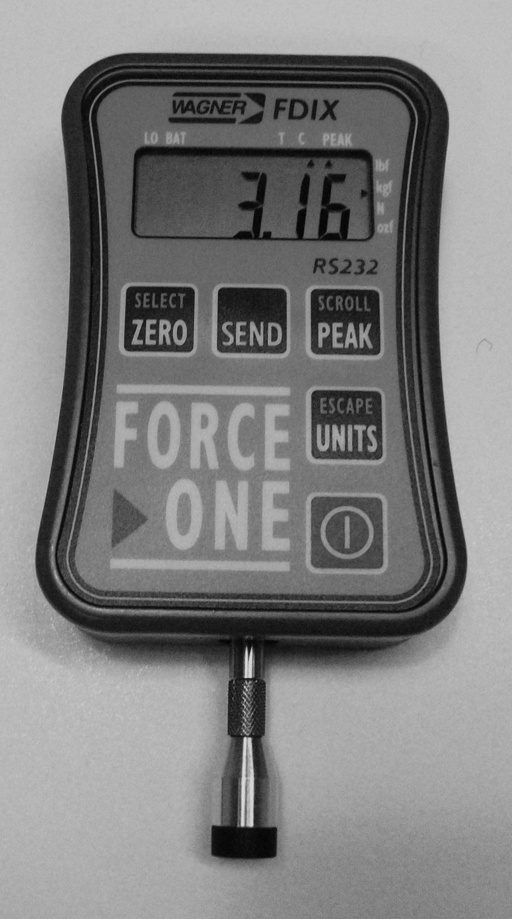
We used a digital pressure algometer (Wagner Force One Model FDIX 50TM, Wagner Instruments; Figure 1), which measures pressure in kg/cm2 with an accuracy of ± 0.2%. Pressure was applied manually to the area of interest using a 1 cm2 rubber tip fixed to the gauge. Nine anatomically well-defined areas were selected for measuring PT. These were glabella, temporomandibular joint, humoral tubercle, proximal radioulnar joint, ulnar styloid process, second metacarpophalangeal joint, medial femoral condyle, anteromedial aspect of tibia, and medial malleolus. Except for the forehead, all areas were tested on both sides, resulting in 17 individual test areas. Negative control sites without relation to arthritis included forehead, and right and left tibia.

The pressure algometer (Wagner Force One Model FDIX 50TM, Wagner Instruments).
Data for single measurement areas were excluded if the area was unavailable for testing because of clothing or bandage; if the instrument slid off during testing; if active arthritis was found in a joint; or if corticosteroids had been given intraarticularly within 1 month prior to testing. One observer (female) tested all children, testing the body areas in the same order for each subject. Pressure was gradually applied at an even rate, about 0.1 kg/s, onto the point of interest. The algometer rubber tip was always held perpendicular to the body area investigated, except for the medial malleolus (45 degrees). Children were seated during testing and blinded from the applied amount of pressure (Figure 2). All children were equally instructed to verbally communicate the initial feeling of pain (PT). As soon as the child indicated the onset of pain, the application of pressure was stopped.

Measuring the pain threshold of the right shoulder.
Based on a pilot study, we had defined 3 different cutoff points depending on the area of interest. For testing in the head region, the cutoff point was 2.00 kg/cm2; for the upper extremity 3.00 kg/cm2; and for the lower extremity, 3.50 kg/cm2. If a child had not indicated the onset of pain when reaching a cutoff point, the pressure application was stopped, and the cutoff point was noted as the PT for that area.
Our study was approved by the local ethics committee and the Danish Data Protection Agency (Record No. 2009-41-3262). The persons responsible for the children gave verbal and written consent.
Statistical analysis
All data were analyzed using PASW (SPSS) statistics version 18. Testing for normality using the Kolmogorov-Smirnov test indicated that variables were not normally distributed. However, considering the relatively large sample size, and the fact that parametric and nonparametric tests yielded very similar results, we decided to report the results of the parametric tests only. Of the 149 participants, 31 had 49 missing data out of a total of 3842 (1.3%). Missing data were imputed using the expectation maximization algorithm25,26.
The internal consistency was calculated on the 17 unique measurements for each individual obtained in the first test of all 149 children, yielding a Cronbach’s alpha coefficient of 0.98. A principal component analysis was conducted on the 17 measurements, revealing a 1-factor solution explaining 77.7% of the variance. Because of the very high level of the Cronbach’s alpha coefficient and the 1-factor solution from the principal component analysis, we found it reasonable to calculate an overall score for each individual (“total mean”) for the further analyses.
The independent-samples t test, the paired-samples t test, and the Pearson correlation coefficient were used when examining pain threshold estimates. The effect size of the independent-samples t test was calculated as Cohen d. Bonferroni corrections were used to control for type I error in the multiple correlation analyses.
We used 1-way between-groups ANOVA when estimating the PT among 3 different age groups. If a significant difference in PT between sexes or age groups was found, a 2-way between-groups ANOVA was conducted to explore a possible interaction effect between the independent variable and the 2 groups on PT.
RESULTS
Characteristics of patients with JIA and controls are shown in Table 1. Neither age nor sex differed significantly between the groups. Compared with the control group, children with JIA were more likely to have taken analgesics or NSAID on the day of testing.
Demographic characteristics of study participants.
The mean of the sum score of the CHAQ-R was 0.3 out of a maximum of 3.0. Sufficient data for calculating JADAS were only available for 36 patients, giving a mean score of 4.85 out of a maximum of 57. Only 1 patient had been given a steroid injection in a joint within 1 month prior to testing.
Forty-four percent of patients with JIA and 71% of the control children rated “no pain” on the Faces Pain Scale for current pain. Mean current pain and “worst pain during the last week” was significantly higher for the JIA group (Table 1).
Pain threshold estimates.
The children with JIA had a total mean PT of 1.33 ± 0.69 kg/cm2, significantly lower than the total mean of 1.77 ± 0.67 kg/cm2 within the healthy control group (p < 0.001). The magnitude of the differences in means (mean difference = 0.44, 0.95% CI: 0.22 to 0.66) was medium (Cohen d = 0.64). Looking individually at all areas tested, we found a similar pattern, including negative control areas normally unaffected in JIA (Table 2).
Results of pressure algometry. Measurements are in kg/cm2 ± SD.
PT was 34% lower in females than in males (p < 0.001). A 2-way between-groups analysis of variance was conducted to explore the effect of sex and the 2 groups (JIA group and control group, respectively) on levels of PT, finding statistically significant main effects for sex (p = 0.0001) and for the 2 groups (p = 0.001) on the level of PT. But the interaction effect of sex and the 2 groups on the level of PT did not reach statistical significance (p = 0.56).
We found no significant difference in total mean PT between age groups below and above median age.
For the entire study population we found a very strong, significant correlation between total mean PT of the dominant and nondominant side (r = 0.98; p < 0.001). The mean difference in PT was 0.01 kg/cm2 (p = 0.27).
Correlations between pain rate, disease activity, disease duration, CHAQ-R, and pain threshold
Current pain correlated well with “worst pain during the past week” (r = 0.58; p < 0.001), but we did not find any correlation between current pain or “worst pain during the past week” and PT total mean.
Neither disease duration nor disease activity correlated with the total mean PT (r = 0.07 and −0.18, respectively; p > 0.30). We found no correlation between CHAQ-R and total mean PT (r = −0.22; p = 0.10).
DISCUSSION
In our study, children with JIA had a significantly lower PT than healthy age-related controls for each of the 17 individual areas tested.
Pressure algometry is a very mobile and ready-to-use method. With a little training, an observer is quickly able to gain information about the pain status of areas all over the body. It is suitable for children because the pain inflicted on the subject is very mild. None of the 149 participating children asked to stop before testing was completed. Kinser, et al showed that with practice, the Wagner Force One FDIX digital pressure algometer was reliable and valid in a laboratory setting19. To our knowledge, our study is the first to use this instrument on humans.
Our findings of a decreased PT in children with JIA are in concordance with other studies on experimental pain in JIA. Investigating cold pressor pain by submerging a hand in ice water, Thastum et al13,16 found a significant difference between the mean pain tolerance in patients with juvenile arthritis compared with healthy controls. However, differences regarding pain threshold did not reach statistical significance. Hogeweg, et al9,27 demonstrated lower PT among children with juvenile chronic arthritis (JCA) for the 4 most frequently affected arthritic joints.
In our study, girls were found to have a substantially lower PT than boys in all areas measured. This opposes the results of other studies finding no difference between pain levels of boys and girls9,12,28. However, some studies observed that girls report more pain than boys11,29,30. The proportion of girls in the JIA group was higher than the proportion in the control group, a difference that might explain why patients with JIA have a lower PT than the control children. However, the results remained the same after controlling for sex. It may be problematic, though, to discuss the difference in PT among sexes because we had only 1 observer (female). Levine and De Simone30 showed that male adult subjects report lower pain to a member of the opposite sex than to another male. Females tended to report higher pain to opposite sex investigators; however, the results did not reach statistical significance. Although their results were based on adults, we cannot discount the possibility that children in our study would have responded differently to a male observer.
A few studies have reported older age to be associated with more severe pain12,31 but most studies do not11. We found no association between age and PT.
The current pain experience correlated with “the worst pain experience during the last week,” but we were surprised to find no association between current pain experience and PT, given that we had expected to find lower PT in children reporting high current pain experience. A possible explanation could be the generally low score of “current pain” among the patients as well as the healthy controls, making it difficult to investigate whether an association should exist. Hogeweg, et al9 reported that the mean PT was negatively correlated to the pain visual analog scale (VAS) and disease activity. However, they did not measure the VAS immediately before the clinical testing and therefore did not investigate the direct association between PT and self-reported pain experience.
The effect of duration of JIA on pain experience and PT has been subject to very little investigation. Thastum, et al16 reported a significant correlation between pain tolerance and disease duration when controlling for age in children with JIA. However, they found no correlation between disease duration and PT, which is in accordance with our results. This was a surprising finding, as we presumed that longer duration of active arthritis would lower the PT. However, if changes involving pain perception occur relatively early after disease onset, this effect may not be visible in our study, because mean disease duration at inclusion in this study was > 5 years.
Previous studies regarding the relationship between disease activity of children with JIA and pain experience have consistently demonstrated limited predictive value for disease activity11. Also, in our study we found no correlation between JADAS score and PT. Thus, the role of disease activity as a predictor of pain in JIA remains uncertain. The children in our study had relatively mild disease (mean JADAS score 4.85) and only 12% of the children with JIA had taken analgesics or NSAID on the day of testing. This may partly be explained by the inclusion criterion of disease duration of at least 6 months, ensuring that no children with recently discovered, untreated JIA were included. Our findings of a significantly lower PT among patients with JIA may therefore be an underestimate, as this may be even lower in children with active disease.
Several studies of pediatric chronic pain conditions support a model of altered pain perception even in areas not affected by disease. Buskila, et al5 reported lower thresholds for tenderness both at control and tender points in 21 children with fibromyalgia (FM) when compared with healthy children. Duarte, et al6 found a lower PT among children with recurrent abdominal pain in all areas measured, including extraabdominal sites. In children with growing pains (GP), Hashkes, et al7 reported decreased PT at tender points characteristic for FM as well as control points when compared with healthy controls. In a 5-year followup, 51% of patients experienced resolution from GP, and the investigators no longer found a difference in PT compared to controls32. They suggested that sensitization of the nervous system is reversible if the underlying condition initially causing pain is removed.
Peripheral and central sensitization may also play a role in JIA. Hogeweg, et al27 found a significantly lower PT of inflamed joints and a number of paraspinal tissues in children with JCA compared with healthy controls. These results are in accordance with our findings, even though our control points were the forehead and the anterior tibia, and not the paraspinal tissues. It is well recognized that pain is multidimensional, and sensitization may not explain all of the pain experience in JIA. Environmental factors, such as parent pain history and family relationships, and cognitive-behavioral factors (e.g., stress, mood, pain coping skills) may also contribute to the pattern of PT13,16.
The subjects were blinded from the pain measurements, but it was not possible to blind the observer because she had to control the speed at which pressure was applied to ensure an even pressure application. Further, the observer was not blinded from the group status of the children (JIA and control group, respectively), because they were not examined at the same locations.
It is important to keep in mind that pain will always be a subjective sensory and emotional experience. Therefore no method can ever be 100% objective. Regardless, pressure algometry may prove to be suitable as a supplement to other pain rating instruments in experimental and clinical settings. The decreased PT in children with JIA as compared to healthy children, even in areas not affected by arthritis, supports the theory of a pain sensitization. It remains uncertain when the sensitization occurs, but considering the fact that we did not find any correlation between disease duration and PT, sensitization may develop relatively soon after the disease erupts. Whether the lower PT in children with JIA has strictly physiological or psychological causes, or a combination, remains to be determined.
Footnotes
-
Support from The Danish Council for Independent Research/Medical Sciences, and The Danish Rheumatism Association.
- Accepted for publication January 21, 2013.


