

Abstract
Objective. Radiographic damage was recently identified as a feature of poor prognosis in polyarticular juvenile idiopathic arthritis (pJIA). However, most radiographic studies did not differentiate pJIA from other subtypes of JIA and little is known about pJIA persisting into adulthood. We describe radiological peripheral involvement in young adults with pJIA compared to patients with rheumatoid arthritis (RA).
Methods. All consecutive patients with pJIA followed in a transition program were included. Age, sex, disease duration, and medical or surgical treatment information was collected. Laboratory tests and standard radiographs of the hands and wrists, feet, and hips were analyzed by 2 independent radiologists blinded to the diagnosis. One RA control group (age < 55 yrs), matched for sex and disease duration, was recruited.
Results. Forty-three patients with pJIA and 59 with RA were included. Radiographs showed hand lesions in 79% of pJIA and 86% of patients with RA, feet lesions in 74% of pJIA and 80% of patients with RA, and hip damage in 35% of pJIA and 17% of patients with RA (p = nonsignificant). Specific to the juvenile forms were lower frequency of proximal interphalangeal joint involvement (51% vs 76%; p = 0.03) and higher risk of bilateral hip damage (86% vs 25%; p < 0.01) than in adult RA.
Conclusion. Structural peripheral damage is as common and as severe in young adults with pJIA as in adults with RA. The main specific feature of pJIA seems to be a high risk of bilateral hip damage. This requires a particular monitoring of pJIA patients with unilateral hip involvement to detect bilateralization.
Juvenile idiopathic arthritis (JIA) is a heterogeneous group of 7 diseases classified by the International League of Associations for Rheumatology (ILAR)1. Two forms with polyarticular onset were identified: rheumatoid factor (RF)-positive and RF-negative polyarticular JIA (pJIA)1. Both diseases are characterized by prolonged synovial inflammation that can lead to destruction of joints2.
Radiographic damage was recently identified as a feature of poor prognosis in cases of pJIA3. Evaluation has for a long time been considered as fundamental to assess disease severity and treatment efficacy in adult rheumatoid arthritis (RA)4. It has recently become a necessity in JIA also with the introduction of effective new therapies2,5,6,7,8,9.
It is commonly believed that pJIA has less destructive potential than adult RA, with the additional possibility of improvement in radiographic joint damage2,10,11,12,13. However, the only study comparing radiographic changes between these 2 groups included a high percentage of oligoarticular forms, which are those with the best outcomes13,14,15. There are, to our knowledge, no controlled studies comparing radiographic damage in a homogeneous pJIA group to that observed in RA.
Further, prevalence of damage in pJIA varies widely from study to study (between 20% and 100%) depending on the population assessed, sites of radiographs, and scoring method used12,13,14,16,17,18,19,20,21,22,23,24. The most common joints involved seem to be wrists, then metacarpophalangeal (MCP) joints, followed by metatarsophalangeal (MTP) joints and hips22,24,25. However, few studies have specifically assessed the prevalence of damage at these sites; and most were performed before the publication of the recent ILAR classification criteria and the biotherapy era19,24,26,27,28,29.
Little is known about radiographic damage in pJIA persisting into adulthood19,30. Understanding the adult outcomes of these pediatric conditions is fundamental to identify patients with poor outcome. Only 1 study assessed hand and hip lesions in juvenile RA persisting into adulthood19. It found a high prevalence of both hand and hip damage, estimated, respectively, at 68% and 41%19. However, that study did not differentiate pJIA from the other subtypes of JIA and was performed 40 years ago, long before the advent of biotherapies19,31. Therefore, the prevalence and characteristics of radiological peripheral involvement in hands, feet, and hips in pJIA persisting into adulthood remain unknown.
The aim of our study was 3-fold: (1) to assess the prevalence and characteristics of structural peripheral involvement in pJIA persisting into adulthood in hands, feet, and hips; (2) to compare damage in pJIA to that observed in a RA control group; and (3) to determine associations between damage in pJIA and characteristics of the disease.
MATERIALS AND METHODS
Clinical and biological study
All unselected consecutive patients with pJIA followed in a transition program in a single tertiary referral center were included in this observational study, performed from June 2009 to December 201032. Because these patients constituted a group with severe disease, it was necessary to have an ultrastructural evaluation at the beginning of this program. All patients had already been registered in the CEMARA database, which was validated by the Commission nationale de l'informatique et des libertés. All patients fulfilled the ILAR classification for pJIA1. Exclusion criteria consisted of a diagnosis of another subtype of JIA and, for detailed analysis [i.e., scoring and distribution of the lesions at each site: wrist, MCP, proximal interphalangeal (PIP), or MTP], a history of surgery at the joint assessed. One RA control group fulfilling the American College of Rheumatology criteria for RA was recruited33. Each patient with pJIA was matched to a control with RA based on sex and disease duration. They were not matched for serology. All RA control patients were < 55 years old to avoid radiographic-confounding lesions of osteoarthritis.
Age, sex, and disease duration information was collected, as well as swollen and tender joint counts and medical [corticosteroids, disease-modifying antirheumatic drugs (DMARD), and/or biological agents] or surgical treatment data. Laboratory tests were performed [erythrocyte sedimentation rate, C-reactive protein, rheumatoid factor (RF), anticitrullinated protein antibodies (ACPA), and antinuclear antibodies (ANA)].
Radiological study
Standard radiographs of the hands and wrists, feet, hip, and cervical spine were performed. Radiographs were analyzed by 2 independent radiologists (RB and VF) who were blinded to the diagnosis. A third reader (AF) established a consensus when required. Structural lesions on the hands and feet were assessed by the modified version of Larsen's scoring method in posteroanterior projection34. This method evaluates 32 joints: 8 PIP, 2 interphalangeal joints of the thumbs, 10 MCP, 2 wrists, and 10 MTP joints. Erosions are defined as a discrete interruption of the cortical surface of the bone and are graded according to the amount of destruction of the joint surface (DJS). Scores for joint space narrowing and erosion in each area range from 0 to 5 [0: intact bony outlines and normal joint space; 1: soft-tissue swelling and/or joint space narrowing and/or subchondral osteoporosis, DJS < 25% (score = 2), DJS 26%–50% (score = 3), DJS 51%–75% (score = 4), and DJS > 75% (score = 5)]. The Larsen score is calculated as the sum of the scores for each area. The hand and foot scores range from 0 to 110 (2 wrists, 10 MCP, 10 PIP) and from 0 to 50 (10 MTP), respectively. The score considered was the mean of the score read by the 2 independent observers. In cases of disagreement, the score was that assigned by the third reader. Hips were assessed in posteroanterior view for the presence of joint space narrowing and/or erosions. Joint damage was defined by joint space narrowing and/or erosions evident on radiographs. The presence of peripheral lesions was considered if there was at least 1 lesion in hands and/or feet and/or hips; i.e., Larsen score ≥ 1 and/or hip damage. Cervical spine radiographs included anteroposterior, lateral with flexion and extension, and open-mouth views.
Statistical analysis
All data analyses were performed using MedCalc® version 9.2.1.0. Data were presented as mean (SD) for continuous variables and numbers (percentages) for categorical variables. Data were statistically analyzed using chi-square tests for differences in frequency and Student's t test for comparison between 2 normally distributed continuous variables. Age and disease duration were presented as medians and interquartile range (IQR) and comparisons were performed by Mann-Whitney U test because distributions were not normal. A p value < 0.05 was considered statistically significant. In cases of p < 0.05, OR estimates and 95% CI were calculated. A multivariate stepwise logistic regression analysis controlling for confounding factors was also performed to determine whether damage was independently affected by juvenile onset, with calculation of OR estimates and 95% CI. Interobserver reliability was determined by comparing the findings obtained by the 2 investigators and by calculating the concordance correlation coefficient κ in cases of binary scores (i.e., presence or absence of peripheral lesions and presence or absence of hip damage). The interobserver agreement for the wrist/hand and foot score was assessed by intraclass correlation coefficient. Concordance between 2 scores was assessed by Spearman's coefficient of rank correlation (rho).
RESULTS
Forty-three patients with pJIA (35 females/8 males) were included in our study; median age was 23.0 years (IQR 20.0–30.8), median disease duration was 13.0 years (IQR 6.3–21.8). pJIA was RF-positive in 23/43 (54%) patients; 16/31 (52%) patients were ACPA-positive. Eleven pJIA patients had ANA; in 9/11 patients, ANA were associated with RF. None had had uveitis. All patients but 2 were treated with DMARD (38/41 received methotrexate), whereas 30/43 (70%) had received biological agents over the disease course (an antitumor necrosis factor agent was used in 90% of the cases). A biological agent was introduced after a mean of 8 years of disease duration. Eleven pJIA patients (26%) had undergone at least 1 previous surgery. Fifty-nine RA control patients (52 females, 7 males) were recruited, with the following characteristics: median age 46.0 years (IQR 32.8–51.0), median disease duration 11.0 years (IQR 8.0–15.8); 79% were RF-positive and 79% were ACPA-positive. Further details are provided in Table 1.
Clinical characteristics of patients with polyarticular juvenile idiopathic arthritis (pJIA) and controls with rheumatoid arthritis (RA). Values are no. (%) unless stated otherwise.
The interobserver concordance kappa coefficient was 0.680 (95% CI 0.597–0.762) between the 2 investigators. For hip score, the kappa coefficient was 0.701 (95% CI 0.583–0.819). The intraclass correlation coefficient was 0.880 (95% CI 0.826–0.918) for hand score and 0.870 (95% CI 0.817–0.910) for foot score. A consensus by the third reader was required in 35 cases (7 for hand score, 18 for foot score, and 10 for hip score).
Prevalence and characteristics of structural damage in pJIA
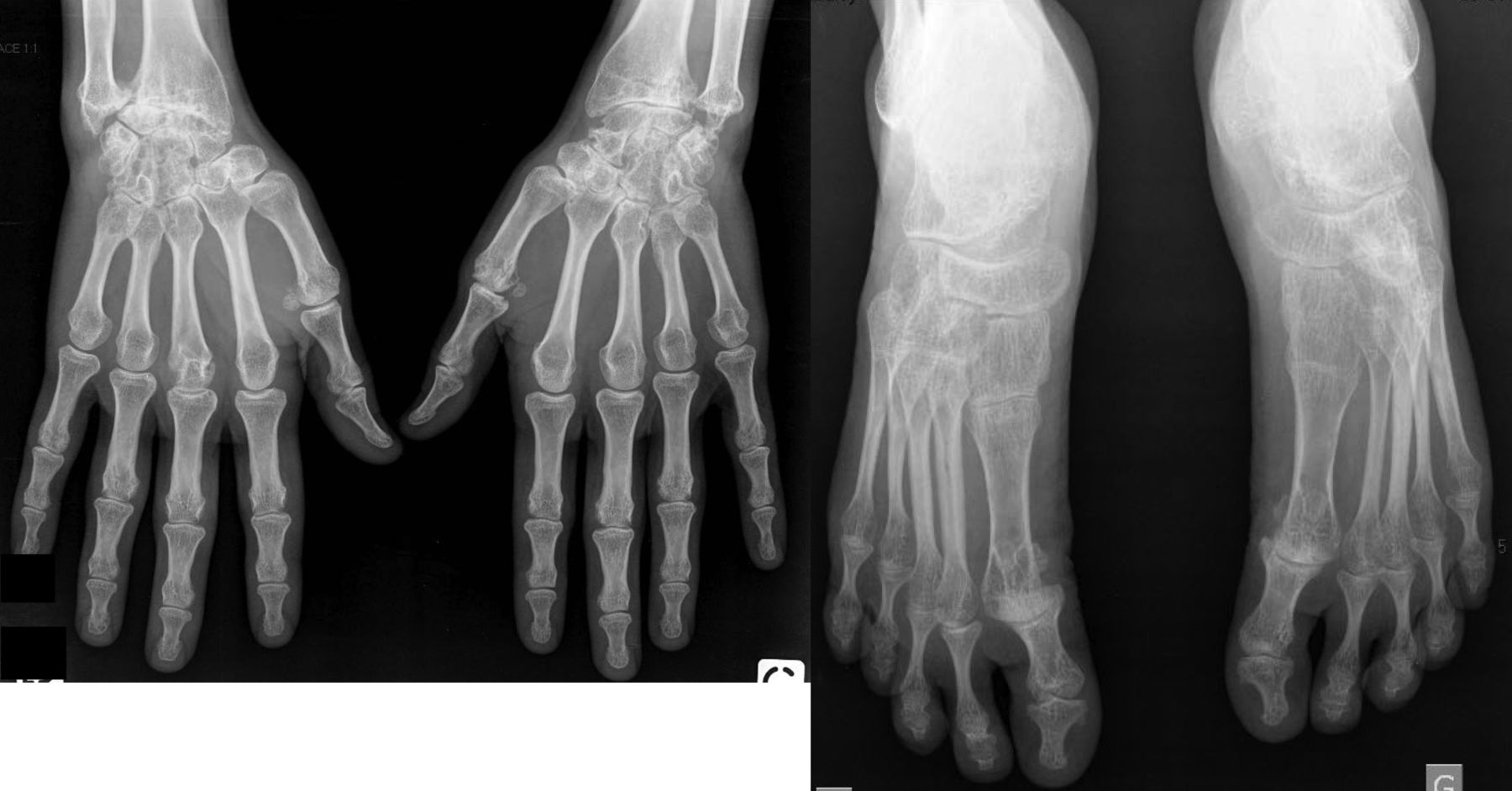
Thirty-six patients with pJIA (84%) had peripheral structural lesions. Radiographs showed hand and foot lesions in 34/43 (79%) and 32/43 (74%) pJIA, respectively. The joints most commonly involved in pJIA were wrists and MCP, in 74% and 77% of patients, respectively. Ninety-seven percent (29/30) of pJIA with hand lesions had carpal involvement, whereas 30/30 (100%) had MCP lesions. Examples of hand and foot radiographs are shown in Figure 1. Hip damage was detected in 14/40 (35%) of pJIA and was bilateral in 12/14 (86%) cases (Figure 2).

Representative structural lesions in hands and feet in polyarticular juvenile idiopathic arthritis (pJIA). A. Hands in a pJIA patient aged 22 years with Larsen score for hands = 22.5. This radiograph is characterized by predominant carpal lesions and involvement of MCP 1, 3, 4, and 5 and PIP 2, 3, 4, and 5. B. Feet in a pJIA patient aged 21 years with Larsen score for feet = 15.5 and with lesions in MTP 1, 2, 3, 4, and 5.
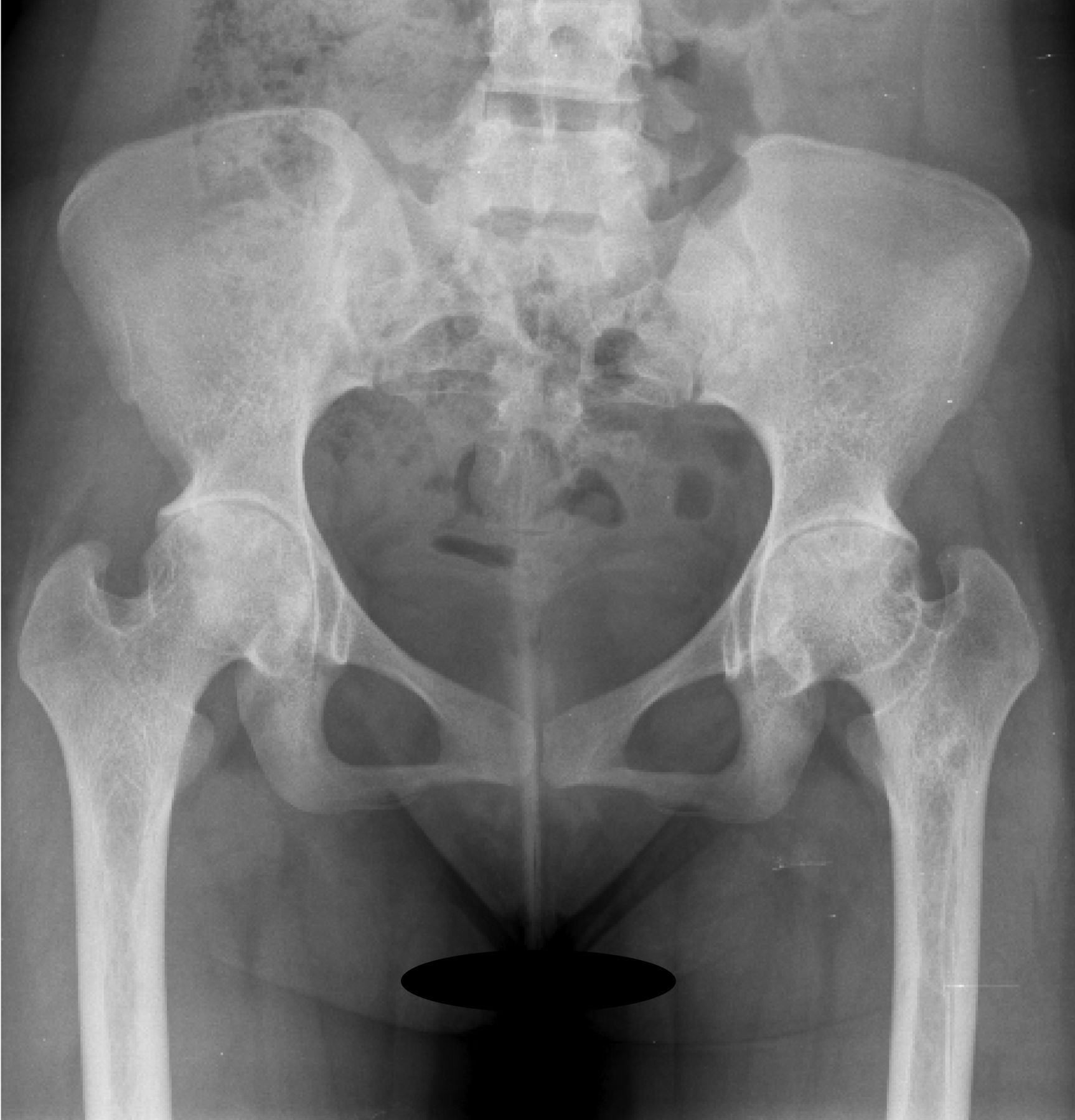
Representative structural lesions in hips in polyarticular juvenile idiopathic arthritis (pJIA). Radiograph shows bilateral hip damage in a pJIA patient of 21 years.
Only 1 patient had hip damage without other peripheral damage (and hip involvement was bilateral). Hip damage led to hip replacement in 5/14 (36%) patients with pJIA and it was bilateral in 3/5 (60%) cases. Other data concerning structural involvement in pJIA are reported in Table 2.
Radiographic lesions observed in pJIA patients and RA controls. Hands and feet were assessed by modified Larsen scoring method. The hand and feet scores range from 0 to 110 and from 0 to 50, respectively. Hips were assessed for presence of hip damage. Values are n (%) unless stated otherwise.
Wrist score correlated with the global Larsen score (rho = 0.8, p < 0.0001). Among the 10 pJIA patients without carpal involvement, only 3 had peripheral radiographic damage: 1 patient had hand (MCP) and foot involvement with a global Larsen score of 6.5, another had only foot involvement with a Larsen score of 8, and 1 patient had hip involvement with bilateral hip lesions.
Comparison with the RA control group
Fifty-six patients with RA (95%) had peripheral structural lesions, not significantly different from patients with pJIA (p = 0.07). Radiographs showed hand and foot lesions in 50/58 (86%) and 47/59 (80%) patients with RA (p = not significant for all comparisons with pJIA patients). Four pJIA and 8 RA patients had undergone previous hand surgery, including wrist arthrodesis (3 pJIA, 4 RA); tenotomy (1 pJIA); synovectomy (1 RA); Sauve-Kapandji procedure combined with synovectomy and tendon transfer (1 RA); wrist arthrodesis and distal ulnar resection (1 RA); and synovectomy, capsulotomy and correction of swan neck deformity (1 RA). Three patients with pJIA and 2 with RA had undergone foot surgery, including foot arthrodesis (1 pJIA, 1 RA), triple arthrodesis (1 pJIA), and hallux valgus surgery (1 pJIA, 1 RA), avoiding detailed radiographic analysis.
Mean global Larsen scores were 28.2 ± 32.5 in pJIA and 25.3 ± 23.9 in RA (p = nonsignificant). Mean hand and foot scores were 21.2 ± 23.9 and 9.6 ± 11.7 in pJIA compared to 18.5 ± 17.6 and 9.8 ± 11.3 in RA, respectively (p = nonsignificant). Hand damage involved wrists and MCP with no statistically significant differences in prevalence or severity, as compared to RA (Table 2). PIP involvement was found more frequently in patients with RA (74%) than in patients with pJIA (51%; p = 0.03; OR 0.37, 95% CI 0.15–0.90), especially in the third (70% vs 38%; p = 0.004; OR 0.27, 95% CI 0.11–0.65) and fourth PIP joints (64% vs 41%; p = 0.03; OR 0.39, 95% CI 0.17–0.93). However, in multivariate analysis, PIP involvement was independently associated with age (p = 0.001) and RF status (p = 0.02), but not with juvenile onset. For the other joints, distribution of the lesions was similar between the 2 groups. Hip damage was detected in 8/47 (17%) of patients with RA (p = nonsignificant compared to pJIA patients). It was more frequently bilateral in pJIA compared to RA patients (p < 0.01; OR 18, 95% CI 2.01–161.05). In multivariate analysis, after adjustment for age, sex, disease duration, RF and ACPA status, bilateral hip damage remained independently associated with juvenile onset (OR 8.80, 95% CI 1.71–45.24). In the RA control group, 3/8 (37%) patients with hip lesions had hip replacement and none was bilateral. Further details are provided in Table 2.
Associations between joint damage and disease characteristics
Demographic characteristics of RF-positive pJIA did not differ from RF-negative disease, except for a higher frequency of ACPA positivity in RF-positive patients. A comparison between RF-positive and RF-negative pJIA showed more frequent hand and foot lesions in RF-positive pJIA [21/23 (91%) vs 13/20 (65%), respectively; p < 0.05; OR 5.65, 95% CI 1.02–31.48; and 21/23 (91%) vs 11/20 (55%); p = 0.01; OR 8.59, 95% CI 1.57–46.89; Table 3]. Detailed analysis was performed in patients without previous hand or foot surgery. Carpal involvement tended to be more frequent in RF-positive polyarthritis (19/22, 86%) than in RF-negative polyarthritis (10/17, 59%; p = 0.06), with a significantly higher carpal score in RF-positive patients (6.5 ± 3.6 vs 3.3 ± 4.0; p = 0.01). Radiographic comparison between RF-positive and RF-negative patients also revealed a trend for less hip damage (4/20 vs 10/20) in the RF-positive subgroup (p = 0.05).
Clinical characteristics and damage observed in RF-positive and RF-negative patients with pJIA. Hands and feet were assessed by modified Larsen scoring method. Hand and foot scores range from 0 to 110 and 0 to 50, respectively. Values are n (%) unless stated otherwise.
The presence of radiographic damage in young adults with pJIA did not correlate with age, sex, disease duration, or RF and ACPA status.
DISCUSSION
The main results of our study are the following: (1) structural peripheral damage is frequent in patients with pJIA who are now adults (84% of our cohort); and (2) peripheral damage observed in young adults with pJIA was similar to that seen in RA except for higher risk of bilateral hip damage.
It is commonly believed that pJIA has a lesser destructive potential than adult RA10,11,12,13. One study found fewer radiographic changes in juvenile patients than in an RA control group matched for sex and disease duration13. However, it included a heterogeneous group of patients with 30% of oligoarticular forms13. Our study is the first to specifically compare damage in young adults with pJIA to that observed in an RA control group matched for sex and disease duration. Our results suggest that damage is as frequent and as severe in pJIA patients who are now adults, as in patients with RA. Only 1 study assessed prevalence of damage in 46 adults who had had juvenile RA (including 28 forms with polyarticular onset) and found results similar to ours (i.e., structural lesions in 78% of the patients and hand erosions in 68%)19.
Consistent with what is observed in pJIA, we found that the most common joints involved in pJIA persisting into adulthood were the wrists and the MCP joints22,24,25,28. The wrist joint has already been identified as the most vulnerable site of radiographic changes in JIA22,25,35,36 and has been suggested as the optimal site for assessing disease progression in pJIA2,15,37. Indeed, wrist disease was frequently associated with involvement of the small joints of the hands2,15. Further, the carpal score correlated closely with the global Larsen score. However, in 3 patients (7% of our cohort), assessment of only the wrist would have led to overlooking damage in other sites, particularly in 1 patient with bilateral hip damage who was symptomatic. Therefore our study suggests that radiographs of both hands and feet should be performed in young adults with pJIA to better assess and specifically to manage damage in each site. Consistently, a previous study demonstrated that radiographs of both hands and feet in RA yielded additional information compared with imaging only a part of these38. In cases with symptoms, structural evaluation should also include radiographs of hips.
Foot lesions were detected in 74% of our cohort. Foot damage was neither less frequent nor less severe compared to RA. Further, in the pJIA group, Larsen scores were similar for foot and hand involvement (data not shown), confirming that foot lesions must be considered along with those of the hands in young adults with pJIA.
Hip damage was found in 35% of our pJIA cohort and showed a trend to be more frequent in juvenile forms. Only 1 study assessed hip involvement in JIA persisting into adulthood, and it found a higher prevalence (41%)19; however, that study was performed 40 years ago, and included patients with a longer disease duration (18 years) and systemic JIA, which are characterized by a high prevalence of hip disease22,39,40,41. Hip involvement was bilateral in 86% of cases, which is in accord with previous studies in pJIA19,29,40,41. But we demonstrated, for the first time, that it is a feature specific to adults with pJIA with an 18-fold risk of developing bilateral hip damage as compared to RA patients. Hip damage led to surgery in 36% of our patients, similar to RA, and hip replacement was bilateral in 60% of cases, whereas it was unilateral in all patients with RA. Thus, this confirms that hip involvement is a severe feature of the disease, associated with a high risk of surgery24,39,40,41,42. Therefore, hip damage should be considered a warning signal, leading to particularly close followup observations and probably a more aggressive treatment plan for adults with pJIA with unilateral hip involvement, to detect progression to bilateral disease and avoid prosthetic surgery.
Surprisingly, despite advances in the management of JIA, the prevalence of hand, foot, and hip damage was not very different in our cohort from that described 25–40 years ago19,24,27. However, our patients were recruited from a tertiary referral center and were characterized by high frequency use of biological agents and surgery. This may result in a bias toward a more severe cohort with more destructive radiographic changes. The higher prevalence of RF-positive polyarthritis could also explain the severity of our patients' disease. There is accumulating evidence that the progression of radiographic damage in JIA occurs early in the course of illness. Prompt control of inflammation early during the disease course is therefore required to avoid progression of structural damage. In our study cohort, biological agents were introduced after a mean of 8 years of disease duration. Therefore, we can hypothesize that our patients were treated later and less aggressively than would be the case in 2012. This could explain the high frequency of structural damage we observed. Comparison of RF-positive and RF-negative disease confirmed that in young adults with pJIA, RF positivity is associated with a more destructive disease, especially in hands and feet12,17,22,23,30,43. Therefore these results confirm that RF-positive JIA should be considered the equivalent of adult RA from a radiographic viewpoint. However, adults with RF-negative pJIA were also characterized by severe structural damage, confirming that it is these severely destructive RF-negative forms that persist more frequently into adulthood.
Our study should be viewed in light of certain limitations: first, it was a cross-sectional study, preventing assessment of the natural history of structural involvement in pJIA. Further, because it was not a prospective study with inception cohorts, we could not determine whether joint damage occurred earlier in RA or pJIA. Second, we did not study lesions specific to juvenile forms, such as growth abnormalities, periostitis, and joint ankylosis10,12,14,20,26,41,44,45. In hips, periarticular osteopenia, cystic changes, and protrusio acetabula were not investigated. However, our main objective was to investigate destructive changes, i.e., erosions and/or joint space narrowing, in adults with pJIA compared to patients with RA. For this purpose, we used a score initially developed in adults with RA, which has been further validated in both diseases15,34,46,47,48, and which correlated closely to the Sharp score15. Moreover, ultrasonography and magnetic resonance imaging (MRI) were not carried out. Some data suggest that in patients with JIA these newer imaging techniques would be more sensitive than conventional radiographs for the detection of bone erosions49,50. However, to date, experience with these techniques in pediatric rheumatology remains very limited2. Therefore, standard radiography is currently the first-line imaging method to assess damage in JIA2,10,11. However, in case of hip involvement, hip MRI should also be performed to investigate persistent disease activity.
Our study also has strengths: (1) our cohort consisted of a homogeneous group of patients with pJIA persisting into adulthood; (2) the study was controlled with RA patients matched for sex and disease duration; (3) the prevalence of radiographic lesions in our patients with RA was concordant with current data13,48; and (4) there was good interexaminer reproducibility for radiograph assessment.
Our results suggest that, in young adults with pJIA, peripheral involvement is frequent (84%). Radiographic changes are similar in frequency and severity to those seen in patients with RA, except for higher risk of hip damage and particularly of bilateral hip damage. Patients with unilateral hip involvement should be carefully followed to detect progression to bilateral disease. Further large prospective studies are warranted to confirm our results, to determine predictive factors of structural severity at disease onset, and to thoroughly investigate the potential benefit of new therapeutic agents.
- Accepted for publication December 20, 2012.


