

To the Editor:
Systemic sclerosis (SSc) is characterized by diffuse fibrosis of the skin and internal organs, whereas localized scleroderma is defined by discrete areas of cutaneous fibrosis without systemic involvement. There are also reports of patients developing exophytic collagenous nodules, which are considered to be a rare variant of localized scleroderma. As in the plaques of localized scleroderma, these collagenous nodules can occur as an isolated skin disease or they can appear in the setting of SSc1,2. Histologically, these nodules can resemble scleroderma: thickened sclerotic collagen bundles without increased spindled fibroblasts intercalating between the collagen, and are therefore termed nodular scleroderma3. In contrast, there have also been reports of patients with scleroderma who develop nodules that histologically have the features of keloid: hypocellular zones of thick glassy hyalinized collagen bundles, which have been referred to as keloidal scleroderma4. We describe 2 cases of SSc, 1 diffuse (dcSSc) and 1 limited (lcSSc), with dramatic eruptive collagenous nodules appearing at different stages of the disease course.
Case 1
A 70-year-old woman presented in June 2003 because of development of Raynaud’s phenomenon and fibrotic skin thickening involving her hands that rapidly progressed to her face, chest, abdomen, and upper and lower extremities [modified Rodnan skin score (mRSS) was 33 (on a scale of 0–51) at her initial visit and peaked to 38 two months later]5. She was started on mycophenolate mofetil and pentoxifylline. By May 2004 the fibrotic areas of skin started to soften (mRSS 30); however, slightly pruritic skin-colored nodules started to appear on her neck and abdomen.
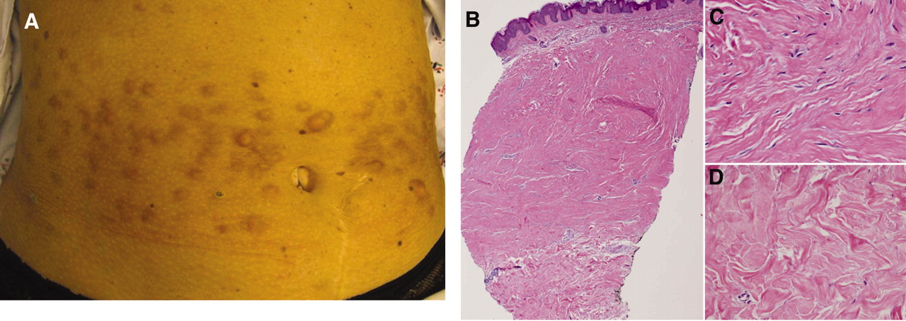
Examination in August 2004 revealed resolving diffuse fibrosis of the skin (mRSS 22) and numerous firm, fixed skin-colored dermal-based nodules ranging from 0.5 to > 1 cm diameter distributed symmetrically on her neck, shoulders, back, buttocks, proximal extremities, chest, and abdomen, with sparing of the face (Figure 1A, 1B). A punch biopsy of a characteristic nodule demonstrated increased sclerotic, thickened wavy collagen fibers with increased number of admixed stellate spindled fibroblasts that spared the papillary dermis but extended into the deep dermis with replacement of the subcutaneous fat (Figure 1C, 1D).

Case 1. A. Neck with numerous firm skin-colored nodules. B. Abdomen with numerous firm skin-colored nodules. C. Scanning view of biopsy from a neck lesion demonstrating increased collagen deposition with replacement of the dermis and portions of subcutaneous fat by sclerotic collagen bundles (H&E, original magnification 2×). D. High-power view of the neck biopsy showing sclerotic, thickened, wavy collagen fibers and increased number of spindled cells with wavy and stellate nuclei (H&E, original magnification 40×).
Over the next few years, her scleroderma skin changes were limited to her fingers (mRSS 5), and pentoxifylline and mycophenolate mofetil were discontinued. The number of collagenous nodules, however, remained unchanged. Surgical removal of several large nodules left hypopigmented (but not hypertrophic) scars.
Case 2
In 2004, a 45-year-old woman first noticed the development of nodules on her abdomen that progressed to involve her neck and upper chest (Figure 2A). She had no history of keloid formation or of trauma to those areas. A punch biopsy revealed increased wavy sclerotic collagen deposition with an increased number of admixed stellate spindled fibroblasts that spared the papillary dermis and extended into the deep dermis with replacement of the subcutaneous fat (Figure 2B, 2C). Beneath this nodule were thickened sclerotic collagen bundles that lacked an associated increased number of fibroblasts — this resembled the typical histologic changes of scleroderma (Figure 2D). She was empirically started on a 4-month trial of hydroxychloroquine, with no response.

Case 2. A. Abdomen with numerous firm skin-colored and hyperpigmented nodules. B. Scanning view of biopsy from the abdomen demonstrating nodular replacement of the reticular dermis by sclerotic collagen bundles associated with a sparse spindle cell population. The underlying dermis and superficial subcutis shows scleroderma-like thickened sclerotic collagen, nearly devoid of spindled cells (H&E, original magnification 2×). C. High-power view of dermal nodular area with spindled cells with dendritic and stellate nuclei interspersed between sclerotic collagen bundles (H&E, original magnification 40×). D. High-power view of lower dermis showing scleroderma-like sclerotic thickened collagen without the spindled cell population (H&E, original magnification 40×).
On presentation to our institution in March 2007, examination revealed telangiectasias involving the cheeks, mucous membranes and palms, abnormal nailfold capillary loops, and sclerodactyly (mRSS of 4) consistent with lcSSc. There were multiple firm, fixed hyperpigmented dermal-based nodules up to 1 cm in diameter distributed symmetrically over her abdomen, neck, upper back, and chest, with sparing of the face and lower extremities.
At last followup, her scleroderma skin changes remained limited to her fingers; however, the number of nodules has continued to increase; she has not undertaken immunosuppressive therapy.
The 2 patients described above developed disseminated collagenous nodules in the setting of SSc. The lesions bore a clinical resemblance to previously reported cases of keloidal or nodular scleroderma in that they were firm, fixed, exophytic nodules. Histologically, our 2 cases are unique in that they both lacked hyalinized collagen alteration, and they showed a pattern of collagen bundles with increased fibroblasts — a pattern typical of neither keloid nor scleroderma. The temporal relationship between the development of lesions and scleroderma was strikingly different in the 2 cases: in Case 1, the diffuse eruption first developed as the patient was recovering from active dcSSc skin changes, whereas in Case 2, the nodular lesions developed around the same time as the onset of her SSc. These cases suggest that a spectrum of acquired collagenous lesions may arise in patients with scleroderma.
Nodular or keloidal scleroderma has been reported as a variant of localized scleroderma that can occur alone, variably referred to as nodular or keloidal morphea6, or in patients with SSc2. Clinically, the skin lesions are characteristically firm, nontender papules that can occur in isolated7, generalized8, and linear3 distributions with a predilection for the trunk9. Most lesions have been reported to develop within the first few months of the onset of SSc symptoms, during a time of clinically active disease8,10.
These cases of dramatic nodular eruption may suggest a unique variant of fibrotic nodules among some patients with scleroderma. Perhaps insight into the profibrotic cytokines that contribute to aberrant fibroblast activity may reveal a common pathogenic thread in the development of SSc, lcSSc, and the acquired collagenous nodules of nodular/keloidal scleroderma11.


