
The anatomy, physiology, and pathophysiology of the retrocalcaneal bursa (RB) have been described well1,2. Ultrasound has been shown to be a valuable tool by enhancing the accuracy of challenging procedures that in the past have been performed blindly, such as aspiration or injection of the RB3,4. We describe ultrasound-assisted corticosteroid injection of this structure.
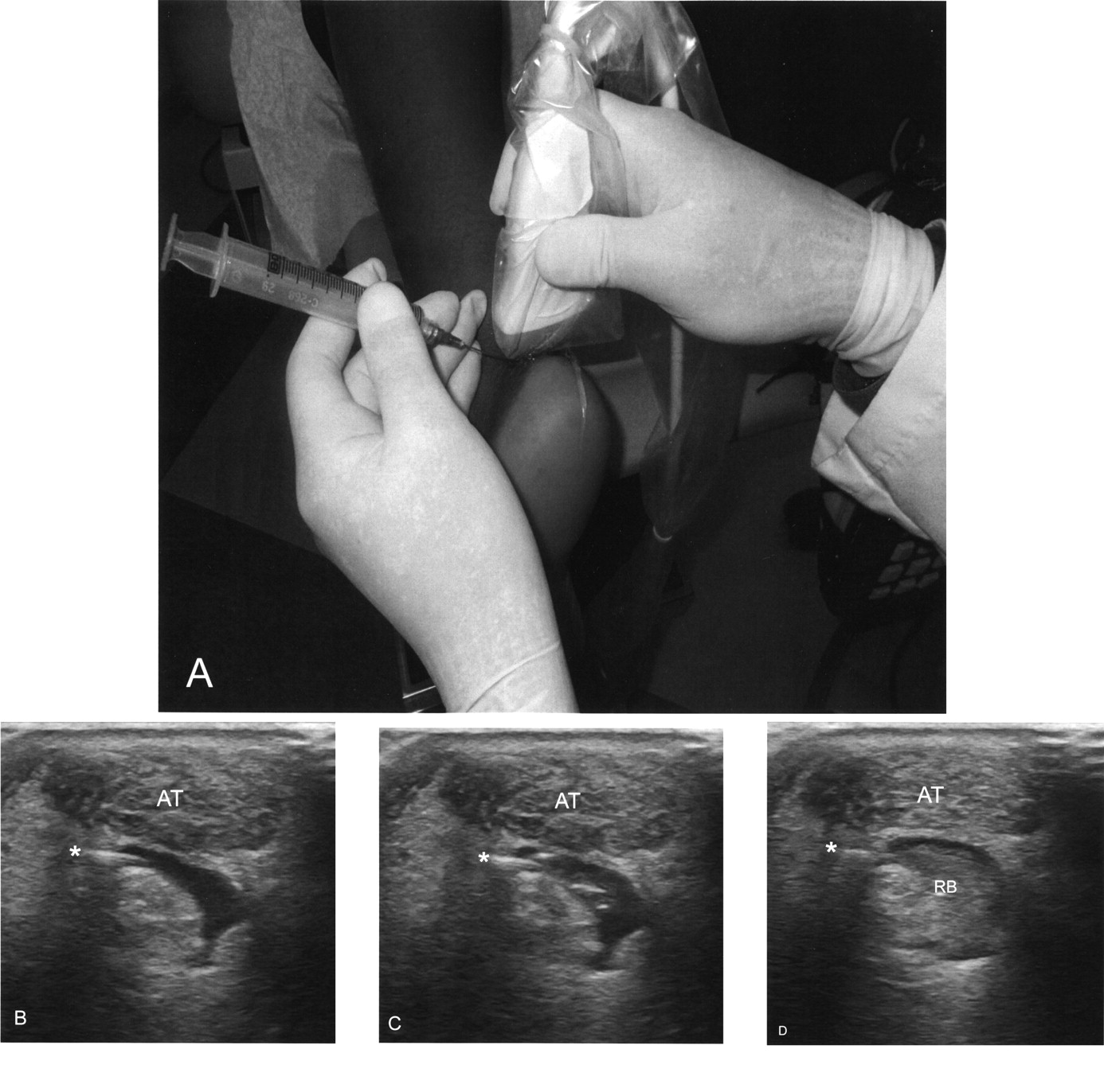
A 55-year-old woman with osteoarthritis presented with left heel pain. On examination she had swelling and tenderness in the left retrocalcaneal area. The ankle joint was unremarkable. A longitudinal sonographic view of the sub-Achilles area (Figure 1A) showed a triangular anechoic area wedged between the Achilles tendon and the calcaneus consistent with retrocalcaneal bursitis. A hyperechoic band (Figure 1A, arrowheads) showed a calcaneal spur. Transverse views (Figures 1B and 1C) illustrate RB at 2 levels (indicated as 1 and 2 of longitudinal view, Figure 1A). A sonographic-assisted injection of 10 mg triamcinolone acetonide was performed in the RB using a lateral approach (Figure 2A). Figures 2B, 2C, and 2D show needle placement (indicated as *) and medication passage into the bursa. No complications of the procedure ensued and the patient’s bursitis had resolved by the time she returned for a 2-month interval visit.

Longitudinal sonographic view (A) of the sub-Achilles area showing a triangular anechoic area between the Achilles tendon (AT) and the calcaneus (Cal), consistent with retrocalcaneal bursitis. Arrowheads indicate a calcaneal spur. B. Transverse view shows retrocalcaneal bursa (RB) at level 1 of view A. C. Transverse view shows RB at level 2 of view A.

A. Injection of 10 mg triamcinolone acetonide into the retrocalcaneal bursa was sonograph-assisted using a lateral approach. B, C, D. Needle placement (*) and medication passage into the bursa.
Ultrasound guidance enhances the accuracy of injection of the retrocalcaneal bursa. The Achilles tendon and retrocalcaneal bursa are easily distinguished sonographically and the needle can be guided into the bursa under direct visualization while avoiding the tendon.
REFERENCES
- 1.
- 2.
- 3.
- 4.