

To the Editor:
Chronic hepatitis C virus infection (HCV) is often associated with extrahepatic manifestations; the majority are immunologically mediated. It has been hypothesized that HCV, which is both a hepato- and a lymphotropic virus, might exert a chronic stimulating effect on the immune system, resulting in B-lymphocyte proliferation1,2. The well established immunologic complications of HCV infection are mixed cryoglobulinemia, membranoproliferative glomerulonephritis, leukocytoclastic vasculitis, porphyria cutanea tarda, sialadenitis, and autoantibody production such as rheumatoid factor (RF). Other possible associations have been reported, like B-cell non-Hodgkin’s lymphomas, polyarteritis nodosa, autoimmune cytopenia, and autoimmune thyroiditis3. We describe a patient with chronic HCV infection who developed acute auricular chondritis (Figure 1A) with scleritis (Figure 1B).
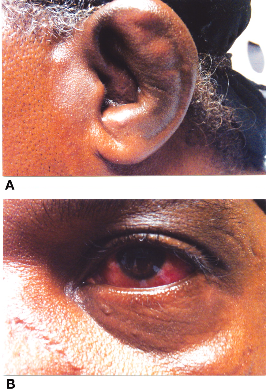
Our patient with chronic HCV infection developed acute auricular chondritis (A) with scleritis (B).
A 56-year-old African American man presented with a 2-week history of increasing pain and swelling of both ears. He also had bilateral pain and redness of eyes associated with photophobia. He denied any sick contacts, trauma, fever, chills, weight changes, difficulty breathing, hoarseness, oral or genital ulcers, joint pain, genital discharge, or rash. He was afebrile with diffuse swelling and erythema of both ears extending into the ear canal but sparing the noncartilagenous part of the ear. Eye examination revealed scleritis with normal visual acuity. C-reactive protein was 3.2 mg/dl and erythrocyte sedimentation rate was 88 mm/h. RF was positive at 23.5 IU/ml; serum protein electrophoresis revealed polyclonal gammopathy with decreased albumin, and IgG3 was elevated at 167 mg/dl. Antibodies to HCV antigen were positive with a viral load of 1,340,000 IU/ml. Antineutrophil cytoplasmic antibody (ANCA) testing was positive by indirect immunofluorescence with a cytoplasmic (C) staining pattern. Testing by enzyme immunoassay demonstrated no antibodies for proteinase 3. The HIV test was negative; anti-collagen type 2 antibodies and cryoglobulins were not detected. Complements were normal. He was felt to have auricular chondritis (Figure 1) and scleritis (Figure 2), presumed to be associated with HCV infection. Treatment was initiated with prednisone 60 mg/day, with good response. Prednisone dose was gradually reduced over several months. He was then referred to the liver clinic for treatment of HCV infection, but declined treatment with pegylated alpha-interferon and ribavirin. He had a recurrence of his symptoms when he discontinued prednisone and has needed moderate doses of prednisone to control the symptoms.
Relapsing polychondritis is a rare disease of unknown etiology and pathogenesis4. The pathogenesis is considered to be autoimmune, based on increased frequency of human leukocyte antigen (HLA)-DR4 in these patients, and detection of antibodies to type II collagen. Histopathology of the involved cartilage reveals inflammation of perichondrium with neutrophils, eosinophils, lymphocytes, and plasma cells. Immunofluorescence studies may show presence of immunoglobulin and C3 complement deposits along the chondrofibrous junction.
We have no direct evidence that the patient’s HCV infection and polychondritis are related, but we postulate that HCV infection may be triggering the polychondritis. Some chronically HCV-infected patients have extrahepatic manifestations including cryoglobulinemia and autoantibody production, such as rheumatoid factors, anti-thyroid antibodies, and other non-organ-specific antibodies. The mechanism leading to the production of these antibodies, although not fully understood, appears to be nonspecific activation of the immune system triggered by the viral infection5. One such mechanism is molecular mimicry between HCV polyprotein and self-antigens, which in turn primes the immune system, leading in time to ever-expanding self-reactivity6.
Chronic HCV infection results in sustained B-cell stimulation. Various mechanisms have been postulated for chronic stimulation of B-cells by HCV. Elevated serum B-lymphocyte activating factor (BAFF) has been found in chronic HCV-infected subjects compared to healthy controls7. HCV-infected subjects with clinical and laboratory features of autoimmunity were found to have the highest levels of BAFF8. It has been postulated that HCV may stimulate B-cells through 3 different pathways: the B-cell receptor recognizing the viral antigens, the CD81 molecule on the surface of B-cells binding to the HCV envelope 2 protein and CD19-CD21 complex engaging the C3d opsonized viral particles, all of them acting in concert to lower the B-cell activation threshold9. The B-lymphocyte stimulator (BlyS) belongs to the human tumor necrosis factor superfamily of cytokines, induces upregulation of CD21 on B-cells, and selectively expands the population of terminally differentiated plasma cells, leading to increased immunoglobulin production. HCV is also implicated in upregulation of BlyS10,11.
That our patient has positive assay for RF and hypergammaglobulinemia with elevated IgG3 suggests that immune activation could have been triggered by HCV, leading in turn to inflammation in the cartilage. The exact pathogenetic mechanism causing polychondritis remains to be determined. We describe an association and cannot prove causality. There is a report describing this association in a patient with mixed cryoglobulinemia12.


