

Tophaceous uric acid deposits are an important differential diagnosis when evaluating benign soft tissue masses, even in the absence of a history of inflammatory arthritis1,2.
A 63-year-old man presented with a slow-growing tumor on the plantar aspect of the right forefoot (Figure 1). The mass was firm, painless, and sessile. There was no history of trauma or previous arthritis. Computed tomography (CT) of the right foot was performed on a dual-source CT scanner at 140 kVp and 80 kVp tube potential. Postprocessing spectral analysis for radiograph attenuation characteristic of uric acid was performed, identifying the tumor as a large tophaceous uric acid deposit3,4 (Figure 2). Uric acid is also seen in several areas of the entire right foot including the plantar fascia, Achilles tendon, anterior tibial tendon, and several joint areas. Aspiration of the first metatarsophalangeal joint confirmed the presence of negatively birefringent crystals.
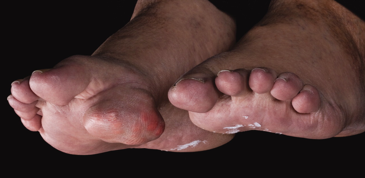
Slow-growing tumor on the right forefoot.

Spectral analysis for radiographic attenuation characteristic of uric acid identified the tumor as a tophaceous uric acid deposit (green areas).


