

Tophaceous gout is a chronic form of gout, characterized by uric acid crystal deposits (tophi) at the articular or periarticular soft tissues. It can lead to a severe deforming arthritis1. Computed tomography (CT) has proven to be useful to identify and localize tophi, to assess tophus volume and the presence of bone erosions, and to distinguish tophaceous deposits from other subcutaneous nodules (e.g., rheumatoid nodules)2–6.
A 62-year-old man with chronic gout, diabetes, and hypertension came to our outpatient department with a 2-year history of recurrent episodes of painful swelling of both wrists and left foot. The clinical examination revealed the presence of multiple nodules on both hands, wrists, the posterior region of the ankle and right elbow, the lateral region of the left ankle, and the great toe of the left foot. Laboratory investigation documented an increased erythrocyte sedimentation rate (75 mm/h), blood cell count (10.4 × 10³/ml), and serum uric acid levels (10.9 mg/dl). A multidetector CT scan with 3-dimensional volume-rendered reconstruction of both hands and wrists was performed. CT images showed multiple tophi on both hands and wrists and, in particular, an extensive tophaceous deposit at the volar side of the left hand (Figure 1).

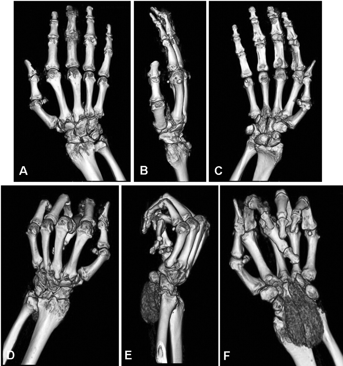
Three-dimensional volume-rendered computed tomography (CT) images of right (A–C) and left (D–F) hands and wrists. Bilateral multiple tophi are visible at the metacarpophalangeal, proximal and distal interphalangeal, radiocarpal, and intercarpal joints; an extensive tophaceous deposit can be observed at the volar side of the left wrist. The attitude of the left hand in flexion is caused by the proximity of the tophi to the flexor tendons of the fingers. CT images obtained using a LightSpeed VCT 64-slice scanner (GE Medical Systems, Milwaukee, WI, USA).
Our report provides visual evidence that multidetector CT allows a detailed morphostructural assessment of involved tissues in patients with tophaceous gout.


