

Abstract
Objective. To study noncalcified coronary plaque (NCP) in systemic lupus erythematosus (SLE).
Methods. Sixty-four-slice coronary multidetector computed tomography (MDCT) was performed in 39 consecutive patients with SLE. MDCT scans were evaluated semiquantitatively by a radiologist using dedicated software. The presence or absence of NCP in each coronary artery was assessed. Patients with mixed plaque (calcified and noncalcified portions) were included in the NCP group.
Results. The patient group was 90% women, 64% Caucasian, 31% African American, 5% other; mean age 50.5 ± 9.6 years. Fifty-four percent (21/39) had NCP. Seventy-six percent (16/21) of those with NCP also had coronary calcium (range 0.7 to 1264.1 Agatston units). In univariate analysis, NCP was associated with age (p = 0.01), current nonsteroidal antiinflammatory drug (NSAID) use (p = 0.04), hormone replacement therapy (p = 0.02), current use of immunosuppressive drugs (p = 0.02), current low serum C3 level (p = 0.07), current physician’s global assessment of activity (PGA; p = 0.05), and low-density lipoprotein cholesterol (p = 0.04). NCP was not associated with other risk factors for atherosclerosis, including total serum cholesterol, high sensitivity C-reactive protein, and lipoprotein(a).
Conclusion. Unlike coronary calcium, which is not associated with SLE activity measures or with active serologies, NCP is more common in patients with SLE with current, 3-, and 6-month activity by PGA. NCP was also associated with the need for current NSAID or immunosuppressive therapy. NCP is an important part of the total atherosclerotic burden in SLE.
Atherosclerosis is a major cause of morbidity and mortality in systemic lupus erythematosus (SLE)1,2. Atherosclerosis in SLE is multifactorial, with immune-mediated damage, traditional cardiovascular risk factors, and prothrombotic factors all playing important roles3–5. Women with SLE aged 35 to 44 years are 50 times more likely to have a myocardial infarction than Framingham controls6. Traditional cardiovascular risk factors such as hypertension, hyperlipidemia, obesity, and smoking are common in SLE, and contribute to the cardiovascular risk7. However, traditional cardiovascular risk factors do not account for the entire risk8.
Coronary calcium is closely associated with atherosclerotic plaque and serves as a surrogate measure of coronary atherosclerosis. A high correlation has been found between coronary calcification and total atherosclerotic burden, based on histopathological findings9. Coronary calcification scores are predictive of future cardiovascular events10–14. In the general population, age and sex are associated with coronary calcium scores15.
A mature atherosclerotic plaque has 2 components: a lipid/macrophage-rich atheromatous material, with overlying fibrous tissue (“fibrous cap”). The atheromatous component can destabilize the plaque, making it vulnerable to rupture. Plaque instability is closely related to the degree of inflammation16. The risk of an acute cardiac event is affected by the plaque composition17. A calcified coronary plaque is known to represent a late stage of atherosclerosis and to be a more stable plaque, and therefore unlikely to be the best measure of the risk of a clinical event. Until recently, only calcified coronary atherosclerosis was readily measured noninvasively, using noninvasive computed tomography (CT).
Noncalcified plaque contributes to the true atherosclerotic burden. In a study by Leber and colleagues18, more noncalcified plaque was found in patients with acute coronary syndrome compared to patients with stable angina pectoris. Ten percent of patients with an acute myocardial infarction had no coronary calcium, but did have more than 2 noncalcified plaques18.
By administering an intravenous contrast agent, noncalcified plaques can now be detected by helical CT. Multislice CT can evaluate both the coronary lumen and calcified/non-calcified coronary plaques19. It can detect plaques without significant luminal stenosis. These coronary plaques can then be identified as calcified, noncalcified, or mixed20–22.
We measured the prevalence of noncalcified plaque in SLE and determined the association with SLE activity measures, traditional cardiovascular risk factors, and other risk factors for atherosclerosis.
MATERIALS AND METHODS
Thirty-nine patients with SLE were enrolled. The study was approved by the Johns Hopkins University School of Medicine Institutional Review Board. All patients gave informed consent. Patients with a creatinine level ≥ 1.2 mg/dl were excluded. Pregnant patients and those allergic to contrast were also excluded.
As part of the Hopkins Lupus Cohort Study, all patients had been seen quarterly since cohort entry for assessment of disease activity [by the physician’s global assessment (PGA), on a 0 to 3 visual analog scale, and the Safety of Estrogens in Lupus Erythematosus National Assessment (SELENA) – Systemic Lupus Erythematosus Disease Activity Index]23,24, laboratory tests (complete blood count, erythrocyte sedimentation rate, serum creatinine, cholesterol, urinalysis, urine protein/creatinine ratio, serum C3 level, serum complement C4 level, and anti-double-stranded DNA), and cardiovascular risk factors, including fasting lipid profile, homocysteine, lipoprotein(a) and fibrinogen.
Image acquisition and evaluation
Coronary calcification was assessed by helical CT with a Siemens Volume Zoom Scanner (Siemens Medical Solutions, Malvern, PA, USA) using the Siemens calcium scoring software as described25,26. Coronary calcification was quantified using a standard Agatston scoring system27.
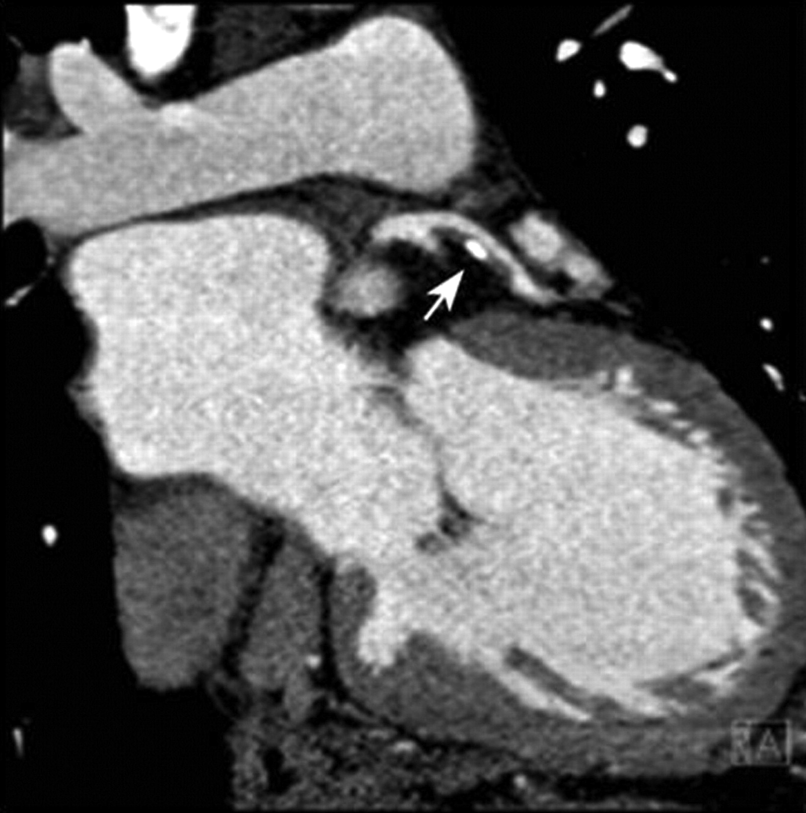
Noncalcified plaque was assessed using 64-slice coronary multidetector CT (MDCT) and evaluated semiquantitatively by a radiologist, who was blinded to the blood test results, using dedicated software (Circulation, Siemens Medical Solutions). For MDCT the standard coronary CT protocol was used, with the administration of intravenous Visipaque 320 at an injection rate of 5 cc/s for 100 cc and an immediate 50 cc saline chaser bolus at the same injection rate28. The presence or absence of noncalcified plaque in each coronary artery was assessed. Included in the noncalcified plaque group (Figure 1) were patients with mixed plaque (Figure 2; calcified and noncalcified portions).

Multidetector computed tomography scan. Arrow indicates noncalcified plaque in a coronary artery.

In this multidetector computed tomography scan, the arrow indicates mixed (calcified and noncalcified) plaque.
Good to moderate reproducibility has been shown in recent studies. Hoffmann and others have shown good to moderate intra- and interobserver variability, not only in detection of coronary artery plaques but also in plaque volumes29. Leber, et al showed an interobserver agreement of 79%, 88%, and 70%, while Ferencik showed 93%, 98%, and 92% for the detection of any plaque, calcified plaque, and noncalcified plaque, respectively30,31.
Statistical analysis
All results for continuous variables were expressed as means ± SD, unless specified otherwise. Continuous variables were analyzed with a 2-sided t test. Categorical variables were compared by the Fisher’s exact test. To assess associations controlling for age, we fit multiple linear or logistic regression models. Statistical analysis was performed using JMP (version 5.0.1, SAS Institute, Cary, NC, USA). A p value ≤ 0.05 was taken as statistically significant.
RESULTS
Data were obtained on 39 subjects with SLE (90% women). The patient group was 64% Caucasian, 31% African American, and 5% other ethnicity. The mean age was 50.5 ± 9.6 years. Cumulative SLE clinical manifestations included malar rash (63%), discoid rash (15%), photosensitivity (60%), oral ulcers (65%), arthritis (80%), serositis (55%), renal disorder (43%), neurological disorder (10%), immunologic disorder (80%), leukopenia (69%), thrombocytopenia (28%), hemolytic anemia (3%), and antinuclear antibody positivity (95%).
Fifty-four percent (21/39) had noncalcified plaque. Seventy-six percent (16/21) of those with noncalcified plaque also had coronary calcium (range 0.7 to 1264.1 Agatston units). Only 1 patient without noncalcified plaque had coronary calcium.
The association of noncalcified plaque with SLE measures and treatment is shown in Table 1. Noncalcified plaque was significantly associated with age (p = 0.01), current nonsteroidal antiinflammatory drug (NSAID) use (p = 0.04), ever use of hormone replacement therapy (p = 0.02), and current immunosuppressive drug use (p = 0.02; Table 1).
Association of noncalcified coronary plaque (NCP) with demographics, SLE variables, inflammatory markers, and treatment.
Table 2 shows the association of noncalcified coronary plaque (NCP) with cardiovascular risk factors. Only low-density lipoprotein (LDL) cholesterol had an association with NCP (p = 0.04, unadjusted). Table 3 shows the association of NCP with other surrogate measures of atherosclerosis. Noncalcified plaque was associated with carotid plaque (p = 0.02, unadjusted) but not after age adjustment (p = 0.23). Age is strongly related to both NCP and carotid plaque, so the observed association between NCP and carotid plaque is due to confounding by age. NCP was also not associated with carotid intima-media thickness (p = 0.35).
Association of noncalcified coronary plaque (NCP) with other cardiovascular risk factors.
Association of noncalcified coronary plaque (NCP) by other measures of atherosclerosis.
Table 4 shows measures of disease activity and their relationship with NCP. We measured these variables at 4 different timepoints: current, last 3 months, last 6 months, and over the previous year. NCP was associated with both current measures of disease activity (PGA of disease activity; p = 0.05), and current treatment (NSAID and immunosuppressive drug use).
Mean (SD) levels of variables and SLE disease activity among those with and without noncalcified coronary plaque, in various periods prior to the measurement of plaque.
In an analysis of 5 patients with NCP alone versus 16 with both NCP and calcified plaque, the serum C4 level was significantly lower (15.5 ± 4.1 vs 21.8 ± 6.5 mg/dl; p = 0.026). C4 was also lower in those with NCP alone than in those with no plaque at all (20.2 ± 7.4). This again supports the hypothesis that disease activity — clinically and/or serologically — is associated with NCP.
DISCUSSION
NCP is very common in SLE. Because only 76% of patients with SLE and NCP also had coronary calcium, our study suggests that the best surrogate measure of atherosclerotic burden in SLE will be a measure of combined plaque (both noncalcified and calcified plaque). For the first time, our study found a surrogate measure of atherosclerosis — noncalcified plaque — that is more common in patients with SLE with measures of currently active lupus, including the PGA of disease activity and current treatment with NSAID or immunosuppressive drugs. However, one of the limitations of our study was the small sample size, and the marginal p values of some of these associations.
Systemic inflammatory processes influence plaque instability, as shown by studies of high-sensitivity C-reactive protein (hs-CRP) and amyloid proteins32–34. Hs-CRP has been shown to predict cardiovascular events in healthy individuals and in those with coronary artery disease35–39. Hs-CRP and its association with coronary calcium in SLE, however, is not clear. Hs-CRP in SLE has been associated with coronary calcium in 1 study40, but we found no association41. In our study, hs-CRP was not associated with noncalcified plaque. In SLE, hs-CRP does not appear to identify patients with SLE who are at risk for atherosclerosis.
NSAID use has been associated with an increased cardiovascular risk42,43. Studies have shown that 73% to 77% of patients with SLE take these agents regularly44,45. Thrombotic complications with NSAID have been observed with connective tissue disorders, including SLE46. In our study, NSAID use was associated with NCP. This could represent a toxicity of NSAID use, or could represent the association of active SLE (for which the NSAID was prescribed) and noncalcified plaque.
The Women’s Health Initiative study found an increased risk of cardiovascular disease, stroke, and venous thromboembolism in patients undergoing hormone therapy47. The SELENA study found an increased risk of total flares (but not severe flares) with hormone replacement therapy in women with SLE48. Our study found an association of NCP with hormone replacement therapy, suggesting further concern about its use in SLE.
In contradistinction to the general population, hyperlipidemia has not been found to be one of the major cardiovascular risk factors in SLE49. In our study, only LDL cholesterol showed an association with noncalcified plaque. In terms of novel risk factors, neither homocysteine nor lipoprotein(a) were associated with noncalcified plaque. However, homocysteine and lipoprotein(a) are both risk factors for stroke in SLE50,51.
The role of complement in atherosclerosis was first highlighted by Geertinger and Sorensen52. In SLE, high (not low) levels of C3 are associated with coronary artery calcification and aortic stiffness53–56. Explanations for the association of high (rather than low) levels of C3 in patients with SLE atherosclerosis include complement as an acute-phase reactant and vascular inflammation leading to an increase in complement synthesis without activation55. In the general population, high C3 levels are correlated with postprandial lipemia and waist-hip circumference56. We have previously shown that high levels of C3 are associated with carotid plaque in SLE57. In our study, low C3 was associated with NCP, although it did not quite reach statistical significance. C4 was significantly lower in those with NCP alone. This supports the hypothesis that current SLE disease activity (clinical and/or serologic) is connected to noncalcified plaque.
This first study of noncalcified plaque in SLE shows that NCP is extremely common in SLE and contributes to overall atherosclerotic burden. It may be especially relevant in that it is more common in those with current disease activity, NSAID use, and immunosuppressive use, identifying ways in which active SLE contributes to accelerated atherosclerosis. However, the measurement of noncalcified plaque is currently available only as a research modality and is not ready or appropriate for routine clinical care.
Footnotes
-
The Hopkins Lupus Cohort is supported by a grant from the National Institutes of Health (NIH AR 43727). Also supported by grant UL1 RR 025005 from the National Center for Research Resources, a component of the NIH, and the NIH Roadmap for Medical Research.
- Accepted for publication October 22, 2009.


