

Systemic sclerosis can give rise to many findings in the abdominal radiograph. Detection of lung base changes together with “sheet-like” soft tissue calcification and large bowel ileus are important clues to a unifying diagnosis of systemic sclerosis.
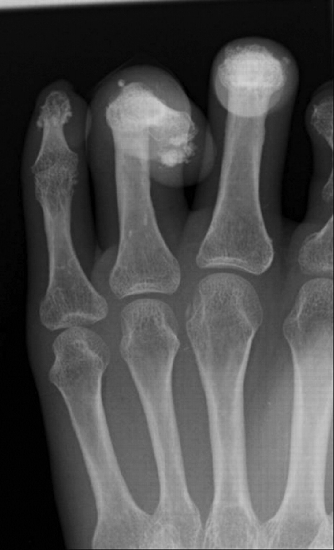
A 51-year-old woman presented with a history of several months’ duration of abdominal distension and constipation. The abdominal radiograph (Figure 1) showed evidence of large-bowel dilatation of the transverse colon, with normal mucosal features and fecal loading in the sigmoid and rectum. Additionally, there was evidence of florid soft-tissue calcification involving bilateral iliacus muscles. The lung bases showed reticular shadowing with pleural thickening, consistent with pulmonary fibrosis. The constellation of signs — muscle calcifications1, pulmonary fibrosis2, and nontoxic megacolon3 — indicates systemic sclerosis, with bowel atony accounting for the patient’s presentation. The hand radiograph (Figure 2) shows the classic appearance of systemic sclerosis — distal phalanx resorption with soft-tissue calcinosis.

The abdominal radiograph shows evidence of dilatation of the transverse colon, with normal mucosal features (arrowheads) and fecal loading.

Hand radiograph shows the classic appearance of systemic sclerosis — distal phalanx resorption with soft-tissue calcinosis.


